. 2013 Mar 19;110(12):4563-8.
doi: 10.1073/pnas.1221602110. Epub 2013 Mar 4.

Heparin-binding domain of fibrin(ogen) binds growth factors and promotes tissue repair when incorporated within a synthetic matrix

Affiliations

Heparin-binding domain of fibrin(ogen) binds growth factors and promotes tissue repair when incorporated within a synthetic matrix

Mikaël M Martino et al. Proc Natl Acad Sci U S A. .

Abstract

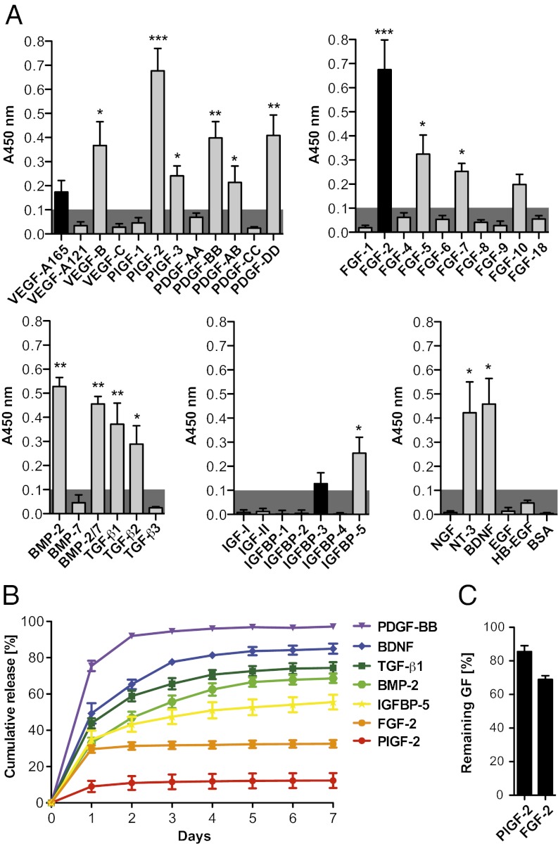
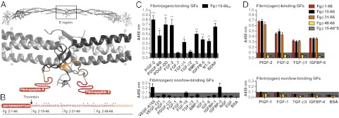
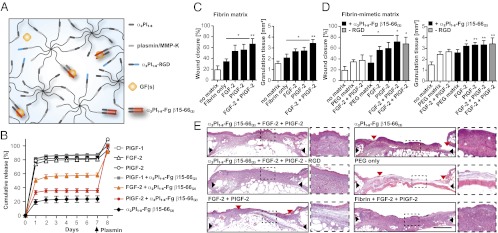
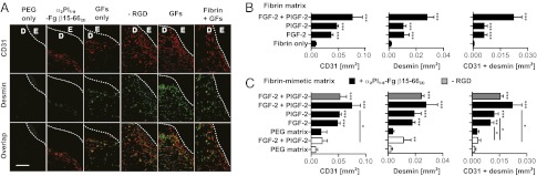
By binding growth factors (GFs), the ECM tightly regulates their activity. We recently reported that the heparin-binding domain II of fibronectin acts as a promiscuous high-affinity GF-binding domain. Here we hypothesized that fibrin, the provisional ECM during tissue repair, also could be highly promiscuous in its GF-binding capacity. Using multiple affinity-based assays, we found that fibrin(ogen) and its heparin-binding domain bind several GFs from the PDGF/VEGF and FGF families and some GFs from the TGF-β and neurotrophin families. Overall, we identified 15 unique binding interactions. The GF binding ability of fibrinogen caused prolonged retention of many of the identified GFs within fibrin. Thus, based on the promiscuous and high-affinity interactions in fibrin, GF binding may be one of fibrin's main physiological functions, and these interactions may potentially play an important and ubiquitous role during tissue repair. To prove this role in a gain-of-function model, we incorporated the heparin-binding domain of fibrin into a synthetic fibrin-mimetic matrix. In vivo, the multifunctional synthetic matrix could fully mimic the effect of fibrin in a diabetic mouse model of impaired wound healing, demonstrating the benefits of generating a hybrid biomaterial consisting of a synthetic polymeric scaffold and recombinant bioactive ECM domains. The reproduction of GF-ECM interactions with a fibrin-mimetic matrix could be clinically useful, and has the significant benefit of a more straightforward regulatory path associated with chemical synthesis rather than human sourcing.


Conflict of interest statement

The authors declare no conflict of interest.

Figures

Fig. 1.
Fig. 1.
GF binding to fibrin(ogen). (A) ELISA plates were coated with GFs or BSA and further incubated with fibrinogen. Bound fibrinogen was detected using an antibody (n ≥ 4; mean ± SEM). A signal significantly greater than 0.1 (gray box) was considered representative of a specific binding. *P < 0.05; **P < 0.01; ***P < 0.001, one-sample Student t test. Binding was highly promiscuous; unique interactions are shown in gray, and previously known interactions are shown in black. (B and C) GF retention in fibrin matrix. Fibrin matrices were produced in the presence of a GF and further incubated in eight volumes of physiological buffer for 7 d. The buffer was changed each day, and released GFs were quantified for each day. (B) Graph showing the cumulative release of GFs over 7 d (n ≥ 3; mean ± SEM). (C) After 7 d, fractions of FGF-2 and PlGF-2 remaining in the matrices were quantified after the gels were digested by plasmin (n = 3; mean ± SEM).
Fig. 2.
Fig. 2.
GF binding to the heparin-binding domain of fibrin(ogen). (A) Ribbon diagram representation of human fibrinogen and its central region E (generated using Swiss-PdbViewer). The coiled-coil domains are in gray; the N terminus portions of the Bβ chain are in yellow (Fg β59–66), and the N terminus parts of the Bβ chain missing in the crystal structure are in red (Fg β1–58). Fibrinopeptide B is highlighted in red (Fg β1–14). (B) Amino acid sequences of the fibrin(ogen) fragments (3). The region within Fg β1–66 with unknown tertiary structure is in red, and the known tertiary structure is in orange. The arrow indicates the thrombin cleavage site to remove fibrinopeptide B, and the stars indicate lysine and arginine residues. (C and D) GF binding to fibrinogen fragments. ELISA plates were coated with GFs or BSA and further incubated with fibrin(ogen) fragments. Bound fibrin(ogen) fragments were detected using an antibody against the tag (n ≥ 4; mean ± SEM). A signal significantly greater than 0.1 (gray box) was considered representative of a relevant binding. For D: *P < 0.05; **P < 0.01; ***P < 0.001, one-sample Student t test.
Fig. 3.
Fig. 3.
Fibrin-mimetic matrix. (A) The matrix comprises two eight-arm PEG-peptide conjugates (PEG-plasmin/MMP-K and PEG-α2PI1–8), a cell-adhesion peptide (α2PI1–8-RGD), α2PI1–8-Fg β15–66(2), and GF(s). Through the transglutaminase factor XIIIa, peptide bounds are formed between the first glutamine in α2PI1–8 (NQEQVSPL) and the lysine in PEG-plasmin/MMP-K, resulting in simultaneous cross-linking and functionalization of the matrix. (B) Retention of fibrin-binding GFs in fibrin-mimetic matrix functionalized with α2PI1–8-Fg β15–66(2). The graph shows the cumulative release of GFs and α2PI1–8-Fg β15–66(2) over 7 d. Fractions of GFs remaining in the matrices were quantified after the matrix was digested by plasmin (n = 3; mean ± SEM). (C–E) Delivering GFs within the fibrin-mimetic matrix functionalized with α2PI1–8-Fg β15–66(2) enhances skin wound healing in diabetic mice. Full-thickness back skin wounds were treated with FGF-2 and/or PlGF-2 (200 ng of each per wound). Twelve groups were tested: no treatment (no matrix); fibrin only or fibrin containing FGF-2 and/or PlGF-2; PEG matrix (with plasmin/MMP-sensitive and RGD peptides) or PEG matrix containing FGF-2 and PlGF-2; PEG matrix functionalized with α2PI1–8-Fg β15–66(2) or PEG matrix functionalized with α2PI1–8-Fg β15–66(2) containing FGF-2 and/or PlGF-2; and PEG matrix functionalized with α2PI1–8-Fg β15–66(2) containing FGF-2 and PlGF-2 but without RGD (−RGD). (C and D) After 10 d, wound closure and granulation tissue area were evaluated by histological analysis. All points are mean ± SEM (n = 8 per matrix). Statistical comparisons were done using ANOVA with Tukey’s test. *P < 0.05; **P < 0.01. (E) Representative histology (H&E staining). Black arrows indicate wound edges; red arrows indicate tips of epithelium tongue. The granulation tissue, stained in pink-violet, is characterized by a large number of cells (granulocytes) with nuclei that stain in dark violet. Muscle under the wounds is stained in red. Fat tissue appears as transparent bubbles. (Scale bar: 1 mm.) To the right are higher-magnification (5×) views of the granulation tissue.
Fig. 4.
Fig. 4.
Angiogenesis within the granulation tissue. Angiogenesis was revealed by staining for endothelial (CD31+) cells and smooth muscle (desmin-positive) cells. (A) Representative images are shown. GFs, FGF-2 + PlGF-2. E, epidermis; D, dermis. The hashed line indicates the basement membrane. (Scale bar: 0.2 mm.) (B and C) The graphs show quantification of stained areas for CD31 and desmin as well as the overlay (n ≥ 4; mean ± SEM). *P < 0.05; **P < 0.01; ***P < 0.001, Student t test.

References

    1. Schultz GS, Wysocki A. Interactions between extracellular matrix and growth factors in wound healing. Wound Repair Regen. 2009;17(2):153–162. - PubMed
    1. Macri L, Silverstein D, Clark RA. Growth factor binding to the pericellular matrix and its importance in tissue engineering. Adv Drug Deliv Rev. 2007;59(13):1366–1381. - PubMed
    1. Martino MM, Hubbell JA. The 12th-14th type III repeats of fibronectin function as a highly promiscuous growth factor-binding domain. FASEB J. 2010;24(12):4711–4721. - PubMed
    1. Smith EM, Mitsi M, Nugent MA, Symes K. PDGF-A interactions with fibronectin reveal a critical role for heparan sulfate in directed cell migration during Xenopus gastrulation. Proc Natl Acad Sci USA. 2009;106(51):21683–21688. - PMC - PubMed
    1. Kricker JA, Towne CL, Firth SM, Herington AC, Upton Z. Structural and functional evidence for the interaction of insulin-like growth factors (IGFs) and IGF binding proteins with vitronectin. Endocrinology. 2003;144(7):2807–2815. - PubMed

Publication types