. 2013 Apr 2;110(14):E1254-62.
doi: 10.1073/pnas.1222458110. Epub 2013 Mar 4.

Chaperone activation by unfolding

Affiliations

Chaperone activation by unfolding

Linda Foit et al. Proc Natl Acad Sci U S A. .

Abstract

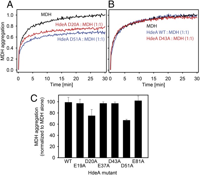
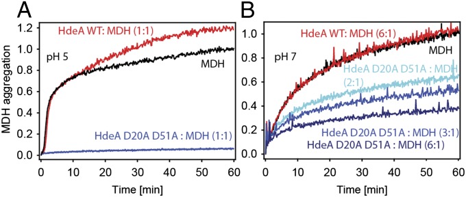
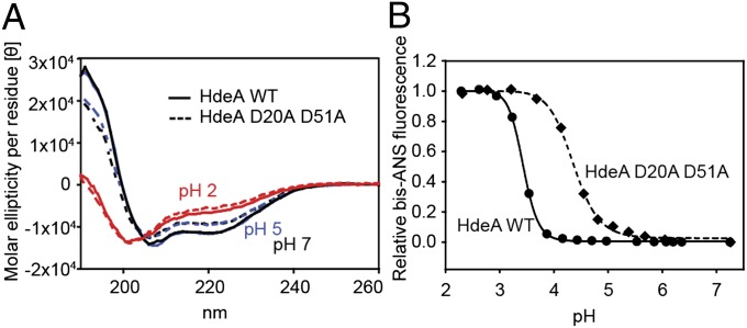
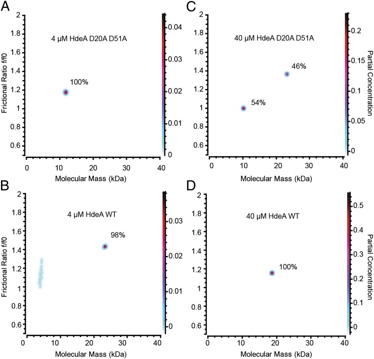
Conditionally disordered proteins can alternate between highly ordered and less ordered configurations under physiological conditions. Whereas protein function is often associated with the ordered conformation, for some of these conditionally unstructured proteins, the opposite applies: Their activation is associated with their unfolding. An example is the small periplasmic chaperone HdeA, which is critical for the ability of enteric bacterial pathogens like Escherichia coli to survive passage through extremely acidic environments, such as the human stomach. At neutral pH, HdeA is a chaperone-inactive dimer. On a shift to low pH, however, HdeA monomerizes, partially unfolds, and becomes rapidly active in preventing the aggregation of substrate proteins. By mutating two aspartic acid residues predicted to be responsible for the pH-dependent monomerization of HdeA, we have succeeded in isolating an HdeA mutant that is active at neutral pH. We find this HdeA mutant to be substantially destabilized, partially unfolded, and mainly monomeric at near-neutral pH at a concentration at which it prevents aggregation of a substrate protein. These results provide convincing evidence for direct activation of a protein by partial unfolding.


Conflict of interest statement

The authors declare no conflict of interest.

Figures

Fig. 1.
Fig. 1.
WebLogo (75) representation of a ClustalW (65) sequence alignment of HdeA sequences. Only the part of the alignment corresponding to the mature E. coli HdeA protein is shown. Numbers indicate residue numbers for mature E. coli HdeA. The residue that is present in the alignment but missing in E. coli HdeA is indicated with “−”. Acidic residues are indicated in red, and basic residues are indicated in blue.
Fig. 2.
Fig. 2.
Predicted effect of protonation of a particular acidic residue on the thermodynamic stability of the dimer interface. Acidic residues in monomer 1 are shown in white, and lysine residues in monomer 2 are shown in blue. Red arrows indicate a destabilization of the dimer interface on protonation of the indicated residue, and green arrows indicate a stabilization of the dimer interface. The length of the arrow indicates the extent of stabilization/destabilization.
Fig. 3.
Fig. 3.
Difference in apparent melting temperature (Tm) for different HdeA mutants (mut) compared with WT HdeA. Apparent Tm values were determined by monitoring the CD signal of 20 μM HdeA protein at 222 nm. (A) Difference in apparent Tm compared with WT. Data are presented as mean ± SD from three independent experiments. (B) Apparent Tm of different HdeA alanine mutants plotted against the predicted effect on the stability of the dimer interface on protonation (Table 1).
Fig. 4.
Fig. 4.
pH-dependent unfolding of HdeA as measured by bis-ANS fluorescence. Binding of 15 μM bis-ANS to 3 μM HdeA was measured in buffer, which was titrated from about pH 7 to pH 2. The midpoints for the pH-induced transition from the state at neutral pH to the state at low pH for the different HdeA mutants tested are 3.4 (WT), 3.8 (D51A), 4.0 (D20A), 3.4 (E19A), 3.5 (E37A), 3.4 (D43A), and 3.5 (E81A).
Fig. 5.
Fig. 5.
Chaperone activity of various HdeA variants at pH 4. (AC) Guanidine denatured MDH was diluted into aggregation buffer to a final concentration of 4 μM. MDH aggregation was measured by monitoring light scattering at 350 nm in the presence or absence of various HdeA variants. (C) Extent of MDH aggregation in the presence of various HdeA variants at pH 4 after 30 min, normalized to the aggregation of MDH in the absence of HdeA. Data are presented as mean ± SD from at least three independent experiments.
Fig. 6.
Fig. 6.
Chaperone activity of HdeA D20A D51A at pH 5 and pH 7. Guanidine denatured MDH was diluted into aggregation buffer at pH 5 (A) or pH 7 (B) to a final concentration of 4 μM. MDH aggregation was measured by monitoring light scattering at 350 nm in the presence or absence of various HdeA variants. Aggregation is normalized to the extent of aggregation of MDH in the absence of HdeA after 60 min.
Fig. 7.
Fig. 7.
Structural characterization of HdeA D20A D51A. (A) Far-UV CD spectra of 20 μM WT HdeA and HdeA D20A D51A at pH 2, pH 5, and pH 7. (B) Binding of 15 μM bis-ANS to 3 μM HdeA was measured in buffer, which was titrated from about pH 7 to pH 2. The midpoints for the pH-induced transition from the state at neutral pH to the state at low pH for the different HdeA mutants tested are 3.4 for WT HdeA and 4.4 for HdeA D20A D51A.
Fig. 8.
Fig. 8.
Analytical ultracentrifugation SV analysis of WT HdeA (B and D) and HdeA D20A D51A (A and C) at pH 5. The molecular mass of WT HdeA is 9.7 kDa (monomer) and 19.5 kDa (dimer). Fractions of the monomeric and dimeric species are indicated. The Kd of dimerization for HdeA D20A D51A at pH 5 is in the range of 30 μM. (A and B) Due to low signal extensity, the fractional ratio and molecular mass are approximate for these very low protein concentrations.

Comment in

  • How bacteria survive an acid trip.
    Hingorani KS, Gierasch LM. Hingorani KS, et al. Proc Natl Acad Sci U S A. 2013 Apr 2;110(14):5279-80. doi: 10.1073/pnas.1303297110. Epub 2013 Mar 25. Proc Natl Acad Sci U S A. 2013. PMID: 23530238 Free PMC article. No abstract available.

References

    1. Smith JL. The role of gastric acid in preventing foodborne disease and how bacteria overcome acid conditions. J Food Prot. 2003;66(7):1292–1303. - PubMed
    1. Gorden J, Small PL. Acid resistance in enteric bacteria. Infect Immun. 1993;61(1):364–367. - PMC - PubMed
    1. Small P, Blankenhorn D, Welty D, Zinser E, Slonczewski JL. Acid and base resistance in Escherichia coli and Shigella flexneri: Role of rpoS and growth pH. J Bacteriol. 1994;176(6):1729–1737. - PMC - PubMed
    1. Hong W, Wu YE, Fu X, Chang Z. Chaperone-dependent mechanisms for acid resistance in enteric bacteria. Trends Microbiol. 2012;20(7):328–335. - PubMed
    1. Zhao B, Houry WA. Acid stress response in enteropathogenic gammaproteobacteria: An aptitude for survival. Biochem Cell Biol. 2010;88(2):301–314. - PubMed

Publication types

MeSH terms