

Prenatal ethanol exposure delays the onset of spermatogenesis in the rat
- PMID: 23488802
- PMCID: PMC3700560
- DOI: 10.1111/acer.12079
Prenatal ethanol exposure delays the onset of spermatogenesis in the rat
Abstract
Background: During late prenatal and early postnatal life, the reproductive system in males undergoes an extensive series of physiological and morphological changes. Prenatal ethanol (EtOH) exposure has marked effects on the development of the reproductive system, with long-term effects on function in adulthood. The present study tested the hypothesis that prenatal EtOH exposure will delay the onset of spermatogenesis.
Methods: Development of the seminiferous tubules and the onset of spermatogenesis were examined utilizing a rat model of fetal alcohol spectrum disorder (FASD). Male offspring from ad libitum-fed control (C), pair-fed (PF), and EtOH-fed (prenatal alcohol exposure [PAE]) dams were terminated on postnatal (PN) days 5, 15, 18, 20, 25, 35, 45, and 55, to investigate morphological changes through morphometric analysis of the testes from early neonatal life through young adulthood.
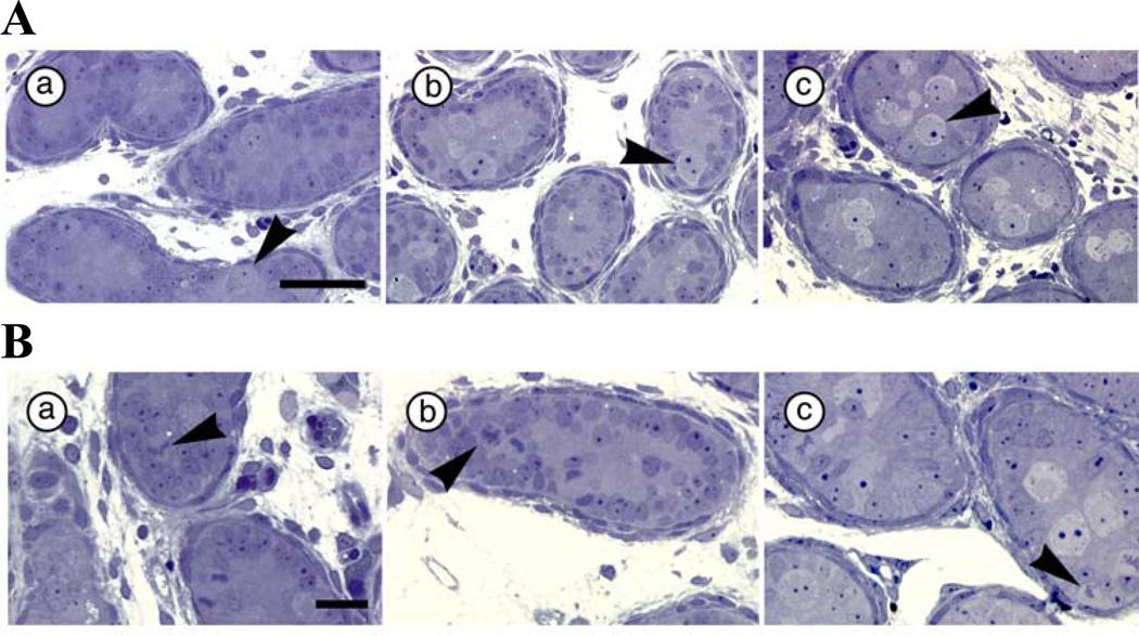
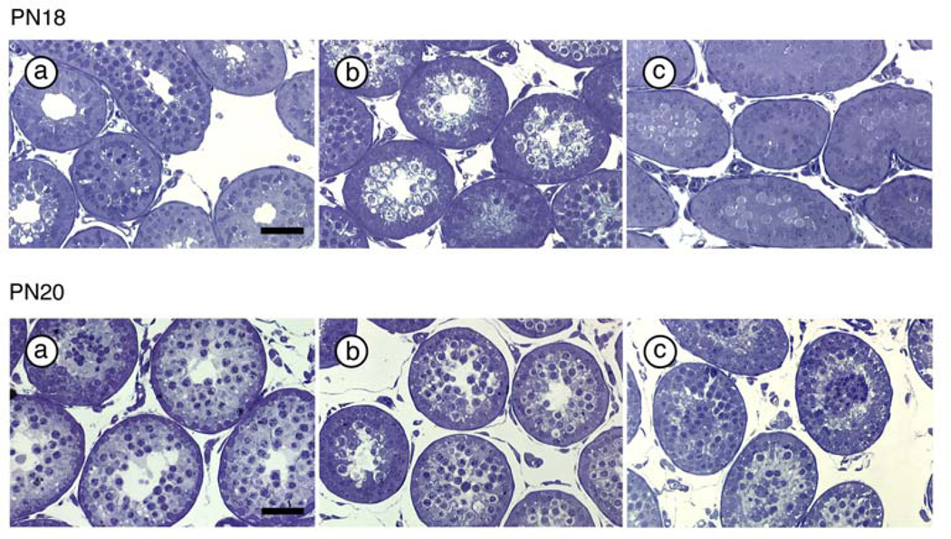
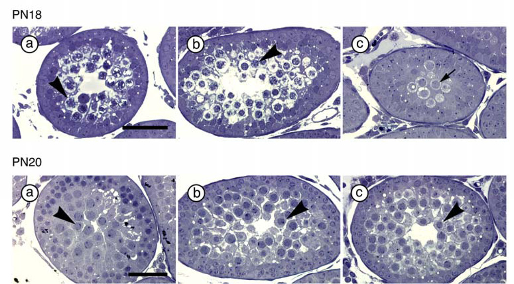
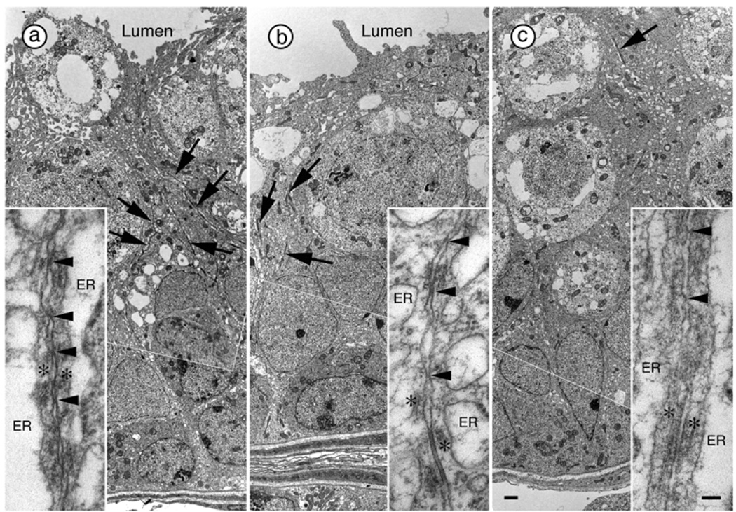
Results: PAE males had lower relative (adjusted for body weight) testis weights compared with PF and/or C males from PN15 through puberty (PN45). In addition, fewer gonocytes (primordial germ cells) were located on the basal lamina on PN5, while more of those touching the basal lamina were dividing in PAE compared with PF and C males, suggesting delayed cell division and migration processes. As well, the percentage of tubules with open lumena was lower in PAE compared with PF and C males on PN18 and 20, and PAE males had fewer primary spermatocytes per tubule on PN18 and round spermatids per tubule on PN25 compared with C males. Finally, the percentage of tubules at stages VII and VIII, when mature spermatids move to the apex of the epithelium and are released, was lower in PAE compared with PF and/or C males in young adulthood (PN55).
Conclusions: Maternal EtOH consumption appears to delay both reproductive development and the onset of spermatogenesis in male offspring, with effects persisting at least until young adulthood.
Keywords: Fetal Alcohol Spectrum Disorder; Prenatal Ethanol Exposure; Reproductive Development; Spermatogenesis.
Copyright © 2013 by the Research Society on Alcoholism.
Figures
References
-
- Abel EL. Effects of ethanol on pregnant rats and their offspring. Psychopharmacology (Berl) 1978;57:5–11. - PubMed
-
- Abel EL, Dintcheff BA. Effects of prenatal alcohol exposure on growth and development in rats. J Pharmacol Exp Ther. 1978;207:916–921. - PubMed
-
- Barron S, Gagnon WA, Mattson SN, Kotch LE, Meyer LS, Riley EP. The effects of prenatal alcohol exposure on odor associative learning in rats. Neurotoxicol Teratol. 1988;10:333–339. - PubMed
-
- Caldwell KK, Sheema S, Paz RD, Samudio-Ruiz SL, Laughlin MH, Spence NE, Roehlk MJ, Alcon SN, Allan AM. Fetal alcohol spectrum disorder-associated depression: evidence for reductions in the levels of brain-derived neurotrophic factor in a mouse model. Pharmacol Biochem Behav. 2008;90:614–624. - PMC - PubMed
Publication types
MeSH terms
Substances
Grants and funding
LinkOut - more resources
Full Text Sources
Other Literature Sources
Research Materials
Miscellaneous
