Tumour necrosis factor receptors and apoptosis of alveolar macrophages during early infection with attenuated and virulent Mycobacterium bovis
- PMID: 23489296
- PMCID: PMC3719067
- DOI: 10.1111/imm.12097
Tumour necrosis factor receptors and apoptosis of alveolar macrophages during early infection with attenuated and virulent Mycobacterium bovis
Abstract
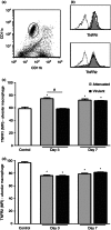
Apoptosis of macrophages has been reported as an effective host strategy to control the growth of intracellular pathogens, including pathogenic mycobacteria. Tumour necrosis factor-α (TNF-α) plays an important role in the modulation of apoptosis of infected macrophages. It exerts its biological activities via two distinct cell surface receptors, TNFR1 and TNFR2, whose extracellular domain can be released by proteolysis forming soluble TNF receptors (sTNFR1 and sTNFR2). The signalling through TNFR1 initiates the majority of the biological functions of TNF-α, leading to either cell death or survival whereas TNFR2 mediates primarily survival signals. Here, the expression of TNF-α receptors and the apoptosis of alveolar macrophages were investigated during the early phase of infection with attenuated and virulent mycobacteria in mice. A significant increase of apoptosis and high expression of TNFR1 were observed in alveolar macrophages at 3 and 7 days after infection with attenuated Mycobacterium bovis but only on day 7 in infection with the virulent M. bovis. Low surface expression of TNFR1 and increased levels of sTNFR1 on day 3 after infection by the virulent strain were associated with reduced rates of apoptotic macrophages. In addition, a significant reduction in apoptosis of alveolar macrophages was observed in TNFR1(-/-) mice at day 3 after bacillus Calmette-Guérin infection. These results suggest a potential role for TNFR1 in mycobacteria-induced alveolar macrophage apoptosis in vivo. In this scenario, shedding of TNFR1 seems to contribute to the modulation of macrophage apoptosis in a strain-dependent manner.
Keywords: Mycobacterium bovis; apoptosis; macrophage; tumour necrosis factor receptor 1; tumour necrosis factor receptor 2.
© 2013 John Wiley & Sons Ltd.
Figures


Similar articles
-
Apoptosis of macrophages during pulmonary Mycobacterium bovis infection: correlation with intracellular bacillary load and cytokine levels.Immunology. 2009 Sep;128(1 Suppl):e691-9. doi: 10.1111/j.1365-2567.2009.03062.x. Epub 2009 Feb 9. Immunology. 2009. PMID: 19740330 Free PMC article.
-
Virulent Mycobacterium tuberculosis strains evade apoptosis of infected alveolar macrophages.J Immunol. 2000 Feb 15;164(4):2016-20. doi: 10.4049/jimmunol.164.4.2016. J Immunol. 2000. PMID: 10657653
-
TNF signalling via the TNF receptors mediates the effects of exercise on cognition-like behaviours.Behav Brain Res. 2018 Nov 1;353:74-82. doi: 10.1016/j.bbr.2018.06.036. Epub 2018 Jun 30. Behav Brain Res. 2018. PMID: 29969604
-
TRAF2 multitasking in TNF receptor-induced signaling to NF-κB, MAP kinases and cell death.Biochem Pharmacol. 2016 Sep 15;116:1-10. doi: 10.1016/j.bcp.2016.03.009. Epub 2016 Mar 16. Biochem Pharmacol. 2016. PMID: 26993379 Review.
-
Tumour necrosis factor in infectious disease.J Pathol. 2013 Jun;230(2):132-47. doi: 10.1002/path.4187. J Pathol. 2013. PMID: 23460469 Review.
Cited by
-
Shedding of the tumor necrosis factor (TNF) receptor from the surface of hepatocytes during sepsis limits inflammation through cGMP signaling.Sci Signal. 2015 Jan 27;8(361):ra11. doi: 10.1126/scisignal.2005548. Sci Signal. 2015. PMID: 25628461 Free PMC article.
-
Immune control of Mycobacterium tuberculosis is dependent on both soluble TNFRp55 and soluble TNFRp75.Immunology. 2021 Nov;164(3):524-540. doi: 10.1111/imm.13385. Epub 2021 Jul 8. Immunology. 2021. PMID: 34129695 Free PMC article.
-
Regulation of Staphylococcus aureus-induced CXCR1 expression via inhibition of receptor mobilization and receptor shedding during dual receptor (TNFR1 and IL-1R) neutralization.Immunol Res. 2019 Jun;67(2-3):241-260. doi: 10.1007/s12026-019-09083-x. Immunol Res. 2019. PMID: 31290001
-
Adventures within the speckled band: heterogeneity, angiogenesis, and balanced inflammation in the tuberculous granuloma.Immunol Rev. 2015 Mar;264(1):276-87. doi: 10.1111/imr.12273. Immunol Rev. 2015. PMID: 25703566 Free PMC article. Review.
-
Metabolic changes enhance necroptosis of type 2 diabetes mellitus mice infected with Mycobacterium tuberculosis.PLoS Pathog. 2024 May 10;20(5):e1012148. doi: 10.1371/journal.ppat.1012148. eCollection 2024 May. PLoS Pathog. 2024. PMID: 38728367 Free PMC article.
References
-
- Cosma CL, Sherman DR, Ramakrishnan L. The secret lives of the pathogenic mycobacteria. Annu Rev Microbiol. 2003;57:641–76. - PubMed
-
- Medina E, Ryan L, LaCourse R, North RJ. Superior virulence of Mycobacterium bovis over Mycobacterium tuberculosis (Mtb) for Mtb-resistant and Mtb-susceptible mice is manifest as an ability to cause extrapulmonary disease. Tuberculosis. 2006;86:20–7. - PubMed
Publication types
MeSH terms
Substances
LinkOut - more resources
Full Text Sources
Other Literature Sources
Medical
Molecular Biology Databases

