. 2013 May;88(4):687-701.
doi: 10.1111/mmi.12206. Epub 2013 Mar 26.

Robust inducible Cre recombinase activity in the human malaria parasite Plasmodium falciparum enables efficient gene deletion within a single asexual erythrocytic growth cycle

Affiliations
Free PMC article

Robust inducible Cre recombinase activity in the human malaria parasite Plasmodium falciparum enables efficient gene deletion within a single asexual erythrocytic growth cycle

Christine R Collins et al. Mol Microbiol. 2013 May.
Free PMC article

Abstract

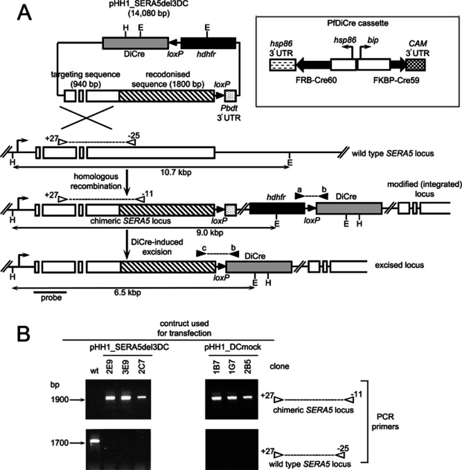
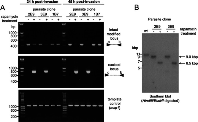
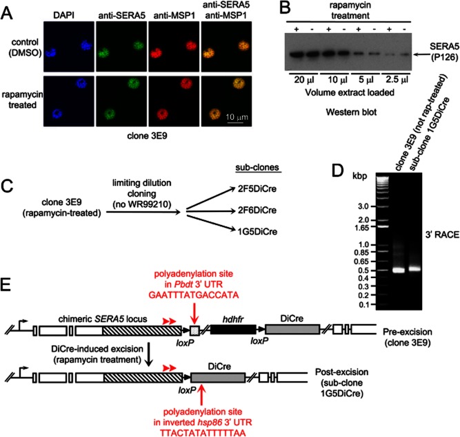
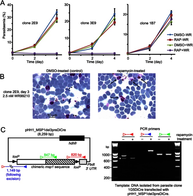
Asexual blood stages of the malaria parasite, which cause all the pathology associated with malaria, can readily be genetically modified by homologous recombination, enabling the functional study of parasite genes that are not essential in this part of the life cycle. However, no widely applicable method for conditional mutagenesis of essential asexual blood-stage malarial genes is available, hindering their functional analysis. We report the application of the DiCre conditional recombinase system to Plasmodium falciparum, the causative agent of the most dangerous form of malaria. We show that DiCre can be used to obtain rapid, highly regulated site-specific recombination in P. falciparum, capable of excising loxP-flanked sequences from a genomic locus with close to 100% efficiency within the time-span of a single erythrocytic growth cycle. DiCre-mediated deletion of the SERA5 3' UTR failed to reduce expression of the gene due to the existence of alternative cryptic polyadenylation sites within the modified locus. However, we successfully used the system to recycle the most widely used drug resistance marker for P. falciparum, human dihydrofolate reductase, in the process producing constitutively DiCre-expressing P. falciparum clones that have broad utility for the functional analysis of essential asexual blood-stage parasite genes.


Figures

Fig. 1
Fig. 1
Design of the DiCre targeting vector, predicted homologous integration and recombinase-mediated excision events, and isolation of transgenic P. falciparum clones. A. The boxed insert shows an expanded view of the PfDiCre expression cassette. Expression of the DiCre components (black block arrows) is driven by the P. falciparumBiP and hsp86 promoters, with transcription termination and polyadenylation being regulated by the 3' UTR sequences from the P. falciparum calmodulin (CAM) and hsp86 genes. Main figure: schematic (not to scale) showing the main features of the pHH1_SERA5del3DC plasmid construct. Targeting sequence derived from the native SERA5 gene was fused in-frame to recodonized sequence (hatched) derived from the SERA5synth gene. A single EcoNI restriction site (E) within the hdhfr cassette (black box), which confers resistance to the antifolate WR99210, is shown. The PfDiCre cassette contains another EcoNI site as well as a unique HindIII site (H). The P. bergheidhfr 3' UTR (Pbdt 3' UTR) lies just downstream of the SERA5synth sequence in the plasmid. Two loxP sites are indicated as black arrowheads in the plasmid. The endogenous SERA5 locus, which contains three introns, is shown below, as is the expected result of homologous integration of the entire plasmid and the architecture of the modified locus relative to flanking HindIII and EcoNI sites. Episomal plasmids are harboured as concatamers in P. falciparum, so integration of more than one copy can occur. However, because the plasmid contains only a partial SERA5 sequence not preceded by a promoter, the only SERA5 copy to be transcribed is the modified chimeric gene directly downstream of the endogenous promoter. DiCre-mediated recombination should result in excision of both the Pbdt 3' UTR from the modified SERA5 locus, and the hdhfr selectable marker, as well as removal of the entire pHH1_SERA5del3DC plasmid backbone. Note that, even if concatamers of pHH1_SERA5del3DC were to integrate, DiCre-mediated excision would still result in removal of all sequence between the duplicated loxP sites, leading to the terminal excised structure shown, in which only a single copy of the modified SERA5 coding sequence remains under the control of its endogenous promoter, and just a single genomic loxP site remains. Positions of primers +27, −11 and −25 used for diagnostic PCR of the expected integration of pHH1_SERA5del3DC and pHH1_DCmock (which is identical aside from the presence of a mock promoter in the place of the hsp86 and BiP promoters within the PfDiCre cassette) are shown by white arrowheads joined by dotted lines. Predicted sizes of the amplicons obtained with primer pairs +27 plus −11, and +27 plus −25, are 1911 bp and 1737 bp respectively. Primer +27 lies just upstream of the targeting sequence in the plasmid, so cannot produce a product from the transfection construct. Positions of primers CAM5‘_R4 (a), hsp86 3'_R1 (b) and sgs5seq5F (c) used for diagnostic PCR analysis of the modified and excised locus are indicated by black arrowheads joined by dotted lines. Predicted sizes of amplicons obtained with primers a plus b, and c plus b are 428 bp and 804 bp respectively. The relative position of the probe used for Southern analysis is indicated. B. Diagnostic PCR analysis of genomic DNA from clones derived by limiting dilution of parasites transfected with constructs pHH1_SERA5del3DC or pHH1_DCmock, confirming integration into the SERA5 locus as expected. Only wild-type (wt) parasite DNA produced a product with primers +27 plus −25, while the amplicon produced with primer pair +27 plus −11, diagnostic of the expected integration event, was obtained only from the transgenic clones.
Fig. 2
Fig. 2
Rapid, regulated and highly efficient DiCre-mediated excision of a floxed genomic P. falciparum sequence. A. PCR analysis showing detection of excision following a 4-h-long treatment of clones 2E9, 3E9 or 1B7 with rapamycin at ring stage. Genomic DNA was prepared from the treated parasites at 24 h and 45 h post invasion. Primer pairs a plus b detect the modified locus prior to excision, while primers c plus b detect the appearance of the rearranged locus resulting from site-directed recombination between the loxP sites (see Fig. 1A). Primers specific for the msp1 gene (MSP1_FOR and MSP1_REV; see Table S1) were used as a control to confirm the presence of genomic DNA in all the template samples. B. Southern blots confirming efficient rapamycin-induced excision in the 2E9 and 3E9 transgenic P. falciparum clones. The genomic DNA used for the Southern blot was prepared at 45 h post invasion. Phosphorimager quantification of the 6.5 kb ‘excised locus’ signals in the rapamycin-treated 2E9 and 3E9 samples, compared with the position in the same tracks corresponding to the non-excised 9.0 kb species (where no band is visible by eye) showed that excision was 97.9% efficient in the case of clone 2E9, and 97.2% efficient in the case of clone 3E9. However, even upon prolonged exposure, no residual ‘non-excised’ signal was visually detectable in the genomic DNA digests from the rapamycin-treated parasites (not shown).
Fig. 3
Fig. 3
DiCre-mediated excision of the Pbdt 3' UTR does not reduce SERA5 expression levels due to the presence of an alternative polyadenylation site within the inverted hsp86 3' UTR. A. IFA analysis of clone 3E9 schizonts derived from ring-stage parasites treated for 4 h with vehicle only (DMSO) or 100 nM rapamycin to induce DiCre-mediated excision of the Pbdt 3' UTR from the modified SERA5 locus (see Figs 1 and 2). Parasites were probed with mAbs specific for SERA5, or MSP1 as a control. No change in SERA5 expression levels was detected following rapamycin treatment. 4,6-Diamidino-2-phenylindol (DAPI) was used to detect parasite nuclei. B. Western blot analysis of extracts of the same schizont populations. Different volumes of an SDS extract of intact schizonts were probed with the anti-SERA5 mAb 24C6.1F1. The results confirm no significant difference in SERA5 expression levels between the control and rapamycin-treated parasites. C. Workflow showing isolation by limiting dilution of parasite subclones from rapamycin-treated P. falciparum clone 3E9, performed in order to obtain genetically homogeneous parasites harbouring the excised locus architecture. Subclone 1G5DiCre was used for subsequent 3' RACE and transfection analysis. D. Agarose gel electrophoresis of 3' RACE products amplified from total RNA of clone 3E9 and subclone 1G5DiCre. A dominant RACE product was obtained in each case. No product was obtained in the absence of reverse transcriptase (not shown). Sizes in kb of DNA marker fragments (left-hand lane) are indicated. E. Schematic (not to scale) showing the overall structure of the modified SERA5 locus in clones 3E9 (prior to excision) and subclone 1G5DiCre (following excision), depicted as in Fig. 1. Relative positions of polyadenylation sites identified by sequencing of the cloned 3' RACE products are indicated in red (see Fig. S1 for the aligned sequence data). Positions of SERA5synth gene-specific forward primers used for the semi-nested 3' RACE are shown (red arrowheads).
Fig. 4
Fig. 4
DiCre-mediated excision successfully recycles the hdhfr selectable marker and produces a P. falciparum recipient clone that constitutively expresses DiCre. A. Typical growth assay showing the results of expanding the control (DMSO-treated) or rapamycin-treated clones 2E9, 3E9 and 1B7 in the presence or absence of WR99210. As predicted based on the efficient excision of the hdhfr cassette, the rapamycin-treated 2E9 and 3E9 clones did not replicate in medium containing WR99210. In contrast, rapamycin treatment of clone 1B7, which harbours the ‘mock’ DiCre cassette, had no effect on growth under any conditions. Day 0 corresponds to the point (∼ 60 h following treatment with rapamycin or DMSO) at which the cultures were transferred to fresh medium ± WR99210. Each data point represents the mean of triplicate microscopic counts, each of at least 500 cells. Error bars, ± 1 SD. B. Giemsa-stained microscopic images of clone 2E9 at day 3 following the start of culture in the presence of WR99210. The appearance of the poorly staining, dysmorphic schizonts (arrowed) in the rapamycin-treated cultures is typical of that usually observed after WR99210 treatment of drug-susceptible wild-type parasites. By day 4, these had completely degenerated and disappeared from the cultures. In contrast, the DMSO-treated control cultures contain numerous healthy schizonts and young ring stages, indicative of normal levels of growth. C. Induction of DiCre-mediated excision in the 1G5DiCre recipient subclone. Shown (left-hand side) is a schematic of reporter plasmid construct pHH1_MSP1del3preDiCre (not to scale) alongside results of PCR analysis of DNA isolated from 1G5DiCre parasites harbouring the plasmid, following treatment ± rapamycin. Rapamycin treatment induced efficient excision of the floxed Pbdt 3' UTR and hdhfr cassette from the plasmid, proving expression of DiCre from the genomic cassette incorporated into the modified SERA5 locus of the 1G5DiCre parasites. No excision was observed in the absence of rapamycin. The relative positions of primers used for the PCR are indicated as coloured arrowheads. Primers used were: 3D7synMSP1_FOR2 (open red); PbDT3'5’R1 (solid red); 3D7endoMSP1_REV4 (blue); 3D7endoMSP1FOR1 (open green); and 3D7synMSP1_REV3 (solid green). See Table S1 for primer sequences. The hatched region in the pHH1_MSP1del3preDiCre schematic indicates the synthetic (recodonized) msp1 sequence, and the expected sizes of the various amplicons are indicated. The extreme left-hand and right-hand tracks on the gel contain DNA size markers. The ∼ 700 bp fragment (asterisked) present in the 3D7synMSP1_FOR2 (open red) plus PbDT3'5’R1 (solid red) PCR tracks is a product of mis-priming, since this was also amplified from 1G5DiCre parasites that had not been transfected with plasmid pHH1_MSP1del3preDiCre (not shown). Rapamycin treatment results in loss of the 820 bp PCR product and the appearance of an excision-specific 1149 bp product. Note that under the PCR conditions used, no product was expected to be amplified from the intact pHH1_MSP1del3preDiCre with primers 3D7synMSP1_FOR2 (open red) and 3D7endoMSP1_REV4 (blue) due to the large size of that predicted product.

Comment in

Similar articles

Cited by

References

    1. Agop-Nersesian C, Pfahler J, Lanzer M, Meissner M. Functional expression of ribozymes in Apicomplexa: towards exogenous control of gene expression by inducible RNA-cleavage. Int J Parasitol. 2008;38:673–681. - PubMed
    1. Andenmatten N, Egarter S, Jackson AJ, Jullien N, Herman JP, Meissner M. Conditional genome engineering in Toxoplasma gondii uncovers alternative invasion mechanisms. Nat Methods. 2013;10:125–127. - PMC - PubMed
    1. Arisue N, Kawai S, Hirai M, Palacpac NM, Jia M, Kaneko A, et al. Clues to evolution of the SERA multigene family in 18 Plasmodium species. PLoS ONE. 2011;6:e17775. - PMC - PubMed
    1. Armstrong CM, Goldberg DE. An FKBP destabilization domain modulates protein levels in Plasmodium falciparum. Nat Methods. 2007;4:1007–1009. - PubMed
    1. Augagneur Y, Wesolowski D, Tae HS, Altman S, Ben Mamoun C. Gene selective mRNA cleavage inhibits the development of Plasmodium falciparum. Proc Natl Acad Sci USA. 2012;109:6235–6240. - PMC - PubMed

Publication types