

Comparison of the effects of topical fusidic acid and rifamycin on wound healing in rats
- PMID: 23489386
- PMCID: PMC7950572
- DOI: 10.1111/iwj.12060
Comparison of the effects of topical fusidic acid and rifamycin on wound healing in rats
Abstract
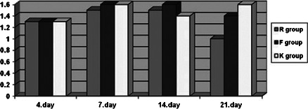
Wound healing is an active and dynamic process that begins from the moment of injury. Any delay in the initiation of the response to injury can prolong the healing process. The aim of this study was to investigate the effects of topically applied fusidic acid and rifamycin on wound healing in a full-thickness wound model. Ten female Sprague-Dawley rats, aged 4 months and weighing 200-250 g, were used. Four rifamycin (R), four fusidic acid (F) and four control (K) areas were generated on their backs by using a 5-mm punch biopsy pen. On the 4th, 7th, 14th and 21st days, biopsies were taken from each wound area of all the rats. Fusidic acid group demonstrated a statistically significant increase of collagen and intensity of fibroblast proliferation on the 21st day of wound healing, whereas in the rifamycin group, healing time was, as expected, similar to physiological wound-healing phases. Despite the limited number of subjects, topical fusidic acid was found to delay wound healing by prolonging fibroblast proliferation.
Keywords: Rifamycin; Topical fusidic acid; Wound healing.
© 2013 The Authors. International Wound Journal © 2013 Medicalhelplines.com Inc and John Wiley & Sons Ltd.
Figures


Similar articles
-
Effects of PerClot® on the healing of full-thickness skin wounds in rats.Acta Histochem. 2012 Jul;114(4):311-7. doi: 10.1016/j.acthis.2011.06.012. Epub 2011 Jul 22. Acta Histochem. 2012. PMID: 21782216
-
Promotion of dermal wound healing by polysaccharides isolated from Phellinus gilvus in rats.J Vet Med Sci. 2005 Jan;67(1):111-4. doi: 10.1292/jvms.67.111. J Vet Med Sci. 2005. PMID: 15699606
-
Topical vitamin K1 promotes repair of full thickness wound in rat.Indian J Pharmacol. 2014 Jul-Aug;46(4):409-12. doi: 10.4103/0253-7613.135953. Indian J Pharmacol. 2014. PMID: 25097279 Free PMC article.
-
Synergistic effect of bismuth subgallate and borneol, the major components of Sulbogin, on the healing of skin wound.Biomaterials. 2003 Aug;24(18):3005-12. doi: 10.1016/s0142-9612(03)00126-1. Biomaterials. 2003. PMID: 12895572
-
Systematic investigation of ethanolic extract from Leea macrophylla: Implications in wound healing.J Ethnopharmacol. 2016 Sep 15;191:95-106. doi: 10.1016/j.jep.2016.06.034. Epub 2016 Jun 14. J Ethnopharmacol. 2016. PMID: 27321280
Cited by
-
Effect of topical rifamycin application on epidural fibrosis in rats.Turk J Phys Med Rehabil. 2019 Jan 31;65(1):24-29. doi: 10.5606/tftrd.2019.2442. eCollection 2019 Mar. Turk J Phys Med Rehabil. 2019. PMID: 31453541 Free PMC article.
-
Locally Used Antibiotics for Spinal Infection Prophylaxis and Their Effects on Epidural Fibrosis: an Experimental Laminectomy Study in Rats Using Rifamycin and Gentamycin.Inflammation. 2019 Apr;42(2):714-720. doi: 10.1007/s10753-018-0929-x. Inflammation. 2019. PMID: 30413905
-
Does antibiotic use accelerate or retard cutaneous repair? A systematic review in animal models.PLoS One. 2019 Oct 10;14(10):e0223511. doi: 10.1371/journal.pone.0223511. eCollection 2019. PLoS One. 2019. PMID: 31600279 Free PMC article.
-
Recent Advances in the Local Drug Delivery Systems for Diabetic Wound Healing: A Comprehensive Review.AAPS PharmSciTech. 2025 Jul 1;26(6):177. doi: 10.1208/s12249-025-03172-x. AAPS PharmSciTech. 2025. PMID: 40593363 Review.
-
Evaluation of burn wound healing activity of novel fusidic acid loaded microemulsion based gel in male Wistar albino rats.Saudi Pharm J. 2020 Mar;28(3):338-348. doi: 10.1016/j.jsps.2020.01.015. Epub 2020 Feb 3. Saudi Pharm J. 2020. PMID: 32194336 Free PMC article.
References
-
- Del Rosso JQ. Wound care in the dermatology office: where are we in 2011? J Am Acad Dermatol 2011;64:1–7. - PubMed
-
- Li W, Dasgeb B, Phillips T, Li Y, Chen M, Garner W, Woodley DT. Wound‐healing perspectives. Dermatol Clin 2005;23:181–92. - PubMed
-
- Singer AJ, Clark RA. Cutaneous wound healing. N Engl J Med 1999;341:738–46. - PubMed
-
- Carter EL. Antibiotics in cutaneous medicine: an update. Semin Cutan Med Surg 2003;22:196–211. - PubMed
-
- Benfer J, Struck H. The effect of rifamycin SV on the wound‐healing process. Arzneimittelforschung 1976;26:1361–4. - PubMed
Publication types
MeSH terms
Substances
LinkOut - more resources
Full Text Sources
Other Literature Sources
Medical
