. 2013 Jun;153(6):771-8.
doi: 10.1016/j.surg.2013.02.002. Epub 2013 Mar 13.

Resection of the primary tumor improves survival in metastatic breast cancer by reducing overall tumor burden

Affiliations

Resection of the primary tumor improves survival in metastatic breast cancer by reducing overall tumor burden

Omar M Rashid et al. Surgery. 2013 Jun.

Abstract

Background: Although many retrospective studies suggest that resection of the primary tumor improves survival in metastatic breast cancer, animal studies suggest that resection induces metastasis. Moreover, there has been no critical evaluation of how well animal studies actually model metastatic breast cancer. We used our newly established orthotopic cancer implantation under direct vision model to evaluate the hypothesis that primary tumor resection improves survival in metastatic breast cancer by reducing overall tumor burden and improving immune responsiveness.

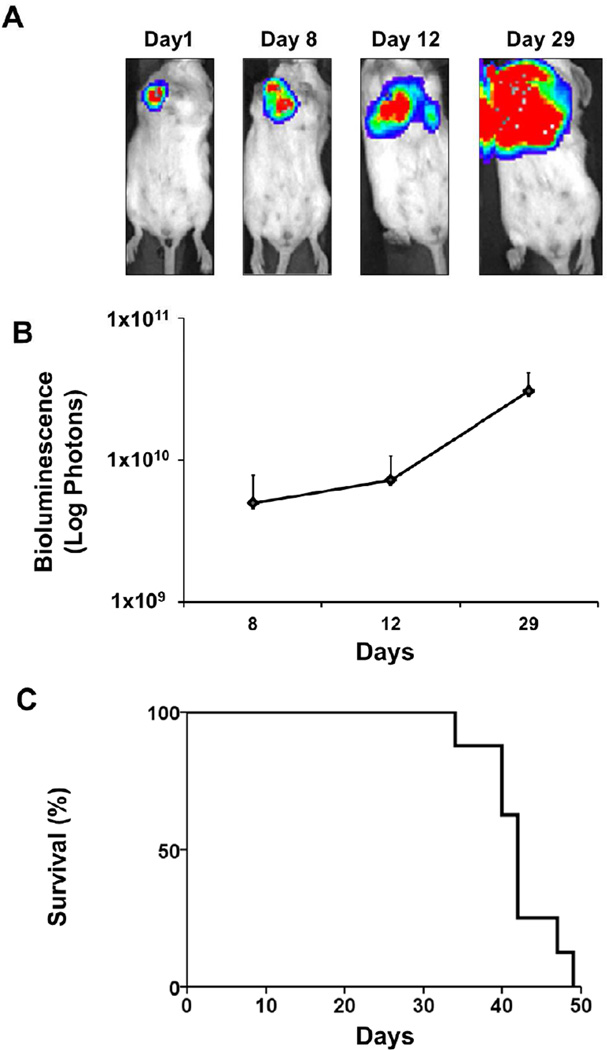
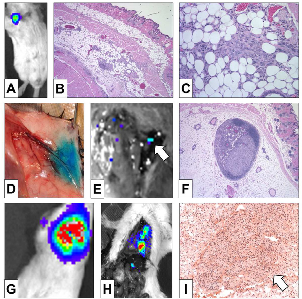
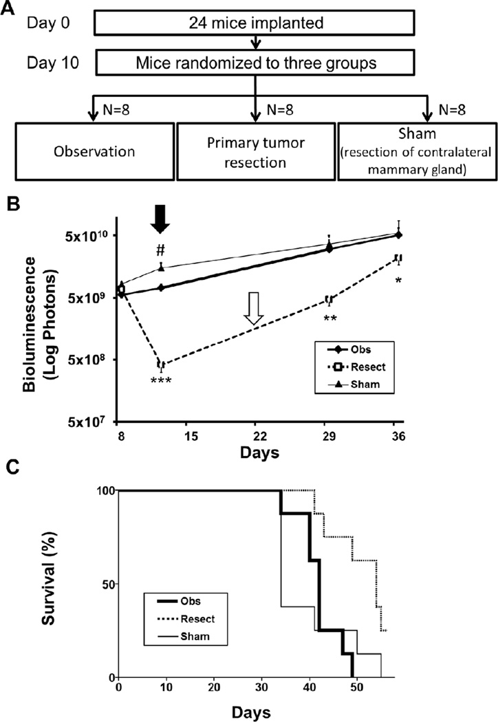
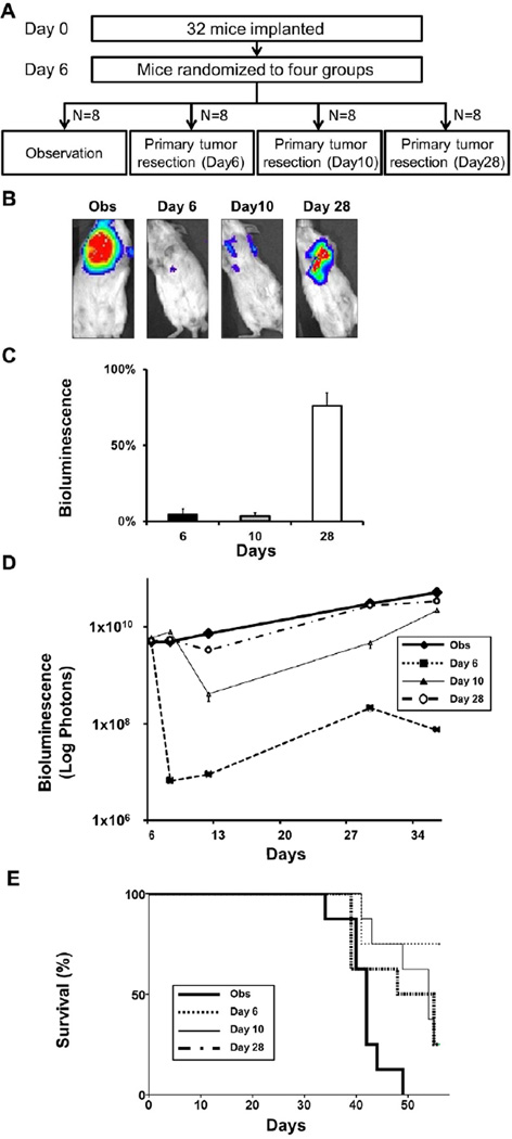
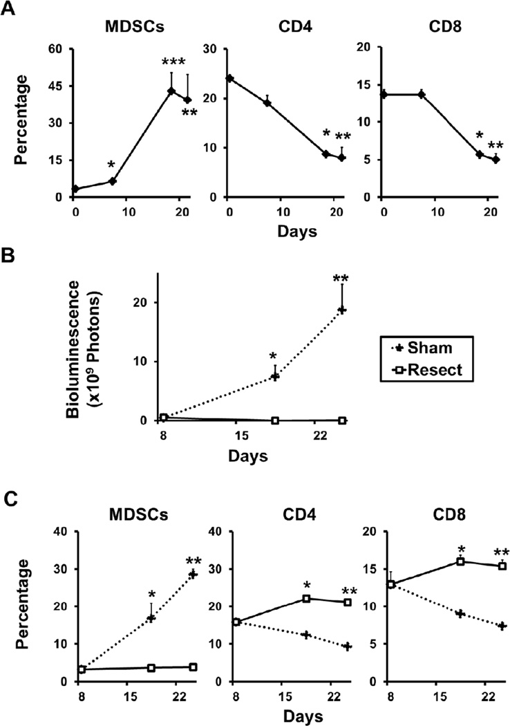
Methods: Murine mammary adenocarcinoma 4T1-luc2 cells that can be visualized by bioluminescence were implanted orthotopically into BALB/c mice under direct vision. Resection of the primary tumors at days 6, 10, and 28 were compared to sham resection of the contralateral normal mammary gland and observation alone. Tumor burden was quantified by bioluminescence. Tumor-draining lymph nodes were identified by intradermal injection of lymphazurin, and primary tumors, lymph nodes, and lungs were examined pathologically. Kaplan-Meier survival analyses were performed. Splenocyte myeloid-derived suppressor cells (MDSCs) and CD4 or CD8 single positive T lymphocytes were quantified by flow cytometry.

Results: Tumors invaded locally, metastasized to regional lymph nodes, and then metastasized to distant organs, with subsequent mortality. Surgical stress increased tumor burden only transiently without affecting survival. When primary tumor resection decreased overall tumor burden substantially, further growth of metastatic lesions did not increase the overall tumor burden compared to observation, and survival was improved, which was not the case when resection did not significantly reduce the overall tumor burden. Decreasing overall tumor burden through resection of the primary tumor resulted in decreased splenic MDSC numbers and increased CD4 and CD8 cells, suggesting the potential for an improved immunologic response to cancer.

Conclusion: Decreasing overall tumor burden through resection of the primary breast tumor decreased MDSCs, increased CD4 and CD8 cells, and improved survival.


Figures

FIGURE 1
FIGURE 1. Our orthotopic implantation model allows for quantification of overall tumor burden and survival analysis
(A) Representative bioluminescent images after orthotopic implantation of 1×10⁵ 4T1-luc2 cells. Bioluminescence images at 1, 8, 12 and 29 days after implantation demonstrate progressive tumor growth and metastatic spread. (B) The total tumor burden is quantified as total photons measured by bioluminescent technology as cancer progressed after implantation. (C) Kaplan-Meier survival curve of this model.
FIGURE 2
FIGURE 2. Our orthotopic implantation model mimics the human pattern of breast cancer progression
Our model produced mammary gland tumors as demonstrated by bioluminescence (A) and confirmed by H&E staining (x40 (B), ×100 (C)). Subdermal injection of Lymphazurin demonstrates lymphatic drainage to the axillary lymph node basin (D), to which 4T1-luc2 tumors regionally metastasized as confirmed by bioluminescence (white arrow in E) and by H&E staining of an axillary lymph node (F). The lack of bioluminescent uptake in the lung fields demonstrates that lymph node metastasis occurred before distant lung metastasis (E). Non-invasive bioluminescence demonstrates lung metastasis after removal of the primary tumor (G), which was confirmed by bioluminescent lesions in the lung on thoracotomy after primary tumor resection (H). H&E staining demonstrates a metastatic lung tumor (white arrow in I).
FIGURE 3
FIGURE 3. Primary tumor resection in metastatic breast cancer significantly prolongs survival despite the observed Fisher and Folkman effects
(A) Study design: Twenty-four mice ten days after implantation with 1×10⁵ 4T1-luc2 cells were randomized to Observation (Obs), Resection of primary tumor (Resect), and Resection of the contralateral mammary gland (Sham) groups (N = 8 per group). (B) Overall tumor burden was quantified by bioluminescence. There was no statistical difference in overall tumor burden between the groups at the time of randomization. Tumor burden was significantly higher in the Sham group (thin line) compared to the Obs group (bold line) on day 12 (Fisher effect (black arrow), #P < .001), but there was no significant difference by day 29. The rate of increase in tumor burden was significantly faster after removal of the primary tumor (Resect group (dotted line); *P < .05, **P < .001, ***P <. 001), with a slope of 11.4 and 4.7 in the Resect group compared to 4.2 and 1.7 in the Obs group (Folkman effect (white arrow)). (C) Kaplan-Meier analysis demonstrates a significant improvement in survival after resection (dotted line) vs. Sham (thin line) (P < .01) and vs. Obs (bold line) (P = .01). No difference in survival was observed between the Sham and Obs groups.
FIGURE 4
FIGURE 4. Only primary tumor resection that significantly reduces overall tumor burden improves survival
(A) Study design: Thirty two mice six days after implantation with 1×10⁵ 4T1-luc2 cells were randomized into 4 groups (N = 8 per group): observation (Obs), primary tumor resection on day 6 (Day 6), tumor resection on day 10 (Day 10), and tumor resection on day 28 (Day 28), again controlling for overall tumor burden. (B) Representative bioluminescence imaging of a mouse from the Obs group, and immediately after resection of primary tumor of Day 6, Day 10, and Day 28 groups. (C) The percentage of remaining overall tumor burden after primary tumor resection for Day 6, Day 10 and Day 28 groups was quantified by bioluminescence. The percentage of overall tumor burden was calculated by dividing the quantification of remaining metastatic lesions by the overall tumor burden before resection (N = 8). (D) Time course of overall tumor burden quantified by bioluminescence for each group. There was no significant difference in overall tumor burden at randomization and only the Day 28 group (bold dotted line) did not reduce overall tumor burden significantly at the time of resection. (E) Kaplan-Meier analysis showed that only Day 28 (bold dotted line) failed to significantly improve survival compared to observation (Obs (bold line) vs. Day 6 (thin dotted line) P < .01, Obs vs. Day 10 (thin line) P < .01).
FIGURE 5
FIGURE 5. Reduction of overall tumor burden by primary tumor resection reverses the immunosuppressive effects of cancer progression
(A) The spleens were harvested untreated mice and from mice 7, 18, and 21 days after implantation (N = 5 per group). Flow cytometry analysis of splenocytes was performed to quantify MDSCs and CD4 and CD8 positive T cells. MDSCs increased (*P < .05, **P<.01, ***P<.05, N = 3), while CD4 (*P < .001, **P < .01) and CD8 positive cells (*P < .01, **P < .01) decreased significantly in the spleen, 18 and 21 days after implantation. (B) After controlling for overall tumor burden 8 days implantation, mice were randomized to 2 groups (N = 5): Resection of primary tumor (Resect), and contralateral mammary gland resection (Sham), both performed on day 10. The Resect group showed significantly reduced overall tumor burden compared to Sham as demonstrated by bioluminescence. (*P < .01, P < 0.01, respectively) (C) Spleens of the Resect and Sham groups were harvested 8, 18 and 25 days after implantation (N = 5 per group). The Resect group suppressed the increase in MDSCs (left panel; *P < .01, **P < .001) and increased CD4 (middle panel; *P < .001, **P < .001) and CD8 (right panel; *P < .001, **P < .001) positive splenocytes compared to Sham.

References

    1. DeSantis C, Siegel R, Bandi P, et al. Breast cancer statistics, 2011. CA Cancer J Clin. 61:409–418. - PubMed
    1. Leung AM, Vu HN, Nguyen KA, et al. Effects of surgical excision on survival of patients with stage IV breast cancer. J Surg Res. 161:83–88. - PubMed
    1. Cady B, Nathan NR, Michaelson JS, et al. Matched pair analyses of stage IV breast cancer with or without resection of primary breast site. Ann Surg Oncol. 2008;15:3384–3395. - PubMed
    1. Rao R, Feng L, Kuerer HM, et al. Timing of surgical intervention for the intact primary in stage IV breast cancer patients. Ann Surg Oncol. 2008;15:1696–1702. - PubMed
    1. Rapiti E. Complete excision of primary breast tumor improves survival of patients with metastatic breast cancer at diagnosis. J Clin Oncol. 2006;24:2743–2749. - PubMed

Publication types