Review
. 2013 May;38(5):243-52.
doi: 10.1016/j.tibs.2013.02.004. Epub 2013 Mar 13.

Protein methylation at the surface and buried deep: thinking outside the histone box

Affiliations
Review

Protein methylation at the surface and buried deep: thinking outside the histone box

Steven G Clarke. Trends Biochem Sci. 2013 May.

Abstract

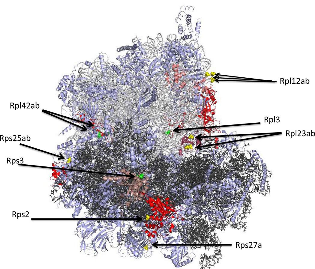
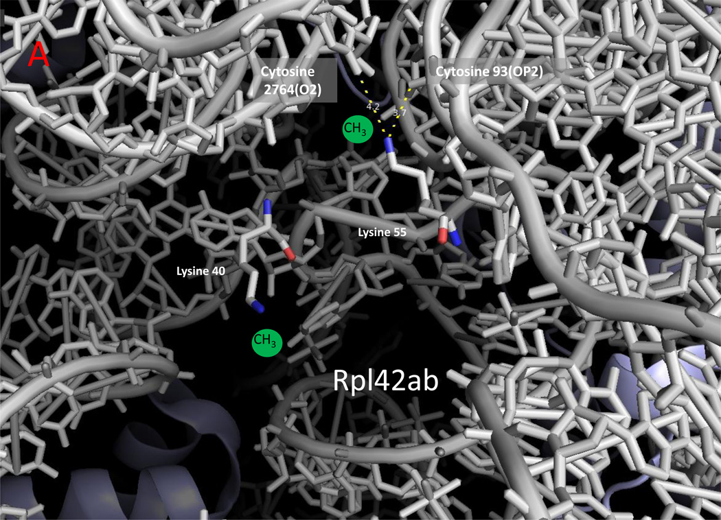
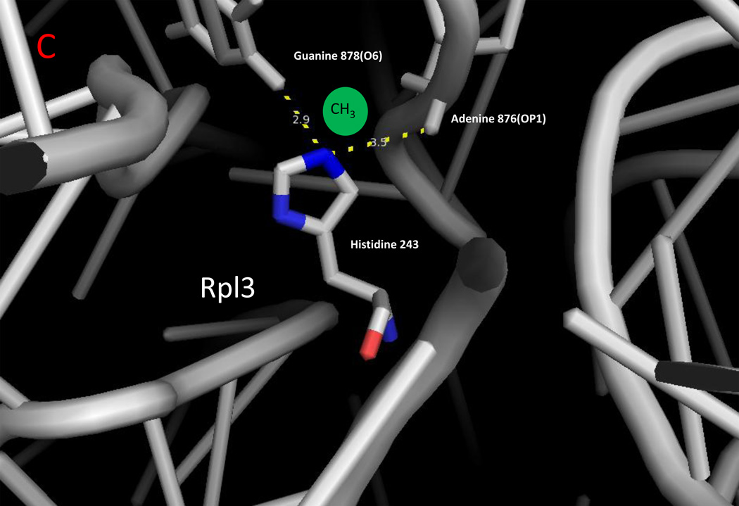
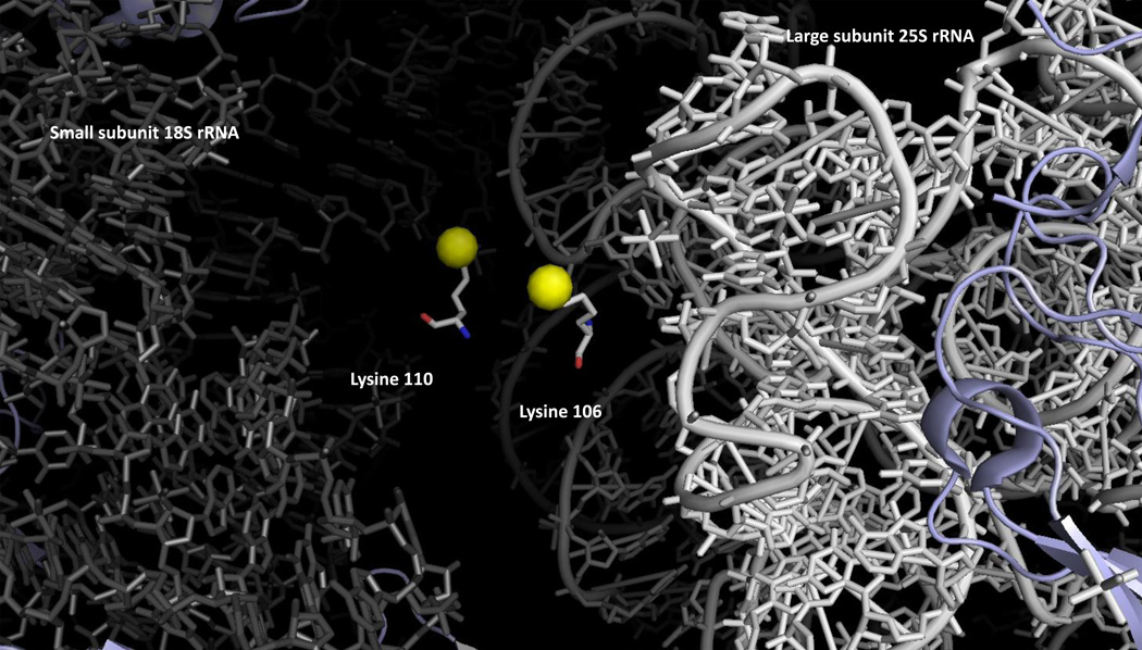
Methylated lysine and arginine residues in histones represent a crucial part of the histone code, and recognition of these methylated residues by protein interaction domains modulates transcription. Although some methylating enzymes appear to be histone specific, many can modify histone and non-histone substrates and an increasing number are specific for non-histone substrates. Some of the non-histone substrates can also be involved in transcription, but a distinct subset of protein methylation reactions occurs at residues buried deeply in ribosomal proteins that may function in protein-RNA interactions rather than protein-protein interactions. Additionally, recent work has identified enzymes that catalyze protein methylation reactions at new sites in ribosomal and other proteins. These reactions include modifications of histidine and cysteine residues as well as the N terminus.


Figures

Figure 1
Figure 1
Surface and buried sites of methylation on cytoplasmic ribosomal proteins in the yeast Saccharomyces cerevisiae. The 25S ribosomal RNA of the large subunit is shown in light gray; the 18S ribosomal RNA of the small subunit is shown in dark gray. Non-methylated proteins are shown in light blue; methylated proteins (Tables 1 and 2) are shown in pink (Rpl12ab, Rpl23ab, Rps27a, Rps3) and red (Rpl3, Rps2, Rps25ab, Rpl42ab). The approximate positions of surface-exposed methyl groups are shown as yellow spheres; buried methyl groups are represented as green spheres. The illustration was made using PyMOL from the PDB structures 3U5F, 3U5G, 3U5H, and 3U5I [45].
Figure 2
Figure 2
Zoomed in view of methylated lysine, arginine, and histidine residues with ribosomal RNA in yeast cytoplasmic ribosomes. The monomethylated residues of Rpl42ab ((a); Lys-39; Lys-54), Rps3 ((b); Arg-145), and Rpl3 ((c); His-242) are shown with nitrogen atoms in blue and oxygen atoms in red [8,14,25]. Ribosomal 25S (Panels A and C) and 18S (Panel B) RNA are shown in gray. Methyl groups were not modeled into these structures; possible locations are suggested by the green spheres. Examples of close contact distances (less than 5 Å) are shown between the methylated atom in the protein and the RNA to emphasize the apposition of the methylated residue to the RNA, although these interactions may differ in a refined structure that includes the methyl groups. The illustration was made using PyMOL and the PDB structures 3U5F, 3U5G, 3U5H, and 3U5I [45].
Figure 2
Figure 2
Zoomed in view of methylated lysine, arginine, and histidine residues with ribosomal RNA in yeast cytoplasmic ribosomes. The monomethylated residues of Rpl42ab ((a); Lys-39; Lys-54), Rps3 ((b); Arg-145), and Rpl3 ((c); His-242) are shown with nitrogen atoms in blue and oxygen atoms in red [8,14,25]. Ribosomal 25S (Panels A and C) and 18S (Panel B) RNA are shown in gray. Methyl groups were not modeled into these structures; possible locations are suggested by the green spheres. Examples of close contact distances (less than 5 Å) are shown between the methylated atom in the protein and the RNA to emphasize the apposition of the methylated residue to the RNA, although these interactions may differ in a refined structure that includes the methyl groups. The illustration was made using PyMOL and the PDB structures 3U5F, 3U5G, 3U5H, and 3U5I [45].
Figure 3
Figure 3
Intrasubunit localization of the methylated lysine residues of Rpl23ab in yeast cytoplasmic ribosomes. Dimethyllysine residues 105 and 109 are shown with the epsilon amino group as a yellow sphere. These residues are positioned at the interface of the small and large ribosomal subunits; 18S rRNA is shown in gray on the left and 25S rRNA in white on the right. The illustration was made using PyMOL from the PDB structures 3U5F, 3U5G, 3U5H, and 3U5I [45].

References

    1. Katz JE, et al. Automated identification of putative methyltransferases from genomic open reading frames. Mol. Cell. Proteomics. 2003;2:525–540. - PubMed
    1. Petrossian TC, Clarke S. Bioinformatic identification of novel methyltransferases. Epigenomics. 2009;1:163–175. - PMC - PubMed
    1. Wlodarski T, et al. Comprehensive structural and substrate specificity classification of the Saccharomyces cerevisiae methyltransferome. PLoS ONE. 2011 - PMC - PubMed
    1. Petrossian TC, Clarke SG. Uncovering the human methyltransferasome. Mol. Cell. Proteomics. 2011 - PMC - PubMed
    1. Schubert HL, et al. Many paths to methyltransfer: a chronicle of convergence. Trends Biochem. Sci. 2003;28:329–335. - PMC - PubMed

Publication types

MeSH terms