

The perinexus: sign-post on the path to a new model of cardiac conduction?
- PMID: 23490883
- PMCID: PMC3686992
- DOI: 10.1016/j.tcm.2012.12.005
The perinexus: sign-post on the path to a new model of cardiac conduction?
Abstract
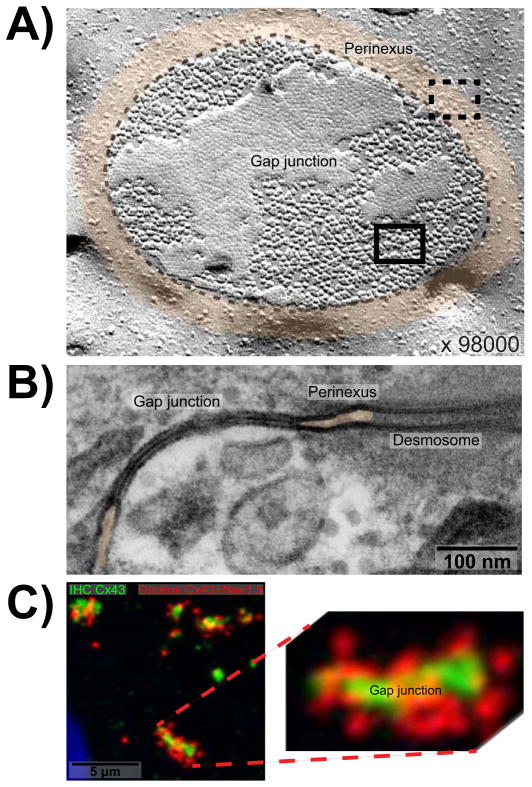
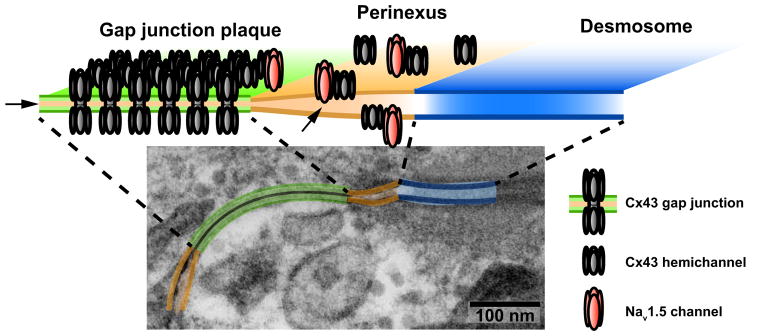
The perinexus is a recently identified microdomain surrounding the cardiac gap junction that contains elevated levels of connexin43 and the sodium channel protein, Nav1.5. Ongoing work has established a role for the perinexus in regulating gap junction aggregation. However, recent studies have raised the possibility of a perinexal contribution at the gap junction cleft to intercellular propagation of action potential via non-electrotonic mechanisms. The latter possibility could modify the current theoretical understanding of cardiac conduction, help explain paradoxical experimental findings, and open up entirely new avenues for antiarrhythmic therapy. We review recent structural insights into the perinexus and its potential novel functional role in cardiac-excitation spread, highlighting presently unanswered questions, the evidence for ephaptic conduction in the heart and how structural insights may help complete this picture.
Copyright © 2013 Elsevier Inc. All rights reserved.
Figures
References
-
- Akar FG, Tomaselli GF. Conduction abnormalities in nonischemic dilated cardiomyopathy: basic mechanisms and arrhythmic consequences. Trends Cardiovasc Med. 2005;15:259–64. - PubMed
-
- Balke CW, Lesh MD, Spear JF, Kadish A, Levine JH, Moore EN. Effects of cellular uncoupling on conduction in anisotropic canine ventricular myocardium. Circ Res. 1988;63:879–92. - PubMed
-
- Beauchamp P, Choby C, Desplantez T, de Peyer K, Green K, Yamada KA, Weingart R, Saffitz JE, Kleber AG. Electrical propagation in synthetic ventricular myocyte strands from germline connexin43 knockout mice. Circ Res. 2004;95:170–8. - PubMed
-
- Callans DJ, Moore EN, Spear JF. Effect of coronary perfusion of heptanol on conduction and ventricular arrhythmias in infarcted canine myocardium. J Cardiovasc Electrophysiol. 1996;7:1159–71. - PubMed
Publication types
MeSH terms
Substances
Grants and funding
LinkOut - more resources
Full Text Sources
Other Literature Sources
Medical
Miscellaneous
