. 2013 Jul;49(1):86-95.
doi: 10.1165/rcmb.2012-0224OC.

X-linked inhibitor of apoptosis regulates lung fibroblast resistance to Fas-mediated apoptosis

Affiliations

X-linked inhibitor of apoptosis regulates lung fibroblast resistance to Fas-mediated apoptosis

Iyabode O Ajayi et al. Am J Respir Cell Mol Biol. 2013 Jul.

Abstract

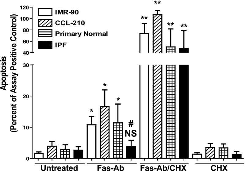
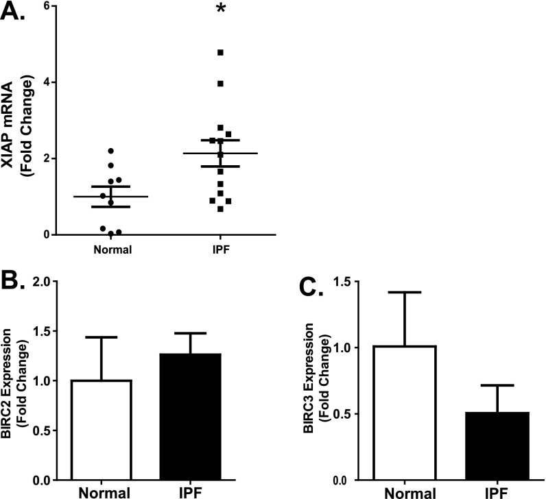
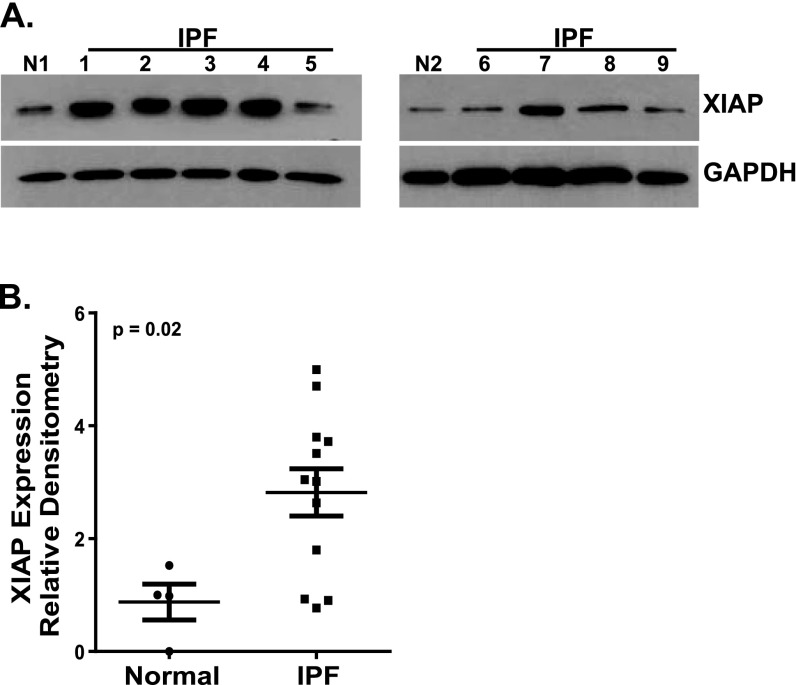
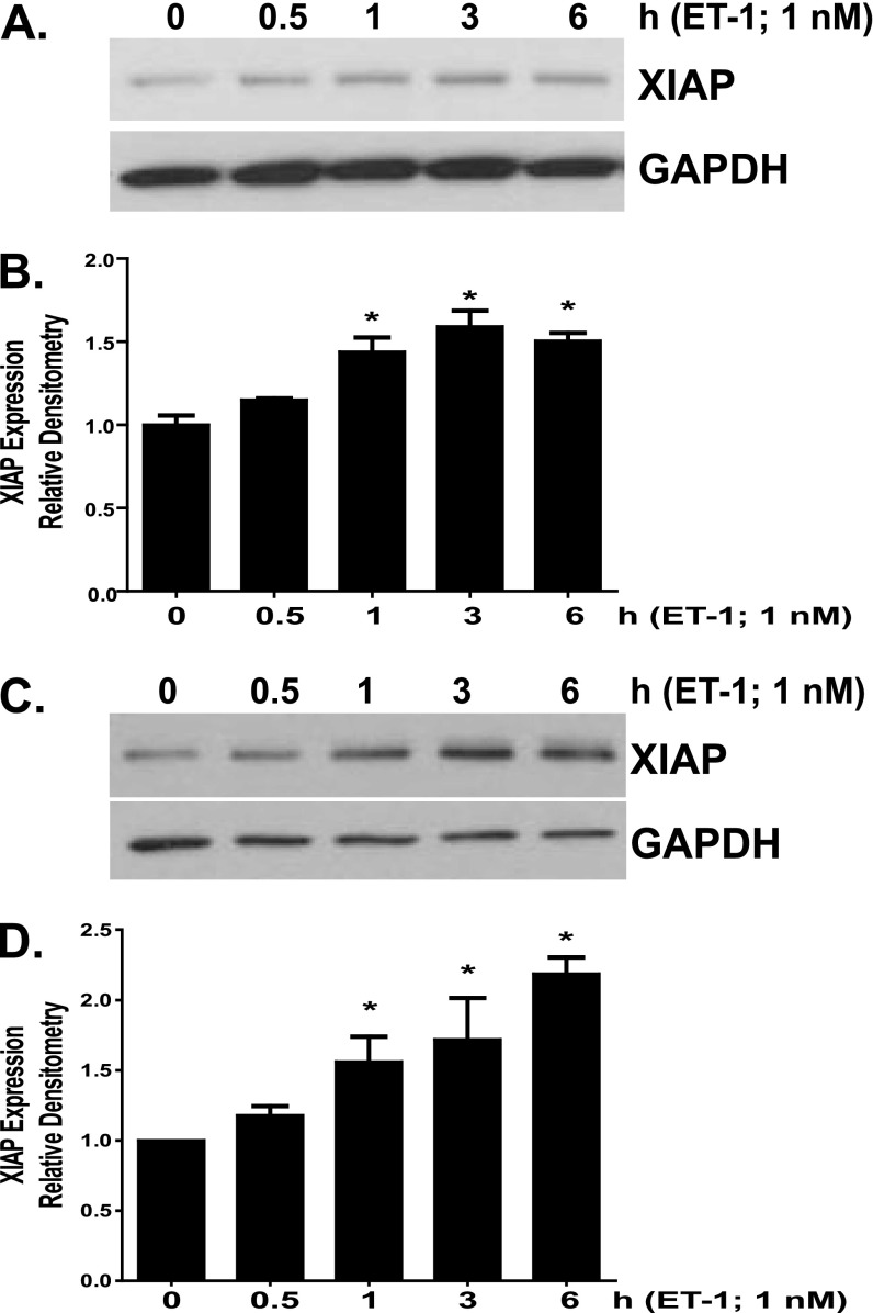
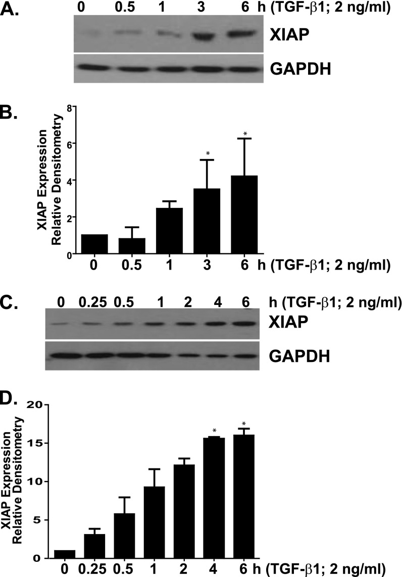
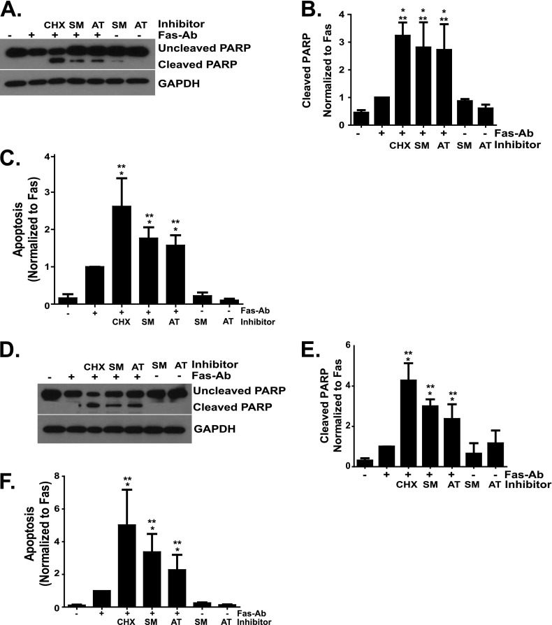
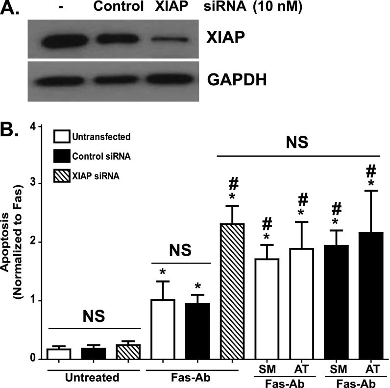
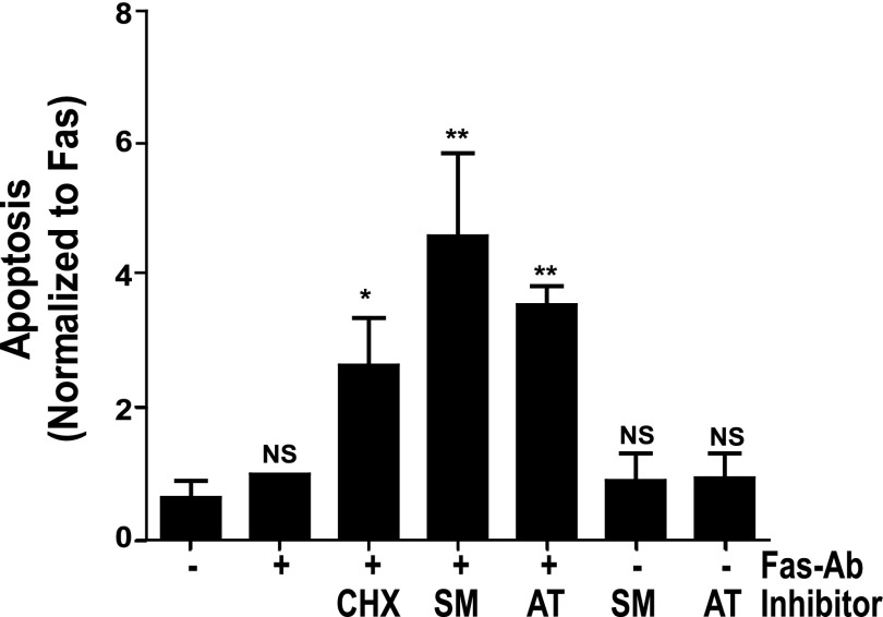
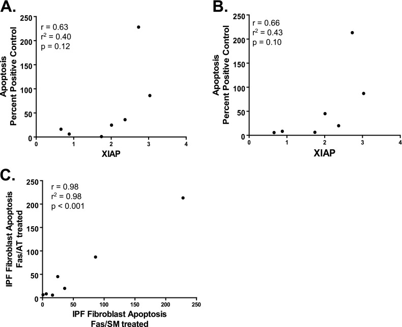
The accumulation of apoptosis-resistant fibroblasts within fibroblastic foci is a characteristic feature of idiopathic pulmonary fibrosis (IPF), but the mechanisms underlying apoptosis resistance remain unclear. A role for the inhibitor of apoptosis (IAP) protein family member X-linked inhibitor of apoptosis (XIAP) has been suggested by prior studies showing that (1) XIAP is localized to fibroblastic foci in IPF tissue and (2) prostaglandin E₂ suppresses XIAP expression while increasing fibroblast susceptibility to apoptosis. Based on these observations, we hypothesized that XIAP would be regulated by the profibrotic mediators transforming growth factor (TGF)β-1 and endothelin (ET)-1 and that increased XIAP would contribute to apoptosis resistance in IPF fibroblasts. To address these hypotheses, we examined XIAP expression in normal and IPF fibroblasts at baseline and in normal fibroblasts after treatment with TGF-β1 or ET-1. The role of XIAP in the regulation of fibroblast susceptibility to Fas-mediated apoptosis was examined using functional XIAP antagonists and siRNA silencing. In concordance with prior reports, fibroblasts from IPF lung tissue had increased resistance to apoptosis compared with normal lung fibroblasts. Compared with normal fibroblasts, IPF fibroblasts had significantly but heterogeneously increased basal XIAP expression. Additionally, TGF-β1 and ET-1 induced XIAP protein expression in normal fibroblasts. Inhibition or silencing of XIAP enhanced the sensitivity of lung fibroblasts to Fas-mediated apoptosis without causing apoptosis in the absence of Fas activation. Collectively, these findings support a mechanistic role for XIAP in the apoptosis-resistant phenotype of IPF fibroblasts.


Figures

<i>Figure 1.</i>
Figure 1.
Growth-arrested IMR-90 (white bar; n = 9), CCL-210 (diagonal cross-hatch bars; n = 3), and primary normal (horizontal cross-hatch bars; n = 3) and idiopathic pulmonary fibrosis (IPF) lung fibroblasts (black bars; n = 9) were treated with or without Fas-activating antibodies (Fas-Ab) (250 ng/ml) in the presence or absence of cycloheximide (CHX) (500 ng/ml) for 16 hours, and apoptosis was assessed by ELISA detection of histone-associated DNA fragments. Relative apoptosis is expressed as a percentage of the assay-positive control that was run on the ELISA plate for each experiment. All samples were run in triplicate for each ELISA. The CHX-alone bars represent a subset of experiments for each fibroblast population, including three IMR-90 experiments, two CCL-210 experiments, two primary normal cell lines, and three of the IPF fibroblast cell lines. *P < 0.05 compared with untreated cells of the same cell line. **P < 0.01 compared with untreated cells of the same cell type. #P < 0.05 compared with Fas-Ab–treated IMR-90, CCL-210, and primary normal lung fibroblasts. NS = no statistically significant difference.
<i>Figure 2.</i>
Figure 2.
RNA isolated from nine different patient-derived normal lung fibroblast lines and 13 different IPF fibroblast lines was reverse transcribed and examined by quantitative real-time PCR for (A) X-linked inhibitor of apoptosis (XIAP), (B) baculoviral inhibitor of apoptosis repeat–containing (BIRC)2, and (C) BIRC3. Each sample was run in triplicate and indexed to the average expression level in the normal lung fibroblasts. *P = 0.025 compared with normal lung fibroblasts.
<i>Figure 3.</i>
Figure 3.
Patient-derived normal lung fibroblasts (n = 4) and IPF fibroblasts (n = 12) were cultured to 60% confluence and growth arrested for 24 hours. XIAP expression was assessed by Western blotting, and all blots were stripped and reprobed for glyceraldehyde-3-phosphate dehydrogenase (GAPDH). Each Western blot contained one normal lung fibroblast line and several IPF lines. (A) Two representative blots demonstrating two different normal fibroblast lines (N1 and N2) and nine different IPF lines (–9). (B) Densitometric analysis was done for each normal and IPF band by determining the XIAP/GAPDH ratio. These ratios were then indexed to the average of the four normals expressed as the fold-change in the IPF cell lines. On the scatterplot, each point represents the average relative density for a different cell line. P = 0.02 for normal versus IPF fibroblasts.
<i>Figure 4.</i>
Figure 4.
IMR-90 (A and B) and CCL-210 (C and D) fibroblasts were treated with endothelin (ET)-1 (1.0 nM). (A and C) Cell lysates were analyzed for XIAP expression by Western blotting, and each blot was stripped and reprobed for GAPDH. (B and D) Each experiment was completed in triplicate; XIAP/GAPDH ratios were indexed to untreated cells for each blot. *P < 0.01 compared with untreated controls.
<i>Figure 5.</i>
Figure 5.
IMR-90 (A and B) and CCL-210 (C and D) fibroblasts were treated with transforming growth factor (TGF)-β1 (2 ng/ml). (A and C) Cell lysates were analyzed for XIAP expression by Western blotting, and each blot was stripped and reprobed for GAPDH. (B and D) Each experiment was completed in triplicate; XIAP/GAPDH ratios were indexed to untreated cells for each blot. *P < 0.01 compared with untreated controls.
<i>Figure 6.</i>
Figure 6.
IMR-90 (AC) and CCL-210 (DF) fibroblasts were treated with or without Fas-Ab (250 ng/ml) in the presence or absence of CHX (500 ng/ml), the Smac/DIABLO mimetic SM164 (100 nM), the Smac/DIABLO mimetic AT406 (1.0 μM), or the Smac/DIABLO mimetics alone for 16 hours. Apoptosis was assessed by Western blotting for cleaved poly-(ADP-ribose) polymerase (PARP) (A and D) and by ELISA detection of histone-associated DNA fragments (C and F). The Western blots were stripped and probed for GAPDH. Relative densitometry was determined by indexing the cleaved PARP/GAPDH ratio for each band to the Fas-Ab treatment (B and E). Densitometry and ELISA data represent at least three independent experiments. *P < 0.01 compared with untreated controls. **P < 0.05 compared with fibroblasts treated with Fas-Ab.
<i>Figure 7.</i>
Figure 7.
(A) XIAP expression was assessed by Western blotting of untransfected IMR-90 fibroblasts and IMR-90 fibroblasts transfected for 24 hours with 10 nM of a nontargeting (control) siRNA or a siRNA-targeting XIAP. The representative blot was stripped and probed for GAPDH. (B) Untransfected IMR-90 fibroblasts and IMR-90 fibroblasts transfected with the control or with XIAP siRNA were treated with or without Fas-Ab (250 ng/ml) with or without the Smac/Diablo mimetics SM164 (100 nM) or AT406 (1.0 μM) for 16 hours, and apoptosis was assessed using ELISA for detection of histone-associated DNA fragments. Data are indexed to the average of the untransfected Fas-Ab–treated cells to allow relative comparisons. Data shown are from five independent experiments with each condition measured in triplicate. *P < 0.01 compared with untransfected cells without treatment. #P < 0.01 compared with untransfected cells treated with Fas-Ab. Additional control conditions (Fas-Ab with cycloheximide, cycloheximide alone, SM164 alone, and AT406 alone) are shown in Figure E3. NS, not significantly different from untreated cells.
<i>Figure 8.</i>
Figure 8.
IPF fibroblasts (n = 7) were treated with or without Fas-Ab in the presence or absence of CHX (250 ng/ml), with the Smac/DIABLO mimetics (SM164, 100 nM or AT406, 1.0 μM), or with the Smac/DIABLO mimetics alone for 16 hours, and apoptosis was assessed by ELISA detection of histone-associated DNA fragments. Each experiment was indexed to Fas-Ab–treated cells in the absence of any inhibitor. *P < 0.01 versus untreated or Fas-Ab–treated cells. **P < 0.001 versus untreated or Fas-Ab–treated cells. NS = not significantly different from untreated cells.
<i>Figure 9.</i>
Figure 9.
Correlation between XIAP protein expression in IPF fibroblasts, as determined by densitometric analysis indexed to the average expression of XIAP in normal lung fibroblasts, and apoptosis (shown as the percent of assay positive control) induced by Fas-Ab combined with (A) SM164 (100 nM; Fas/SM) or (B) AT406 (1.0 μM; Fas/AT). (C) Correlation between apoptosis in IPF fibroblasts treated with Fas-Ab in combination with SM164 (Fas/SM) or AT406 (Fas/AT). For AC, each point represents a distinct IPF fibroblast cell line.

References

    1. Hecker L, Jagirdar R, Jin T, Thannickal VJ. Reversible differentiation of myofibroblasts by MyoD. Exp Cell Res. 2011;317:1914–1921. - PMC - PubMed
    1. Hinz B, Phan SH, Thannickal VJ, Prunotto M, Desmouliere A, Varga J, De Wever O, Mareel M, Gabbiani G. Recent developments in myofibroblast biology: paradigms for connective tissue remodeling. Am J Pathol. 2012;180:1340–1355. - PMC - PubMed
    1. Hinz B, Gabbiani G. Fibrosis: recent advances in myofibroblast biology and new therapeutic perspectives. F1000 Biol Rep. 2010;2:78. - PMC - PubMed
    1. Tomasek JJ, Gabbiani G, Hinz B, Chaponnier C, Brown RA. Myofibroblasts and mechano-regulation of connective tissue remodelling. Nat Rev Mol Cell Biol. 2002;3:349–363. - PubMed
    1. Desmouliere A, Redard M, Darby I, Gabbiani G. Apoptosis mediates the decrease in cellularity during the transition between granulation tissue and scar. Am J Pathol. 1995;146:56–66. - PMC - PubMed

Publication types

MeSH terms