. 2013 Jul;49(1):78-85.
doi: 10.1165/rcmb.2012-0107OC.

Differential regulation of pulmonary vascular cell growth by hypoxia-inducible transcription factor-1α and hypoxia-inducible transcription factor-2α

Affiliations

Differential regulation of pulmonary vascular cell growth by hypoxia-inducible transcription factor-1α and hypoxia-inducible transcription factor-2α

Aftab Ahmad et al. Am J Respir Cell Mol Biol. 2013 Jul.

Abstract

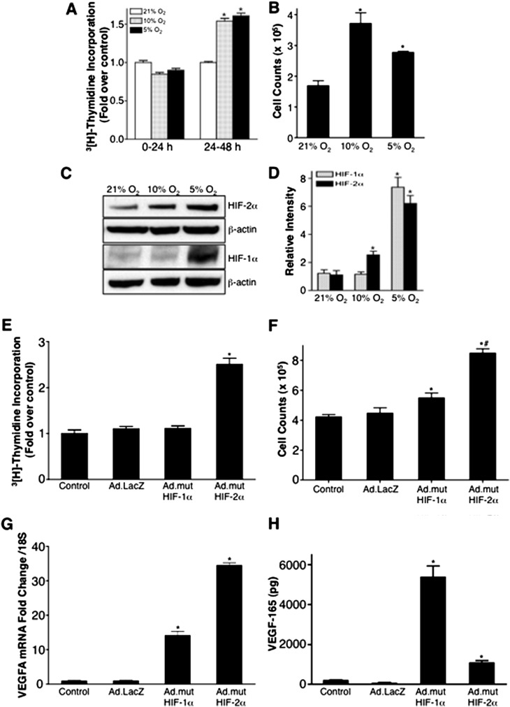
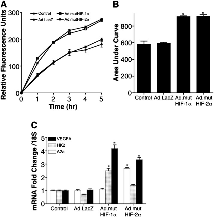
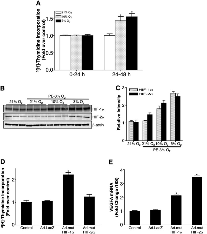
Hypoxia-inducible transcription factors HIF-1α and HIF-2α can contribute to pulmonary hypertension and vascular remodeling, but their mechanisms remain unknown. This study investigated the role of HIF-1α and HIF-2α in pulmonary artery endothelial and smooth muscle cells. The exposure of human pulmonary artery endothelial cells (HPAECs) to hypoxia (10% O₂ or 5% O₂) increased proliferation over 48 hours, compared with cells during normoxia (21% O₂). The adenovirus-mediated overexpression of HIF-2α that is transcriptionally active during normoxia (mutHIF-2α) increased HPAEC proliferation, whereas the overexpression of HIF-1α, which is transcriptionally active during normoxia (mutHIF-1α), exerted no effect. The knockdown of HIF-2α decreased proliferation during both hypoxia and normoxia. Both HIFs increased migration toward fibrinogen, used as a chemoattractant. In an angiogenesis tube formation assay, mutHIF-2α-transduced cells demonstrated increased tube formation, compared with the mutHIF-1α-transduced cells. In addition, the tubes formed in HIF-2α-transduced cells were more enduring than those in the other groups. In human pulmonary artery smooth muscle cells (HPASMCs), chronic exposure to hypoxia increased proliferation, compared with cells during normoxia. For HPASMCs transduced with adenoviral HIFs, HIF-1α increased proliferation, whereas HIF-2α exerted no such effect. Thus, HIF-1α and HIF-2α exert differential effects in isolated cells of the human pulmonary vasculature. This study demonstrates that HIF-2α plays a predominant role in the endothelial growth pertinent to the remodeling process. In contrast, HIF-1α appears to play a major role in pulmonary smooth muscle growth. The selective targeting of each HIF in specific target cells may more effectively counteract hypoxic pulmonary hypertension and vascular remodeling.


Figures

<i>Figure 1.</i>
Figure 1.
Effects of hypoxia and hypoxia-inducible transcription factors (HIFs) on proliferation of human pulmonary artery endothelial cells (HPAECs). (A) HPAECs were exposed to 21% O2, 10% O2, or 5% O2 for the indicated times. 3[H]-thymidine was added 24 hours before harvest, and the incorporated radioactivity was measured. (B) HPAECs were similarly exposed to 21% O2, 10% O2, or 5% O2 for 48 hours, and cells were counted after trypsinization. (C) Immunoblots with nuclear lysates from HPAECs were exposed similarly for 48 hours. Blots were probed with anti–HIF-2α, anti–HIF-1α, and β-actin antibodies. (D) For densitometry, blots were imaged using a Bio-Rad Gel Doc system (Bio-Rad, Hercules, CA), and band-quantitated using Image Lab software (Media Cybernetics). (E) HPAECs were transduced with control Ad.LacZ or adenovirus vectors encoding constitutively active HIF-1α (Ad.mutHIF-1α) or constitutively active HIF-2α (Ad.mutHIF-2α) at a multiplicity of infection of 10 plaque-forming units/cell. 3[H]-thymidine was added 6 hours after transduction, and proliferation was assessed 24 hours after transduction. (F) Cells were also counted after adenoviral transductions. (G) In parallel experiments, RNA was isolated, and real-time–PCR was performed for human vascular endothelial growth factor (VEGF)–A. (H) In adenoviral-transduced cells, total VEGF-165 was measured in media and cell lysates. *P < 0.05, compared with the 21% O2 control cells (A and B) or control or Ad.LacZ-treated cells (D, E, and H). #P < 0.05, compared with the Ad.LacZ-treated cells (F).
<i>Figure 2.</i>
Figure 2.
Effects of HIF knockdown on the proliferation of HPAECs. HPAECs were transfected with control siRNA (siCtr) or siRNA against HIF-1α (siHIF-1α) or HIF-2α (siHIF-2α). Twenty-four hours after transfection, cells were exposed to 21% O2 or 10% O2. (A) 3[H]-thymidine was added after 24 hours of exposure, and proliferation was assessed at 48 hours. (B) In parallel experiments, RNA was isolated and RT-PCR was performed for human HIF-1α and HIF-2α. (C) In parallel experiments, an immunoblot of nuclear lysates of cells transfected with siRNAs was probed with anti–HIF-2α, anti–HIF-1α, and β-actin antibodies. Ctr, control. *P < 0.05 versus normoxia-exposed control or siCtr cells. #P < 0.05 versus respective control cells during normoxia or hypoxia.
<i>Figure 3.</i>
Figure 3.
Effects of HIFs on the migration of HPAECs. HPAECs were transduced with control Ad.LacZ, Ad.mutHIF-1α, or Ad.mutHIF-2α. Cells were stained with DilC12 (3) (BD Biosciences, Bedford, MA), and 300,000 cells were plated on FluoroBlok inserts (BD Biosciences). Plates were incubated at 37°C in a cell-culture incubator, and the migration toward fibrinogen (50 μg/ml) was recorded using a fluorescence plate reader. (A) Changes in fluorescence with time and (B) areas under the curve were calculated. (C) RT-PCR of the migrated cells. *P < 0.05 versus control or Ad.LacZ-transduced cells.
<i>Figure 4.</i>
Figure 4.
Effects of HIFs on the angiogenic activity of HPAECs. HPAECs were either untreated or transduced with an empty adenovirus vector (Ad.Empty Vector), or the constitutively active HIF-1α (Ad.mutHIF-1α) or the constitutively active HIF-2α (Ad.mutHIF-2α) adenoviral constructs at a multiplicity of 10 plaque-forming units/cell. Eighteen hours after transduction, HPAECs were split and plated at 100,000 cells/well in a 24-well plate coated with Matrigel (BD BioSciences). After 24 hours, images were taken at ×4 magnification. (A) Representative images. An average of 25 images per condition was analyzed, using Image Pro Premier software (Media Cybernetics). The average number of branch points per field (B), the average number of tubes per field (C), and the average area (in μm2) covered by tubes per field (D) were calculated and plotted. *P < 0.05, compared with nontransduced control cells. #P < 0.5, compared with the empty vector control cells. $P < 0.05, compared with Ad.mutHIF-1α.
<i>Figure 5.</i>
Figure 5.
Effects of hypoxia and HIFs on the proliferation of human pulmonary artery smooth muscle cells (HPASMCs). (A) HPASMCs were pre-exposed (PE) for 4 days to 3% O2 and replated. Twenty-four hours after plating, cells were exposed to 21% O2, 10% O2, or 3% O2 for the indicated times. 3[H]-thymidine was added 24 hours before harvest, and the incorporated radioactivity was measured. (B) Immunoblot with nuclear lysate from HPASMCs exposed similarly. Blots were probed with anti–HIF-1α, stripped, and reprobed with anti–HIF-2α, followed by stripping and probing with β-actin antibodies. (C) For densitometry, blots were imaged using a Bio-Rad Gel Doc system, and band-quantitated using Image Lab software (Media Cybernetics). (D) HPASMCs were transduced with control Ad.LacZ, Ad.mutHIF-1α, or Ad.mutHIF-2α constructs. 3[H]-thymidine was added 6 hours after transduction, and proliferation was assessed 24 hours after transduction. (E) In parallel experiments, RNA was isolated, and RT-PCR was performed for human VEGF-A. *P < 0.05 versus normoxia-exposed cells (A) or control and Ad.LacZ-treated cells (D and E).

References

    1. Pak O, Aldashev A, Welsh D, Peacock A. The effects of hypoxia on the cells of the pulmonary vasculature. Eur Respir J. 2007;30:364–372. - PubMed
    1. Yu L, Hales CA. Hypoxia does neither stimulate pulmonary artery endothelial cell proliferation in mice and rats with pulmonary hypertension and vascular remodeling nor in human pulmonary artery endothelial cells. J Vasc Res. 2011;48:465–475. - PMC - PubMed
    1. Presberg KW, Dincer HE. Pathophysiology of pulmonary hypertension due to lung disease. Curr Opin Pulm Med. 2003;9:131–138. - PubMed
    1. Brusselmans K, Compernolle V, Tjwa M, Wiesener MS, Maxwell PH, Collen D, Carmeliet P. Heterozygous deficiency of hypoxia-inducible factor–2alpha protects mice against pulmonary hypertension and right ventricular dysfunction during prolonged hypoxia. J Clin Invest. 2003;111:1519–1527. - PMC - PubMed
    1. Yu AY, Shimoda LA, Iyer NV, Huso DL, Sun X, McWilliams R, Beaty T, Sham JS, Wiener CM, Sylvester JT, et al. Impaired physiological responses to chronic hypoxia in mice partially deficient for hypoxia-inducible factor 1alpha. J Clin Invest. 1999;103:691–696. - PMC - PubMed

Publication types

MeSH terms

Substances