. 2013 Apr;24(5):727-43.
doi: 10.1681/ASN.2012070700. Epub 2013 Mar 14.

Vps34 deficiency reveals the importance of endocytosis for podocyte homeostasis

Affiliations

Vps34 deficiency reveals the importance of endocytosis for podocyte homeostasis

Wibke Bechtel et al. J Am Soc Nephrol. 2013 Apr.

Abstract

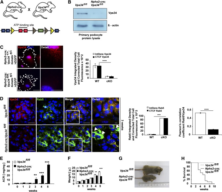
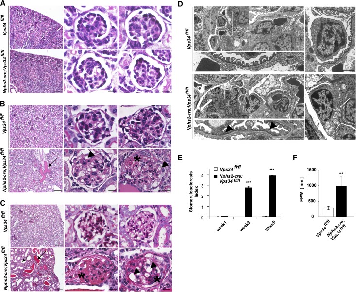
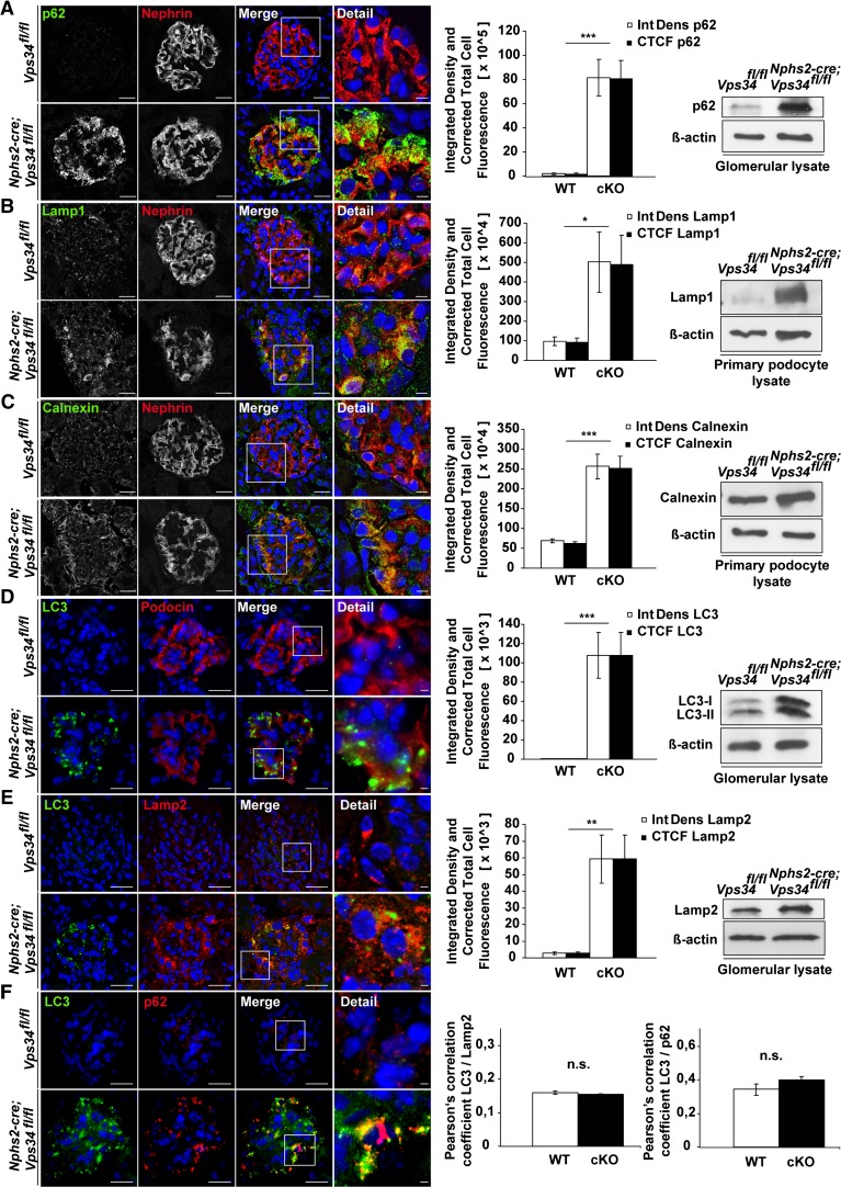
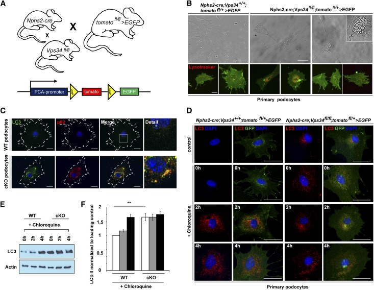
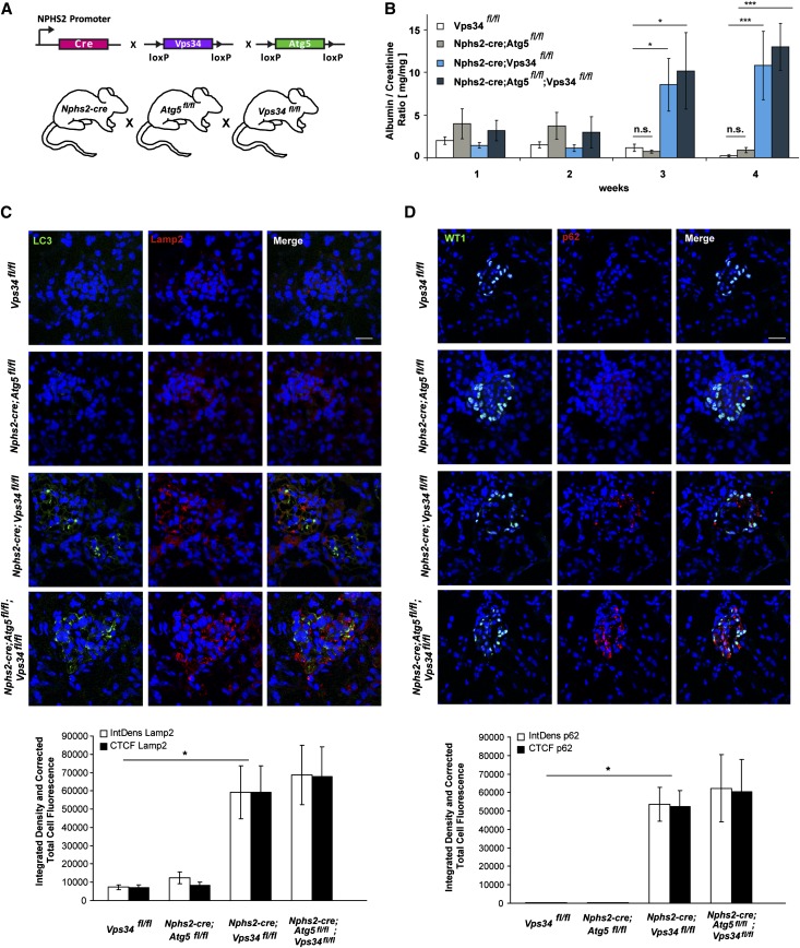
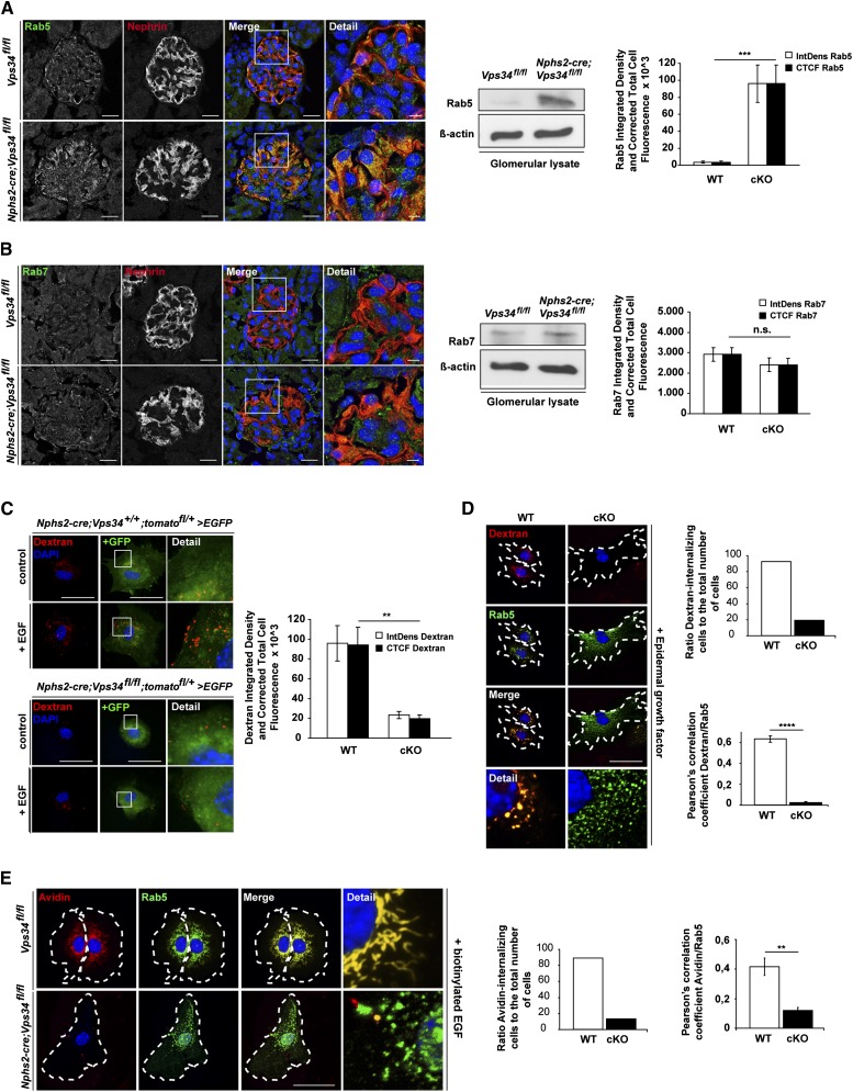
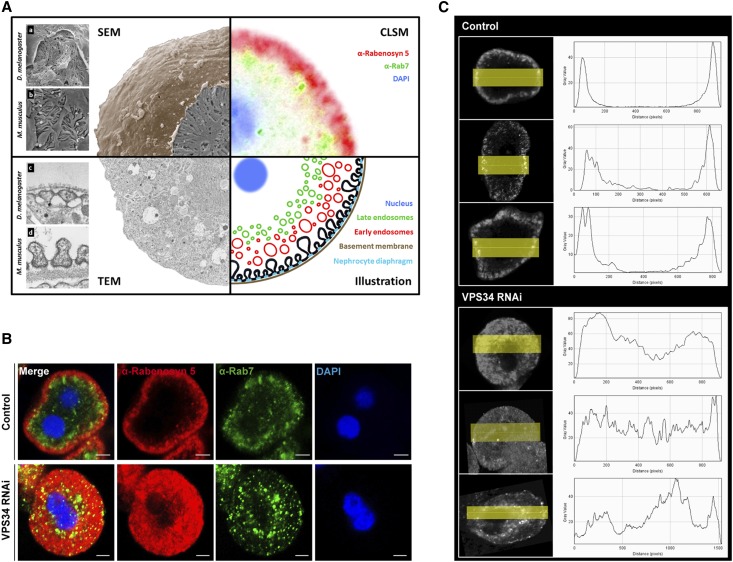
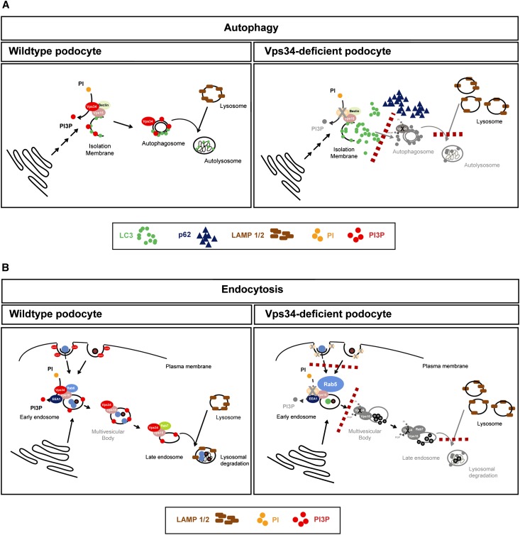
The molecular mechanisms that maintain podocytes and consequently, the integrity of the glomerular filtration barrier are incompletely understood. Here, we show that the class III phosphoinositide 3-kinase vacuolar protein sorting 34 (Vps34) plays a central role in modulating endocytic pathways, maintaining podocyte homeostasis. In mice, podocyte-specific conditional knockout of Vps34 led to early proteinuria, glomerular scarring, and death within 3-9 weeks of age. Vps34-deficient podocytes exhibited substantial vacuolization and foot process effacement. Although the formation of autophagosomes and autophagic flux were impaired, comparisons between podocyte-specific Vps34-deficient mice, autophagy-deficient mice, and doubly deficient mice suggested that defective autophagy was not primarily responsible for the severe phenotype caused by the loss of Vps34. In fact, Rab5-positive endosomal compartments, endocytosis, and fluid-phase uptake were severely disrupted in Vps34-deficient podocytes. Vps34 deficiency in nephrocytes, the podocyte-like cells of Drosophila melanogaster, resulted in a block between Rab5- and Rab7-positive endosomal compartments. In summary, these data identify Vps34 as a major regulator of endolysosomal pathways in podocytes and underline the fundamental roles of endocytosis and fluid-phase uptake for the maintenance of the glomerular filtration barrier.


Figures

Figure 1.
Figure 1.
Conditional Vps34 depletion in mouse podocytes induces massive proteinuria and early lethality. (A) Mice expressing cre recombinase under control of the podocyte-specific Nphs2 promoter were crossed to Vps34fl/fl mice to generate podocyte-specific Vps34 knockout mice. (B) Western blot analysis of freshly isolated podocyte protein lysates confirmed significant reduction of Vps34 in podocytes of Nphs2-cre;Vps34fl/fl conditional knockout mice. β-actin was used as loading control. (C) Quantification of immunofluorescence microscopy confirms efficient knockout of Vps34 in primary isolated podocytes. Scale bars, 20 µm; 5 µm in detail. (D) Colabeling immunofluorescence staining with Rab5 and Vps34 on kidney sections of conditionally Vps34-deficient mice and wild-type controls. Increased Rab5 expression in Vps34-deficient podocytes at 3 weeks of age (white arrowheads). ****P<0.0001. (E) Nphs2-cre;Vps34fl/fl mice develop early proteinuria. Albumin/creatinine ratios are significantly increased from 3 weeks of age in Vps34fl/fl;Nphs2-cre mice (n=10 per group; ***P<0.001, **P<0.01; two-tailed t test, mean values ± SEM are shown). (F) Weight curve of Vps34fl/fl control and Nphs2-cre;Vps34fl/fl conditional knockout mice; significant differences in weight can be observed by week 4 (n=10 per group; ***P<0.001, **P<0.01, two-tailed t test, mean values ± SEM are shown). (G) Nphs2-cre;Vps34fl/fl mice exhibit significant growth impairment at 5 weeks of age. (H) Kaplan–Meyer survival curve of Vps34fl/fl control and Nphs2-cre;Vps34fl/fl mice (n=35 per group). All Nphs2-cre;Vps34fl/fl mice died within 9–10 weeks after birth; the first death was observed at 3 weeks of age.
Figure 2.
Figure 2.
Podocyte-specific Vps34 deficiency causes rapid podocyte degeneration and early-onset glomerulosclerosis. (A–C) PAS staining of kidney sections of 1-, 3-, and 9-week-old Nphs2-cre;Vps34fl/fl mice and littermate controls. (A) By 1 week after birth, kidney sections do not display significant differences in glomerular architecture. (B) Kidney sections of 3-week-old Nphs2-cre;Vps34fl/fl mice show first signs of tubular protein accumulation (arrow), vacuolization (arrowheads), and segmental sclerosis (asterisk). (C) At 9 weeks of age, kidney sections of Nphs2-cre;Vps34fl/fl mice show undulated renal surface, widespread tubular protein casts with tubular dilatation (arrows), and severe glomerular sclerosis (asterisks). Scale bars, 200 µm in left panels; 10 µm in center and right panels. (D) By 3 weeks of age, electron microscopy of Nphs2-cre;Vps34fl/fl mice showed extensive vacuolization of podocytes (white arrow) and focal foot process fusions (black arrowheads). Scale bars, 5 µm in upper left panels; 2 µm, upper center panel; 1 µm, upper right panel; 500 nm, lower panels. (E) Glomerulosclerosis index of glomerular sections of 1-, 3-, and 9-week-old Nphs2-cre;Vps34fl/fl mice and wild-type controls. (F) Foot process widths of Nphs2-cre;Vps34fl/fl mice and wild-type controls (***P<0.001, two-tailed t test, mean values ± SEM are shown).
Figure 3.
Figure 3.
Autophagy is impaired in Nphs2-cre;Vps34fl/fl mice. Immunofluorescence staining of kidney sections of 3-week-old Nphs2-cre;Vps34fl/fl and littermate control mice. (A–E) Quantification of confocal immunofluorescence microscopy and Western blot analyses of glomerular or primary podocyte protein lysates displayed significant accumulation of the autophagy marker p62, the lysosomal markers Lamp1 and -2, the ER stress marker Calnexin, and the autophagy marker LC3 in podocytes of Nphs2-cre;Vps34fl/fl mice. (E) Confocal microscopy showed no significant colocalization of LC3 and the lysosomal marker Lamp2 in Vps34-deficient podocytes. (F) Colocalization of LC3 and p62 in Vps34-deficient podocytes in confocal microscopy. *P<0.05, **P<0.01, ***P<0.001. Scale bars, 20 µm; 5 µm in detail.
Figure 4.
Figure 4.
Autophagic flux is abrogated in primary Vps34-deficient podocytes. (A) Nphs2-cre;tomatofl/+>EGFP mice were crossed with Vps34fl/fl and Vps34+/+ mice to obtain Vps34-deficient, GFP-positive podocytes and GFP-positive control podocytes. (B) Primary podocytes at passage 1 were observed by phase/contrast microscopy for 9 days. Vps34-deficient podocytes showed substantial perinuclear vacuolization. Counterstaining with Lysotracker Red was performed to determine if the vacuoles seen in Vps34-deficient podocytes represent accumulation of lysosomal acidic vesicles. Scale bars, 20 µm. (C) Immunofluorescence staining showed massive accumulation of LC3 and p62 in Nphs2-cre;Vps34fl/fl;tomatofl/+>EGFP podocytes. Scale bars, 20 µm. (D–F) Autophagic flux was impaired in Nphs2-cre;Vps34fl/fl;tomatofl/+>EGFP primary podocytes. Serum deprivation induced LC3 accumulation in wild-type GFP-positive primary podocytes that was further enhanced by blockade of autophagosomal degradation with the lysosomal inhibitor chloroquine (10 µM). In contrast, LC3 was already accumulated in Vps34-deficient primary GFP-positive podocytes with no further increase on serum starvation or disruption of autophagosomal–lysosomal fusion by chloroquin. **P<0.01. Scale bars, 50 µm.
Figure 5.
Figure 5.
Abrogated autophagic flux is not causative for the severe podocyte phenotype of Vps34-deficient podocytes. (A) Vps34fl/fl mice were crossed to Nphs2-cre;Atg5fl/fl mice to obtain Nphs2-cre;Atg5fl/fl;Vps34fl/fl conditional double knockout mice. (B) Albumine/creatinine ratios were significantly increased from 3 weeks of age in Vps34fl/fl;Nphs2-cre and Nphs2-cre;Atg5fl/fl;Vps34fl/fl conditional double knockout mice compared with Nphs2-cre;Atg5fl/fl mice and littermate controls (n=10 per group, *P<0.05, ***P<0.01, two-tailed t test, mean values ± SEM are shown). (C) Confocal microscopy showed accumulation of LC3 and Lamp2 in podocytes of Nphs2-cre;Vps34fl/fl single mutant mice and Nphs2-cre;Atg5fl/fl;Vps34fl/fl conditional double knockout mice. No significant colocalization of LC3 and Lamp2 could be observed. Nphs2-cre; Atg5fl/fl single conditional knockout mice did not show increase of LC3 or Lamp2 in podocytes at 3 weeks of age. Scale bars, 20 µm. (D) p62 accumulates in podocytes of Nphs2-cre;Vps34fl/fl single conditional knockout mice and Nphs2-cre;Atg5fl/fl;Vps34fl/fl conditional double knockout mice. Nphs2-cre;Atg5fl/fl mice did not show increase of p62 in podocytes at 3 weeks of age. Scale bars, 20 µm.
Figure 6.
Figure 6.
Ablation of Vps34 in mouse podocytes leads to impaired endocytosis. (A and B) Confocal immunofluorescence microscopy of kidney sections of 3-week-old Nphs2-cre;Vps34fl/fl and littermate control mice. (A) Rab5, a marker for early endosomes and activator of Vps34, was strongly increased in Nphs2-cre;Vps34fl/fl podocytes at 3 weeks of age. Scale bars, 5 µm. (B) Rab7, a marker for the late endosome, showed no significant differences in podocytes of Nphs2-cre;Vps34fl/fl and Vps34fl/fl mice. Scale bars, 5 µm. (A and B) Western blot analysis of glomerular protein lysates of 3-week-old mice shows that the early endosomal marker Rab5 is accumulated in glomerulus lysates of Nphs2-cre;Vps34fl/fl mice compared with littermate controls. No changes in the late endosomal marker Rab7 can be observed. β-actin was used as loading control. (C and D) Immunofluorescence and quantification of fluid-phase uptake in primary podocytes. Stimulation with 20 nM EGF for 5 minutes induced uptake of dextran in GFP-positive primary control podocytes. In contrast, Vps34-deficient primary GFP-positive podocytes showed impaired uptake of dextran, indicating a blockade in endocytosis. Scale bars, 50 µm. (D) Costaining with Rab5 revealed significant overlap in wild-type podocytes, whereas in Vps34-deficient primary podocytes, Rab5 accumulation did not colocalize with dextran. (E) Immunofluorescence and quantification of streptavidin uptake in primary podocytes. Stimulation of GFP-positive primary wild-type podocytes with 20 nM biotinylated EGF induced uptake of streptavidin coupled to Alexa Fluor 555. In contrast, Vps34-deficient primary GFP-positive podocytes showed impaired uptake of streptavidin, indicating a blockade in receptor-mediated endocytosis. Scale bars, 50 µm. Costaining with Rab5 revealed significant overlap in wild-type podocytes, whereas in Vps34-deficient primary podocytes, Rab5 accumulation did not colocalize with streptavidin. **P<0.01, ***P<0.001, ****P<0.0001.
Figure 7.
Figure 7.
Vps34 is an important regulator of the early endosomal compartment in the podocyte-like Drosophila cell—the nephrocyte. (A) Combined schematic of electron microscopy and confocal immunofluorescence microscopy of GCNs. (a–d) Structural comparison of (a) GCN with (b) podocytes and (c) nephrocyte diaphragm with (d) slit diaphragm. The rough surface of the GCN underneath the (a) basement membrane is formed by the (c) nephrocyte diaphragm. In contrast to the mammalian slit diaphragm, which is formed between (b) neighboring podocytes, (a) the Drosophila nephrocyte diaphragm is formed within one GCN. The illustration shows the endocytosis process by formation of early endosomes from invaginations of the nephrocyte diaphragm and their maturation to late endosomes. (B) Confocal immunofluorescence microscopy of Vps34-deficient and wild-type control GCNs. The control shows the thin outer ring of Rabenosyn5-positive early endosomes (red) and the inner ring of Rab7-positive late endosomes (green). On expression of Vps34RNAi, this pattern is disrupted, and the whole cytoplasm is filled with Rabenosyn5-positive endosomes. Control: prospero GAL4/+(2); Vps34RNAi: prospero-GAL4/upstream activating sequence-Vps34-RNAi(2). Scale bars, 5 µm. (C) Distribution profile of the early endosomal marker Rabenosyn5.
Figure 8.
Figure 8.
Schematic illustration of Vps34 deficiency in podocytes. (A) Vps34 deficiency leads to incomplete formation of the autophagosomal membrane, resulting in deficient autophagy and autophagosomal fusion. LC3 can still be converted from LC3-I to -II, but no functional autophagosome is formed. It leads to an accumulation of nondegraded LC3 and p62 as well as an accumulation of vacant lysosomes and accumulation of Lamp1/2. The absence of PI(3)P causes a blockade in autophagosomal formation and autophagosomal–lysosomal fusion. (B) Endosomal trafficking is blocked in Vps34-deficient podocytes. Lack of PI(3)P production inhibits fluid-phase uptake, receptor-mediated endocytosis, and maturation of the early endosome to the late endosome, resulting in an accumulation of Rab5. Rab7, a marker for the late endosome, is not affected. Lamp1/2, markers for the lysosome, are upregulated, indicating unused lysosomes because of insufficient endolysosomal fusion.

References

    1. Farquhar MG, Vernier RL, Good RA: An electron microscope study of the glomerulus in nephrosis, glomerulonephritis, and lupus erythematosus. J Exp Med 106: 649–660, 1957 - PMC - PubMed
    1. Farquhar MG, Palade GE: Segregation of ferritin in glomerular protein absorption droplets. J Biophys Biochem Cytol 7: 297–304, 1960 - PMC - PubMed
    1. Farquhar MG, Palade GE: Glomerular permeability. II. Ferritin transfer across the glomerular capillary wall in nephrotic rats. J Exp Med 114: 699–716, 1961 - PMC - PubMed
    1. Farquhar MG, Wissig SL, Palade GE: Glomerular permeability. I. Ferritin transfer across the normal glomerular capillary wall. J Exp Med 113: 47–66, 1961 - PMC - PubMed
    1. Caulfield JP, Farquhar MG: The permeability of glomerular capillaries to graded dextrans. Identification of the basement membrane as the primary filtration barrier. J Cell Biol 63: 883–903, 1974 - PMC - PubMed

Publication types

Substances

LinkOut - more resources