. 2013 Mar 14;4(3):e546.
doi: 10.1038/cddis.2013.74.

Olig2/Plp-positive progenitor cells give rise to Bergmann glia in the cerebellum

Affiliations

Olig2/Plp-positive progenitor cells give rise to Bergmann glia in the cerebellum

S-H Chung et al. Cell Death Dis. .

Abstract

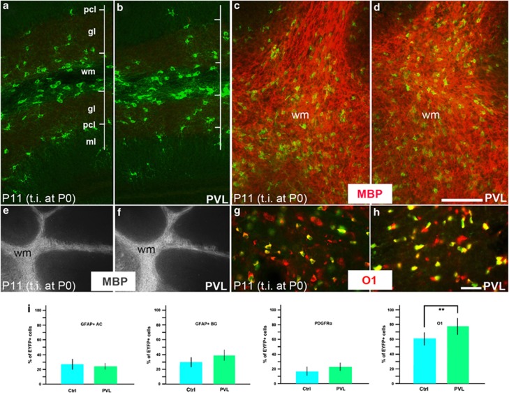
NG2 (nerve/glial antigen2)-expressing cells represent the largest population of postnatal progenitors in the central nervous system and have been classified as oligodendroglial progenitor cells, but the fate and function of these cells remain incompletely characterized. Previous studies have focused on characterizing these progenitors in the postnatal and adult subventricular zone and on analyzing the cellular and physiological properties of these cells in white and gray matter regions in the forebrain. In the present study, we examine the types of neural progeny generated by NG2 progenitors in the cerebellum by employing genetic fate mapping techniques using inducible Cre-Lox systems in vivo with two different mouse lines, the Plp-Cre-ER(T2)/Rosa26-EYFP and Olig2-Cre-ER(T2)/Rosa26-EYFP double-transgenic mice. Our data indicate that Olig2/Plp-positive NG2 cells display multipotential properties, primarily give rise to oligodendroglia but, surprisingly, also generate Bergmann glia, which are specialized glial cells in the cerebellum. The NG2+ cells also give rise to astrocytes, but not neurons. In addition, we show that glutamate signaling is involved in distinct NG2+ cell-fate/differentiation pathways and plays a role in the normal development of Bergmann glia. We also show an increase of cerebellar oligodendroglial lineage cells in response to hypoxic-ischemic injury, but the ability of NG2+ cells to give rise to Bergmann glia and astrocytes remains unchanged. Overall, our study reveals a novel Bergmann glia fate of Olig2/Plp-positive NG2 progenitors, demonstrates the differentiation of these progenitors into various functional glial cell types, and provides significant insights into the fate and function of Olig2/Plp-positive progenitor cells in health and disease.


Figures

Figure 1
Figure 1
Expression pattern of Plp-expressing NG2+ cells in the mouse cerebellum: Plp-expressing NG2+ cells primarily generate oligodendrocytes. (a) Sagittal sections of the postnatal day 60 (P60) Plp-Cre-ERT2/Rosa26-EYFP (PCE/R) double-transgenic mouse cerebellum immunofluorescence stained with anti-GFP after tamoxifen injection (t.i.) at P7. Strong and specific EYFP-positive cells were observed throughout the cerebellum. (a') A higher magnification view of a rectangle area in (a). (b) Sagittal sections of the PCE/R double-transgenic mouse cerebellum immunofluorescence stained with anti-GFP. Tamoxifen was injected at embryonic day 19.5 (E19.5) and sections analyzed at P15. Specific immunoreactivity encompassing both somata and cellular processes was seen in the molecular and the Purkinje cell layers. (c and d): EYFP-positive cells were present in the white matter of the cerebellum at P60 (c) and P15 (d). (e and f) Double immunofluorescence with anti-GFP and the immature oligodendrocyte marker, anti-NG2 showed a colocalization in the white matter of the cerebellum. (g) Quantification of the percentile of EYFP-positive cells immunoreactive for NG2. As the distribution of oligodendrocyte lineage cells was concentrated in the white matter region of the cerebellum, the counting was performed in the white matter. (h and j): P15 (h) and P60 (j) PCE/R cerebellar sagittal sections were double immunofluorescence stained with anti-GFP and anti-O1. Tamoxifen was injected at E19.5 or P7, respectively. (i and k): P15 (i) and P60 (k) cerebellar sagittal sections were double immunofluorescence stained with anti-GFP and anti-Olig2. Tamoxifen was injected at E19.5 or P7, respectively. (l and m): P15 (l) and P60 (m) cerebellar sagittal sections were double immunofluorescence stained with anti-GFP and anti-PDGFRα. Tamoxifen was injected at E19.5 or P7, respectively. (n) Quantification of the percentile of EYFP-positive cells in the white matter that were immunoreactive for Olig2, O1, or PDGFRα. Abbreviations: CN, cerebellar nuclei; gl, granule cell layer; ml, molecular layer; pcl, Purkinje cell layer; PPEP, Plp-expressing progenitor cells; t.i., tamoxifen injection; wm, white matter. Scale bars: (a and b)=150 μm; (a', c, and d)=50 μm; (f, e, k, h–m)=25 μm
Figure 2
Figure 2
Plp-expressing NG2+ cells also give rise to astrocytes and Bergmann glia (Part 1). (a) Double-immunofluorescence labeling of P15 sections with anti-GFP and anti-GFAP in the PCE/R mouse cerebellum showed that EYFP+ cells were able to give rise to astrocytes. Tamoxifen was injected at E19.5. (a' and a'') represent the rectangular area of (a). (b) Double immunofluorescence with anti-GFP and the pan-neuronal marker anti-NeuN showed no colocalization in the molecular and the Purkinje cell layers of the cerebellum. (c and d) Double immunostaining with anti-GFP and anti-GFAP (red) showed that EYFP+ cells gave rise to astrocyte and Bergmann glia at P15 cerebellum (t.i. at E19.5). (e) EYFP+ Bergmann glia was obvious in the lateral part of cerebellar lobule X. (e' and e'') represent the rectangular area of (e). (f and h) Double immunofluorescence staining with anti-GFP and anti-GFAP showed that EYFP+ Bergmann glia expressed GFAP. (g) EYFP+ Bergmann glia was more abundant when tamoxifen was injected at P7 and sections analyzed at P60. Abbreviations: gl, granule cell layer; ml, molecular layer; pcl, Purkinje cell layer; PPEP, Plp-expressing progenitor cells; t.i., tamoxifen injection; wm, white matter. Scale bars: (a)=50 μm; (b)=150 μm; (e)=250 μm; (f and h)=100 μm; (g)=125 μm
Figure 3
Figure 3
Plp-expressing NG2+ cells also give rise to astrocytes and BG (Part 2). (a and b) Quantification of the percentile of EYFP+GFAP+ cells out of the whole GFAP+ cells in the white matter. The number of EYFP+ BG glia was significantly increased at P60 (t.i. at P7) compared with P15 (t.i. at E19.5) (**P<0.01) (b) (c) Double immunostaining with anti-GFP and anti-NeuN at later tamoxifen injection (at P7) and analysis (at P60) showed that GFP-positive BG were not neurons in the cerebellum. (d) Sagittal sections double immunofluorescence stained with anti-GFP and anti-Olig2. Olig2 immunoreactivity was not observed in EYFP-positive BG, but present in EYFP-positive cells in the granular layer. (e) The Purkinje cell marker calcium binding protein (CaBP) staining at P15 showed no specific EYFP expression in the Purkinje cells in the cerebellum. Abbreviations: AC, astrocyte; BG, Bergmann glia; gl, granule cell layer; ml, molecular layer; pcl, Purkinje cell layer; t.i., tamoxifen injection; wm, white matter. Scale bars: (c and d)=100 μm; (e)=25 μm
Figure 4
Figure 4
Olig2-expressing NG2+ cells primarily generate oligodendrocytes in the mouse cerebellum. (a and b) Single immunofluorescence labeling for anti-GFP in the Olig2-Cre-ER/Rosa26-EYFP (OCE/R) mouse cerebellum. Tamoxifen was injected at P5 and sections analyzed at P8 (a). Tamoxifen was injected at P6 and analyzed at P11 (b). In both cases, strong EYFP-positive cells were observed throughout the cerebellum. (b' and b'') High-power images of the rectangular region of (b). EYFP-positive Bergmann glia-like cells were obvious in the molecular and the Purkinje cell layers of the cerebellum (b'). There were abundant EYFP-positive profiles in the white matter (b''). (c and -d) Double immunofluorescence labeling with anti-GFP (green) and anti-O1 (red) showed that some EYFP-positive cells coexpressed O1. Quantification of the percentile of O1+ and PDGFRα+ oligodendrocyte lineage cells in the white matter (d). Abbreviations: gl, granule cell layer; ml, molecular layer; pcl, Purkinje cell layer; t.i., tamoxifen injection; wm, white matter. Scale bars: (a and b)=100 μm; (c)=25 μm
Figure 5
Figure 5
Olig2-expressing NG2+ cells also give rise to astrocytes and BG. (a–d) Double immunofluorescence labeling of P11 sections with anti-GFP and anti-GFAP in the OCE/R mouse cerebellum. There were double-labeled cells immunopositive for EYFP and GFAP confirming that they were astrocytes (a and a'). Tamoxifen was injected at P6. (a') A higher magnification and separate view of the region indicated in (a). (b–d) Double immunostaining with anti-GFP (green) and anti-GFAP (red) showed that Olig2-Cre expression was present in BG at the P11 cerebellum. (e–g) Double immunofluorescence labeling of P11 sections with anti-GFP and anti-Olig2 showed that Olig2+ cells were not colabeled with EYFP+ BG. (h and i) Quantification of the percentile of EYFP+ and GFAP+ astrocytes (h) and BG (i) out of the whole GFAP+ cells (*P<0.05). Abbreviations: AC, astrocyte; BG, BG; gl, granule cell layer; ml, molecular layer; pcl, Purkinje cell layer; t.i., tamoxifen injection. Scale bars: (a, b, and e)=100 μm
Figure 6
Figure 6
NG2+ cells do not give rise to neurons in the OCE/R mouse cerebellum, and a very small subset of cerebellar nuclei neurons in the PCE/R mouse cerebellum ectopically express EYFP. (a–c) Immunofluorescence labeling with the neuronal marker NeuN revealed that EYFP-immunopositive cells did not express NeuN in the granular layer and in the white matter of the OCE/R cerebellum. (d–f) Sagittal sections of P15 PCE/R mouse cerebellum immunofluorescence stained with anti-GFP and anti-NeuN after t.i. at E19.5. A few EYFP-positive cells were colabeled with anti-NeuN representing a very small subset of ectopically EYFP-labeled cerebellar nuclei neurons. (g) Double immunofluorescence labeling of P60 sections with anti-GFP and anti-NeuN in PCE/R mouse cerebellum showed no double-labeled cells. (h and i) Ectopic EYFP staining 24 h after tamoxifen injection in the PCE/R mouse cerebellum. (h) Similar to those seen in (d–f), ectopic expression of few EYFP+ neurons could only be found in the cerebellar nuclei areas. (i) No ectopic EYFP expression in any cell types in the cortex and white matter of the cerebellum. Abbreviations: gl, granule cell layer; ml, molecular layer; pcl, Purkinje cell layer; t.i., tamoxifen injection; wm, white matter. Scale bars: (b, d–h)=50 μm; (i)=125 μm
Figure 7
Figure 7
MK-801 treatment impairs the normal development of BG. (a and b) Single immunofluorescence labeling with anti-GFP in the PCE/R mouse cerebellum after treatment of NBQX. Tamoxifen was injected at P0 and sections analyzed at P24. General distribution profile and pattern of EYFP-positive cells were normal for both the control (a) and NBQX treatment (b). (c and d) Single immunofluorescence labeling with anti-GFP in the PCE/R mouse cerebellum after MK-801 treatment. Tamoxifen was injected at P0 and sections analyzed at P13 and P24. The general morphology of EYFP-positive BG was impaired (d) compared with the control (c). (e–g) The number of EYFP+ BG was significantly decreased in the MK-801 treatment group (f and g). (h and i) Quantification of the number of EYFP+/GFAP+ BG after NBQX or MK-801 treatment (***P<0.001). Abbreviations: BG, Bergmann glia; gl, granule cell layer; ml, molecular layer; pcl, Purkinje cell layer; t.i., tamoxifen injection. Scale bars: (a and b)=100 μm; (c–g)=125 μm
Figure 8
Figure 8
Hypoxic–ischemic injury does not result in fate changes in the OCE/R mouse cerebellum, but the number of oligodendrocyte lineage cells is increased. (a–f) Immunofluorescence labeling of EYFP and MBP in the OCE/R mouse cerebellum after induction of neonatal brain injury using a mouse model of PVL. Tamoxifen was injected at P0 and sections analyzed at P11. The number and anatomical topography of EYFP+ cells were not significantly changed between the control (a) and the hypoxic–ischemic cerebellum (b). The topography of MBP-positive oligodendrocytes in the cerebellum was relatively normal after hypoxic–ischemic injury (c and e: control; d and f: PVL). (g and h) Double immunostaining with anti-GFP and anti-O1 showed abundant double-labeled cells in the granular layer. (i) Quantification of the percentile of EYFP+ cells that expressed GFAP+ for astrocytes and BG, PDGFRα for oligodendroglial precursor cells, and O1 for oligodendroglial cells in the cerebellum after PVL induction. The number of EYFP+/O1+ oligodendroglial cells was increased after PVL induction (**P<0.01). Abbreviations: AC, astrocyte; BG, Bergmann glia; Ctrl, control; gl, granule cell layer; ml, molecular layer; pcl, Purkinje cell layer; PVL, periventricular leukomalacia; t.i., tamoxifen injection; wm, white matter. Scale bars: (a–d)=100 μm; (h and g)=125 μm; (j and i)=250 μm

Similar articles

Cited by

References

    1. Levine JM, Reynolds R, Fawcett JW. The oligodendrocyte precursor cell in health and disease. Trends Neurosci. 2001;24:39–47. - PubMed
    1. Stallcup WB. The NG2 proteoglycan: past insights and future prospects. J Neurocytol. 2002;31:423–435. - PubMed
    1. Raff MC, Miller RH, Noble M. A glial progenitor cell that develops in vitro into an astrocyte or an oligodendrocyte depending on culture medium. Nature. 1983;303:390–396. - PubMed
    1. Kondo T, Raff M. Oligodendrocyte precursor cells reprogrammed to become multipotential CNS stem cells. Science. 2000;289:1754–1757. - PubMed
    1. Belachew S, Chittajallu R, Aguirre AA, Yuan X, Kirby M, Anderson S, et al. Postnatal NG2 proteoglycan-expressing progenitor cells are intrinsically multipotent and generate functional neurons. J Cell Biol. 2003;161:169–186. - PMC - PubMed

Publication types

MeSH terms