. 2013:3:1462.
doi: 10.1038/srep01462.

A novel human endogenous retroviral protein inhibits cell-cell fusion

Affiliations

A novel human endogenous retroviral protein inhibits cell-cell fusion

Jun Sugimoto et al. Sci Rep. 2013.

Abstract

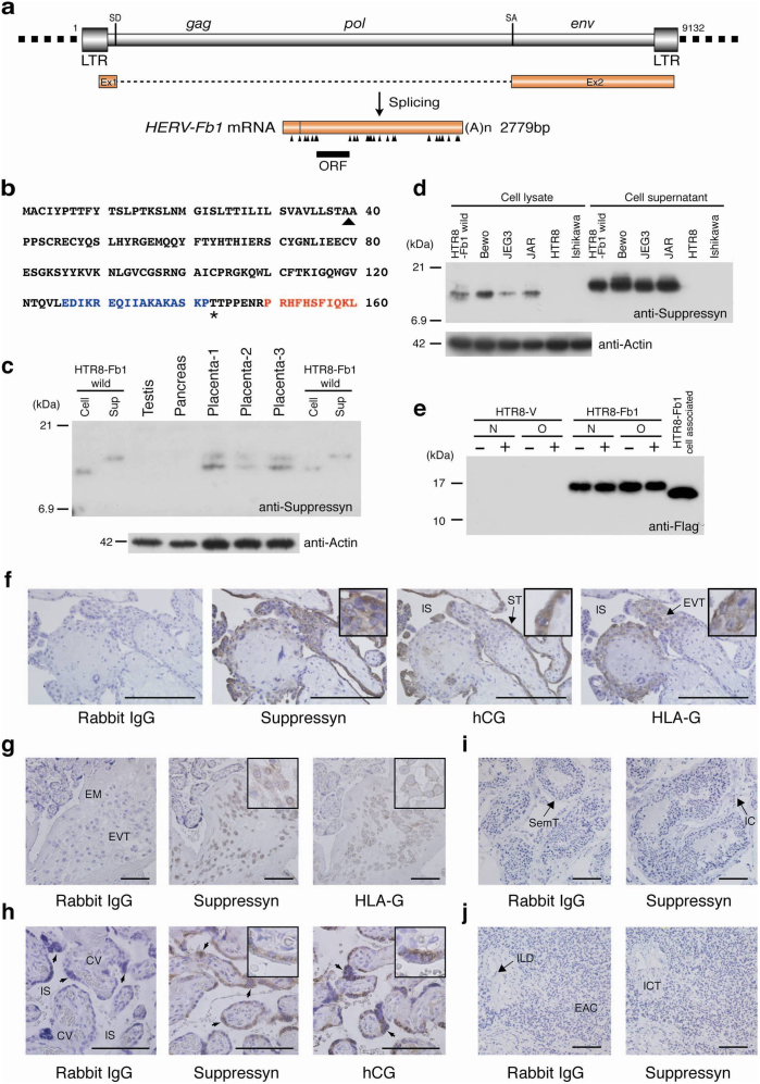
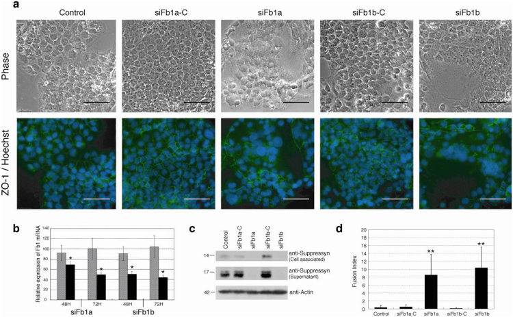
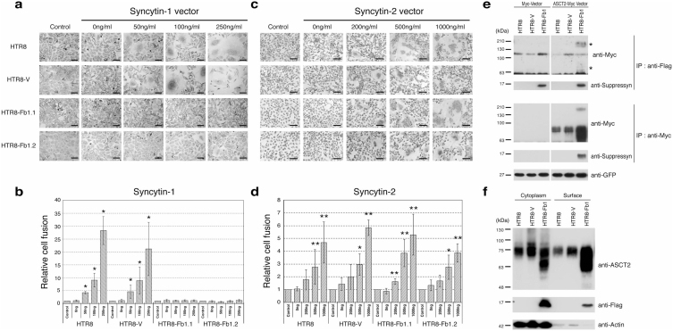
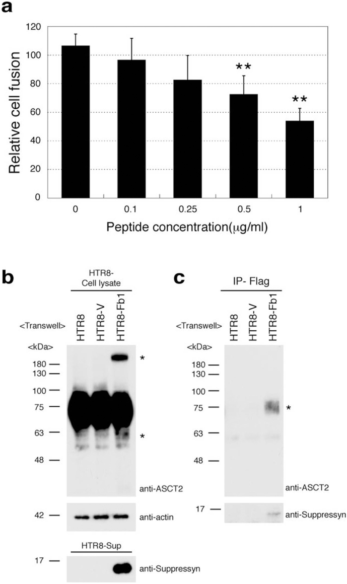
While common in viral infections and neoplasia, spontaneous cell-cell fusion, or syncytialization, is quite restricted in healthy tissues. Such fusion is essential to human placental development, where interactions between trophoblast-specific human endogenous retroviral (HERV) envelope proteins, called syncytins, and their widely-distributed cell surface receptors are centrally involved. We have identified the first host cell-encoded protein that inhibits cell fusion in mammals. Like the syncytins, this protein, called suppressyn, is HERV-derived, placenta-specific and well-conserved over simian evolution. In vitro, suppressyn binds to the syn1 receptor and inhibits syn1-, but not syn2-mediated trophoblast syncytialization. Suppressyn knock-down promotes cell-cell fusion in trophoblast cells and cell-associated and secreted suppressyn binds to the syn1 receptor, ASCT2. Identification of the first host cell-encoded inhibitor of mammalian cell fusion may encourage improved understanding of cell fusion mechanisms, of placental morphogenesis and of diseases resulting from abnormal cell fusion.


Figures

Figure 1
Figure 1. Human suppressyn structure, coding sequence and protein expression.
(a) Splicing pattern of the HERV-Fb1 gene and its transcriptional product. Arrowheads, stop codons; LTR, long terminal repeat; SD, splice donor; SA, splice acceptor. (b) Primary human suppressyn sequence. Arrowhead, predicted signal sequence cleavage site; Asterisk, predicted O-glycosylation site. The peptide sequence used to raise the polyclonal antibody is indicated in blue, that for the monoclonal in red. (c) Northern analysis has demonstrated placental specificity for Fb1; here western analysis using a monoclonal antibody against suppressyn confirms protein expression and demonstrates cell-associated and secreted (sup) forms in cell lines and in human tissues. Lanes 5–7: three independent human placental samples from term gestations; Lane 1 and 8, 2 and 9: cell-associated and secreted suppressyn from HTR8 cells stably-transfected with a suppressyn-expressing vector (positive controls). (d) Suppressyn protein expression in cell lines. Positive controls as in (c). (e) Soluble, tagged suppressyn was purified over Flag-M2 agarose and treated with or without N- and O-glycosidase. No differences in migration were seen between treated and untreated preparations. (f) Immunohistochemical detection of suppressyn using a polyclonal antibody demonstrates suppressyn localization to hCG-expressing syncytiotrophoblast (ST) and to the HLA-G positive extravillous trophoblast (EVT) in early gestation placenta. All negative tissue controls use rabbit IgG as primary antibody; HLA-G detection in EVT used a monoclonal anti-human MEMG/1 antibody; hCG detection in syncytiotrophoblast used a polyclonal antibody against hCG. (g–j) Localization of suppressyn protein in human third trimester placental tissues using a polyclonal antibody; (g) extravillous trophoblast and (h) syncytiotrophoblast. Negative control tissues exposed to the polyclonal anti-suppressyn antibody are depicted in (i) testis and (j) pancreas. EVT, extravillous trophoblast; EM, extracellular matrix; IS, intervillous space; CV, chorionic villi (fetal tissue); syncytiotrophoblast (arrow heads); EAC, exocrine acinar cells; ICT, interlobular connective tissue; ILD, interlobular duct; IC, interstitial cells; SemT, seminiferous tubule. Magnifications of positively–stained sections in the upper right corners of select images are to aid in discrimination of subcellular localization. Scale bar indicates 100 μm.
Figure 2
Figure 2. Fb1 knock-down increases cell fusion in syn1 expressing cells.
Fb1-specific mRNA knock-down in BeWo choriocarcinoma cells using distinct siRNA (siFb1a and siFb1b) or their respective scrambled siRNA controls. Fb1 knock down induced a significant increase in cell fusion (a, d). (a) ZO-1-FITC and Hoechst 33342 immunocytochemistry was performed 72 hours after siRNA exposure and images are depicted in the lower row. The upper row shows matched, phase-contrast images. Scale bar represents 100 μm. Quantitative RT-PCR, immunoprecipitation and immunoblotting confirm decreases in (b) Fb1 mRNA (50–60% knock-down) and (c) protein (cell-associated and secreted) in siRNA treated Bewo cells. Expression (b) is normalized to unexposed samples cultured for similar time periods (48 or 72H). (d) Mean fusion indices for siRNA-exposed and control cells (error bars represent standard deviations; n = 4 × 5 fields). Cell fusion was assessed using phase contrast microscopy and quantitated using cell fusion indices. All observations were performed at a final magnification of 200× and total nuclei were counted per field using Leica MetaMorph image analyzing software. The number of fused syncytial aggregates and the number of nuclei in each aggregate was counted manually and fusion indices were defined as [(N-S)/T] × 100. N is the number of nuclei in syncytia, S is the number of syncytia, and T is the total number of nuclei counted. The fusion index quantitates the percentage of fusion events in a cell population. Black bars- Fb1 siRNA exposed; hatched bars-control siRNA exposed. Data in (b, d) are representative of three independent experiments performed in duplicate. *p < 0.05 and **p < 0.01 compared to matched siRNA control. Statistical comparisons used Kruskal-Wallis and Mann-Whitney U-testing with (d) or without (b) Bonferroni corrections.
Figure 3
Figure 3. Suppressyn inhibits syn1- but not syn2-induced trophoblast cell fusion and suppressyn binds to the syn1 receptor ASCT2.
HTR8 trophoblast cells were stably transfected with vectors driving the expression of Fb1 (HTR8-Fb1.1 and 1.2) or vector alone (HTR-V). Cells were then transiently transfected with increasing amounts of a vector driving expression of syn1 (a) or syn2 (c), counterstained with hematoxylin and analyzed using phase contrast microscopy. To calculate fusion indices, cells were resuspended and analyzed by flow cytometry (b and d). Relative cell fusion was defined as the number of fused syn1- or syn2-transfected cells (gated by flow cytometry) divided by the number of fused cells in parent (HTR8) and in stable vector- or Fb1-transfected (HTR8-V, HTR8-Fb1.1, HTR8-Fb1.2), but syncytin non-transfected, cells cultured under identical conditions (indicated as controls for each cell line, *p < 0.05 **p < 0.01 when compared to non-transfected controls). Statistical comparisons were made using Kruskal-Wallis and Mann-Whitney U-testing with Bonferroni corrections. Data in (b) and (d) are representative of three independent experiments performed in duplicate. (e) HTR8 parental cells were stably transfected with vectors driving expression of flag-tagged Fb1 and then transiently transfected with a myc-vector or with a vector driving the expression of myc-tagged ASCT2. For the transfection internal control, the pAcGFP1-C1 vector was co-transfected with the myc tag vector and GFP protein expression quantitated using a monoclonal anti-GFP antibody. Lysates were immunoprecipitated with an anti-myc tag antibody and protein-G agarose or anti-flag-M2 agarose. Immunoprecipitants were separated by PAGE and immunoblotted with a myc-specific antibody. *Co-precipitated high and low molecular weight ASCT2. (f) HTR8, HTR8-V and HTR8-Fb1 cells were surface biotinylated and proteins purified using the Pierce cell surface protein isolation kit. Equal amounts of cytoplasmic and biotinylated surface protein were subsequently analyzed with standard SDS-PAGE and immune detection. Data in (e) and (f) are representative of three independent experiments.
Figure 4
Figure 4. Secreted suppressyn binds ASCT2 and inhibits syn1- induced trophoblast cell fusion.
(a) Syn1 transfected HTR8 cells were cultured in the presence of increasing amounts of recombinant secreted suppressyn for 24 hours prior to fusion assessment. Relative cell fusion was assessed by flow cytometry as in Fig 3b but was defined as the number of fused cells (gated by flow cytometry) in suppressyn peptide-exposed cells normalized to the number of fused cells in syn1-transfected HTR8 cells exposed to a control peptide. **p < 0.01 compared to 0 μg/ml sample. Statistical comparisons were made using Kruskal-Wallis and Mann-Whitney U-testing with Bonferroni corrections. Data are representative of three independent experiments performed in duplicate. (b, c) Transwell (0.4 μm; Becton Dickenson 353493) cell culture models (upper chamber; HTR8-V or HTR8-Fb1, lower chamber; HTR8 parent cell) were used to assess the effects of secreted suppressyn on non-transfected ASCT2-expressing target cells. Structural changes (*high and low molecular weight suppressyn forms) were observed in HTR8 cells after exogenous exposure to suppressyn (b). Coprecipitation experiments demonstrate direct binding of ASCT2 and suppressyn (c). Data in (b) and (c) are representative of three independent experiments. *Co-precipitated ASCT2.

Similar articles

Cited by

References

    1. Abmayr S. M. & Pavlath G. K. Myoblast fusion: lessons from flies and mice. Development 139 (4), 641–656 (2012). - PMC - PubMed
    1. Soe K. et al. Involvement of human endogenous retroviral syncytin-1 in human osteoclast fusion. Bone 48 (4), 837–846 (2011). - PubMed
    1. Frendo J. L. et al. Direct involvement of HERV-W Env glycoprotein in human trophoblast cell fusion and differentiation. Mol Cell Biol 23 (10), 3566–3574 (2003). - PMC - PubMed
    1. Mi S. et al. Syncytin is a captive retroviral envelope protein involved in human placental morphogenesis. Nature 403 (6771), 785–789 (2000). - PubMed
    1. Blaise S., de Parseval N., Benit L. & Heidmann T. Genomewide screening for fusogenic human endogenous retrovirus envelopes identifies syncytin 2, a gene conserved on primate evolution. Proc Natl Acad Sci U S A 100 (22), 13013–13018 (2003). - PMC - PubMed

MeSH terms

Associated data