. 2013:3:1463.
doi: 10.1038/srep01463.

Deriving dopaminergic neurons for clinical use. A practical approach

Affiliations

Deriving dopaminergic neurons for clinical use. A practical approach

Rodolfo Gonzalez et al. Sci Rep. 2013.

Abstract

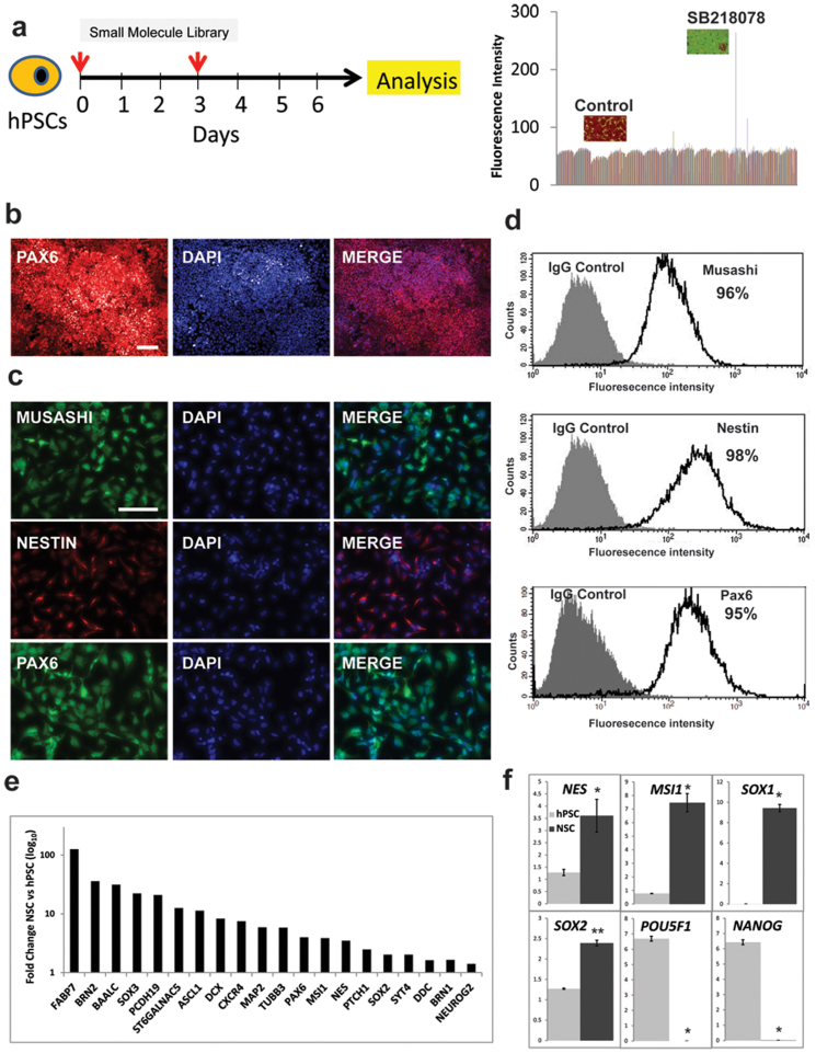
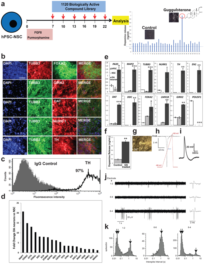
New small molecules that regulate the step-wise differentiation of human pluripotent stem cells into dopaminergic neurons have been identified. The steroid, guggulsterone, was found to be the most effective inducer of neural stem cells into dopaminergic neurons. These neurons are extensively characterized and shown to be functional. We believe this new approach offers a practical route to creating neurons of sufficient quality to be used to treat Parkinson's disease patients.


Conflict of interest statement

R.G., I.G., A.O., T.A., G.W., A.N., J.C.; R.S. are employees and stockholders of International Stem Cell Corporation. M.B. is an employee and stockholder of Axion Biosystems. The authors have no other relevant affiliations or financial involvement with any organization or entity with a financial interest in or financial conflict with the subject matter or materials discussed in the manuscript apart from those disclosed.

Figures

Figure 1
Figure 1. Generation of hPSC derived NSCs through small molecules.
(a) Diagram showing small molecule screening strategy for the neural induction of hPSCs. (b) hPSCs treated with SB218078 and DMH1 for seven days and stained for PAX6. Scale bar is 100 μm. (c) Expanded hPSC-NSCs stained for Nestin, Musashi, and PAX6. Scale bar is 100 μm. (d) FACS analyses of hPSC-NSC population stained for Musashi, Nestin, PAX6, and OCT4. (e) Gene expression microarray analysis showing fold expression induction of neural markers from hPSCs to NSCs; n = 2. (f) Gene expression analysis by RT-PCR of hPSC and hPSC-NSCs. Mean ± s.e.m., two-tailed Student's t-test: n = 3–5; α = 0.05; **P < 0.01; *P < 0.05.
Figure 2
Figure 2. Guggulsterone promotes generation of DA neurons.
(a) Diagram showing small molecule screening strategy for the differentiation of hPSC derived NSCs (hPSC-NSC) into DA neurons. (b) Staining for dopaminergic markers after 30 days of guggulsterone (GS) treatment. Scale bar is 100 μm. (c) FACS analysis of derived DA neurons stained for TH. (d) Fold induction of dopaminergic markers from NSCs to DA neurons as determined by gene expression microarray analysis; n = 2 (e) Relative gene expression by RT-PCR in DA neurons, NSCs and undifferentiated hPSCs. Mean ± s.e.m., one-factor ANOVA with Dunnett test comparing expression of DA neurons and NSCs against hPSC controls: n = 3–5; α = 0.05; ***P < 0.001; **P < 0.01; *P < 0.05. (f) Dopamine release assay of GS-treated neurons versus control cells (NSCs treated for 1 week with 100 ng/ml FGF8 and 2 μM purmorphamine and 2 weeks with 0.1% DMSO instead of GS). Mean ± s.e.m., two-tailed Student's t-test: n = 3; α = 0.05; **P < 0.01. (g) Representative phase-contrast image of a whole-cell patch-clamped GS-treated neuron. Scale bar is 10 μm. (h) Sodium current elicited with application of voltage ramp from −60 to +50 mV to cell in (g) in voltage clamp mode. Ramp parameters and waveform are shown in red superimposed on the current trace. The blue arrow indicates the point at which the stimulation ramp crossed the 0 mV value. (i) Action potential elicited from cell in (g) by injection of 500 pA for 200 ms in current clamp mode. Action potentials were found in 7 out of 20 cells patch-clamped. (j) Extracellular microelectrode array (MEA) recordings of GS-treated neurons, showing 10 s voltage traces for three representative electrodes. A single spike for each electrode is expanded at right. (k) Interspike interval histograms for electrodes in (j) showing: (↓↓) peaks associated with intervals between spikes within a burst and; (↓) those for longer intervals between bursts, or between individual spikes on non-bursting electrodes. Similar results were obtained with 2 MEA dishes.

References

    1. Kriks S. et al. Dopamine neurons derived from human ES cells efficiently engraft in animal models of Parkinson's disease. Nature 480, 547–551 (2011). - PMC - PubMed
    1. Hargus G. et al. Differentiated Parkinson patient-derived induced pluripotent stem cells grow in the adult rodent brain and reduce motor asymmetry in Parkinsonian rats. Proc Natl Acad Sci USA 107, 15921–15926 (2010). - PMC - PubMed
    1. Ganat Y. M. et al. Identification of embryonic stem cell-derived midbrain dopaminergic neurons for engraftment. J Clin Invest. 122, 2928–2939 (2012). - PMC - PubMed
    1. Parish C. L. et al. Wnt5a-treated midbrain neural stem cells improve dopamine cell replacement therapy in parkinsonian mice. J Clin Invest. 118, 149–160 (2008). - PMC - PubMed
    1. Redmond D. E. Jr. et al. Behavioral improvement in a primate Parkinson's model is associated with multiple homeostatic effects of human neural stem cells. Proc Natl Acad Sci USA 104, 12175–12180 (2007). - PMC - PubMed

Publication types

MeSH terms

Associated data