. 2013 May;6(3):535-43.
doi: 10.1161/CIRCHEARTFAILURE.112.000080. Epub 2013 Mar 14.

Tumor necrosis factor receptor-associated factor 2 signaling provokes adverse cardiac remodeling in the adult mammalian heart

Affiliations

Tumor necrosis factor receptor-associated factor 2 signaling provokes adverse cardiac remodeling in the adult mammalian heart

Vijay G Divakaran et al. Circ Heart Fail. 2013 May.

Abstract

Background: Tumor necrosis factor superfamily ligands provoke a dilated cardiac phenotype signal through a common scaffolding protein termed tumor necrosis factor receptor-associated factor 2 (TRAF2); however, virtually nothing is known about TRAF2 signaling in the adult mammalian heart.

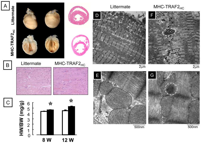
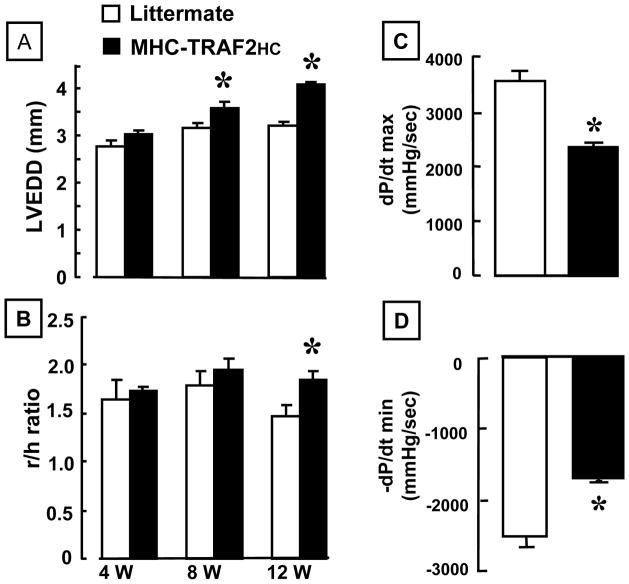
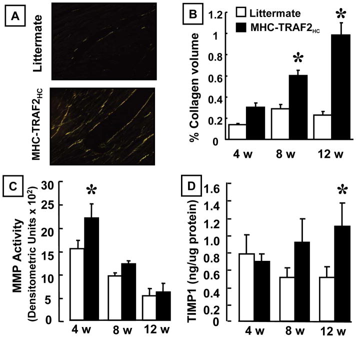
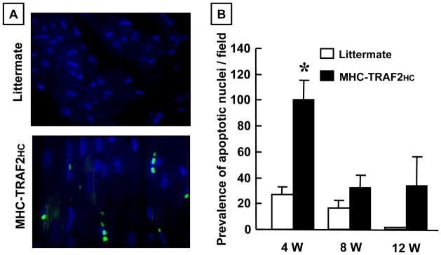
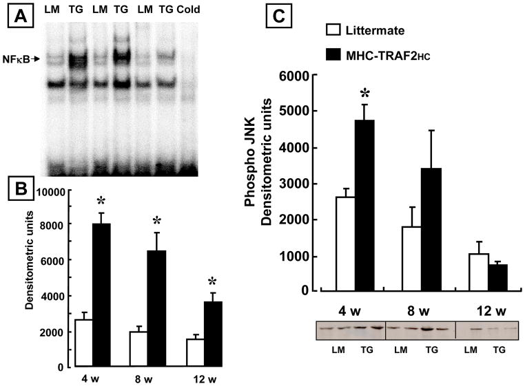
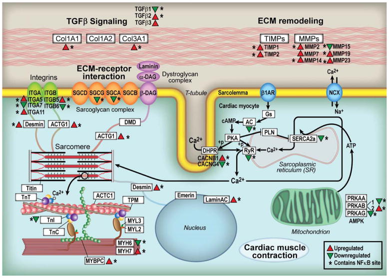
Methods and results: We generated multiple founder lines of mice with cardiac-restricted overexpression of TRAF2 and characterized the phenotype of mice with higher expression levels of TRAF2 (myosin heavy chain [MHC]-TRAF2(HC)). MHC-TRAF2(HC) transgenic mice developed a time-dependent increase in cardiac hypertrophy, left ventricular dilation, and adverse left ventricular remodeling, and a significant decrease in LV+dP/dt and LV-dP/dt when compared with littermate controls (P<0.05 compared with littermate). During the early phases of left ventricular remodeling, there was a significant increase in total matrix metalloproteinase activity that corresponded with a decrease in total myocardial fibrillar collagen content. As the MHC-TRAF2(HC) mice aged, there was a significant decrease in total matrix metalloproteinase activity accompanied by an increase in total fibrillar collagen content and an increase in myocardial tissue inhibitor of metalloproteinase-1 levels. There was a significant increase in nuclear factor-κB activation at 4 to 12 weeks and jun N-terminal kinases activation at 4 weeks in the MHC-TRAF2(HC) mice. Transciptional profiling revealed that >95% of the hypertrophic/dilated cardiomyopathy-related genes that were significantly upregulated genes in the MHC-TRAF2(HC) hearts contained κB elements in their promoters.

Conclusions: These results show for the first time that targeted overexpression of TRAF2 is sufficient to mediate adverse cardiac remodeling in the heart.

Keywords: TNF receptor–associated factor 2; dilated cardiomyopathy; inflammation; tumor necrosis factor superfamily.


Figures

Figure 1
Figure 1
Characterization of MHC-TRAF2HC transgenic mice. (A) Photographs of whole hearts, coronal and sagittal sections of both LM and MHC-TRAF2HC mouse hearts (12 weeks). (B) Representative hematoxylin-eosin stained cross sections at the level of the papillary muscles (400x). (C) Heart-weight-to-body-weight ratio (mg/g) of LM and MHC-TRAF2HC at 8 and 12 weeks (n= 6–8 mice/group/time point) (*= p< 0.05 vs. LM at the indicated time point) (D–G) Representative transmission electron micrographs from 12 week MHC-TRAF2HC transgenic mice and LM at low (x7500, D,F) and high (x 60,000, E,G) magnification. Protein aggregates are enclosed by the circles.
Figure 2
Figure 2
LV structure and function. (A) 2-D echocardiographic assessment of left-ventricular end diastolic dimension (LVEDD) and (B) r/h (radius/wall thickness) ratio of 12 week LM and MHC-TRAF2HC at 4, 8 and 12 weeks (n= 6–8 mice/group/time point). (*= p< 0.05 vs. LM at the indicated time point) (C) LV +dP/dt and (D) LV −dP/dt assessed in 12 week in LM and MHC-TRAF2HC (n= 6 mice/group) (*= p< 0.05 vs. LM at 12 weeks).
Figure 3
Figure 3
Myocardial fibrillar collagen content, MMP activity and TIMP levels. (A) Representative picrosirius red staining (400x) for myocardial fibrillar collagen content in LM and MHC-TRAF2HC mice. (B) Group data for % collagen volume for LM control and MHC-TRAF2HC mice at 4, 8 and 12 weeks (n=5 mice/group/time point). (*= p< 0.05 vs. LM at the indicated time point) (C) Group data for total MMP zymographic activity (arbitrary densitometric units) in LM control and MHC-TRAF2HC at 4, 8 and 12 weeks of age weeks (n=5–6 mice/group/time point). (*= p< 0.05 vs. LM at the indicated time point) (D) TIMP-1 levels in LM control and MHC-TRAF2HC control mice at 4, 8 and 12 weeks of age. (n= 6–8 mice/group/time point). (*= p< 0.05 vs. LM at the indicated time point)
Figure 4
Figure 4
Prevalence of apoptosis. (A) Fluorescence microscopic (400x) images of TUNEL staining in LM control and MHC-TRAF2HC mice at 4 weeks of age. TUNEL stained apoptotic nuclei appear green. DAPI staining (blue) was used to determine the total nuclei per high power field. (B) Group data for prevalence of myocyte apoptosis in LM and MHC-TRAF2HC mice at 4, 8 and 12 weeks (n=5 mice/group/time point). (* p < 0.05 vs. LM at 4 weeks)
Figure 5
Figure 5
NF-κB activation and JNK activity. (A) Representative electromobility shift assay of NF-κB binding in nuclear extracts in 12 week littermate control (LM) and MHC-TRAF2HC (TG) mouse hearts. To determine the specificity of DNA-protein binding, nuclear extracts from MHC-TRAF2HC hearts were treated with a 20x excess of unlabeled oligonucleotide, and by supershift assays using polyclonal antibody directed against the p50 component of NF-κB (B) Densitometric quantification of NF-κB activity in 4, 8 and 12 week old LM and MHC-TRAF2HC mice. (n=3–5 mice per group per time point) (* p<0.05 vs. LM controls). (*= p< 0.05 vs. LM at 4,8 and 12 weeks) (C) Densitometric quantification of phospho-JNK activity using an in gel kinase assay in LM and MHC-TRAF2HC mice at 4, 8 and 12 weeks of age. (n=4 mice per group per time point) (* p < 0.05 vs. LM controls at 4 weeks).
Figure 6
Figure 6
Transcriptional profiling. KEGG functional analysis identified significant changes in gene expression in pathways implicated in both hypertrophic and dilated cardiomyopathy in the MHC-TRAF2HC compared to LM control hearts. The diagram illustrates the specific changes in cardiac hypertrophy/dilated cardiomyopathy-related genes in relation to their anatomic location within the cardiac myocyte in MHC-TRAF2HC hearts vs. LM controls at 12 weeks. Identification of potential κB sites within significantly dysregulated genes was performed using UCSC_TFBS option in the DAVID functional annotation tool. (Key: red triangles denote increased gene expression; green triangles denote decreased gene expression; * denotes genes containing predicted κB sites in the promoter)

References

    1. Sivasubramanian N, Coker ML, Kurrelmeyer K, DeMayo F, Spinale FG, Mann DL. Left ventricular remodeling in transgenic mice with cardiac restricted overexpression of tumor necrosis factor. Circulation. 2001;2001:826–831. - PubMed
    1. Kubota T, McTiernan CF, Frye CS, Slawson SE, Koretsky AP, Demetris AJ, Feldman AM. Dilated cardiomyopathy in transgenic mice with cardiac specific overexpression of tumor necrosis factor-alpha. Circ Res. 1997;81:627–635. - PubMed
    1. Bryant D, Becker L, Richardson J, Shelton J, Franco F, Pechock RM, Thompson M, Giroir BP. Cardiac Failure in transgenic mice with myocardial expression of tumor necrosis factor-a (TNF) Circulation. 1998;97:1375–1381. - PubMed
    1. Ueland T, Aukrust P, Damas JK, Gullestad L, Yndestad A. The tumor necrosis factor superfamily in heart failure. Future Cardiol. 2006;2:101–111. - PubMed
    1. Jain M, Jakubowski A, Cui L, Shi J, Su L, Bauer M, Guan J, Lim CC, Naito Y, Thompson JS, Sam F, Ambrose C, Parr M, Crowell T, Lincecum JM, Wang MZ, Hsu YM, Zheng TS, Michaelson JS, Liao R, Burkly LC. A novel role for tumor necrosis factor-like weak inducer of apoptosis (TWEAK) in the development of cardiac dysfunction and failure. Circulation. 2009;119:2058–2068. - PMC - PubMed

Publication types

Substances