. 2005 Fall;4(1):A27-33.
Epub 2005 Oct 15.

Demonstrating the temperature sensitivity of synaptic transmission in a Drosophila mutant

Affiliations

Demonstrating the temperature sensitivity of synaptic transmission in a Drosophila mutant

Jacob L Krans et al. J Undergrad Neurosci Educ. 2005 Fall.

Abstract

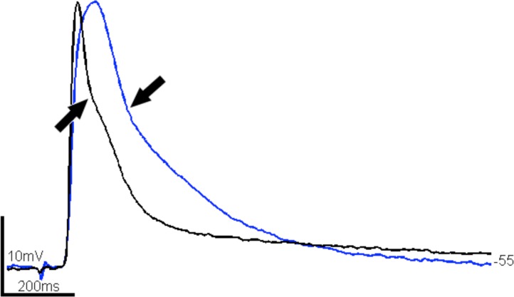
We describe exercises that illustrate the temperature sensitivity of synaptic transmission. The temperature dependence of synaptic transmission is demonstrated by cooling the larval Drosophila melanogaster preparation and recording excitatory junction potentials. Vesicle recycling is explored by utilizing a mutation of the shibire gene. This shibire mutant shows a robust reduction in synaptic vesicle recycling when temperature exceeds a known threshold (∼29° C). Students gain proficiency with the Drosophila larval neuromuscular junction preparation while investigating principles of vesicle release, vesicle recycling, synaptic facilitation and synaptic depression. We show that the viability of the larval preparation is prolonged in vitro with moderate cooling, which is particularly important when introducing the preparation as a novel exercise.

Keywords: Drosophila; excitatory junction potential (ejp); neuromuscular junction (NMJ); shibireTS1 mutant.


Figures

Figure 1.
Figure 1.
Still images captured from instructional videos of the larval dissection and electrode placement and stimulation. Screen shots are taken from videos that are available for download (Supplemental Material, www.funjournal.org/materialsKrans.asp). A: Two pins are placed at the anterior and posterior ends of the larva. Arrows indicate reflective tracheas that span the dorsal larva. Panel B shows all six pins in place and four abdominal segments have been outlined (A2:A5). The anterior end is to the left as evidenced by the pigmented mouth parts (arrow). The upper left portion of panel C shows the filleted larval preparation through a dissection microscope with transverse illumination (anterior at bottom). Arrows indicate the extracellular electrode – for stimulation (bottom arrow) - and the intracellular electrode, for muscle recording. Muscles 7, 6, 13, and 12 are outlined in yellow boxes (left to right). The upper right portion of panel C shows the physiology rig with two electrodes positioned around the preparation stand and dish. A compound ejp is depicted in the bottom of panel C.
Figure 2.
Figure 2.
Junction potentials of the larval D. melanogaster preparation over time, with cooling. Excitatory potentials recorded in muscle 12 of the fifth abdominal segment at two different times at ∼18° C. A and C were recorded upon initial setup of the preparation. B and D were recorded two hours later (times given at far left). Spontaneous potentials (C and D) maintained their monophasic shape and amplitude, though evoked potentials (stimulus = arrowhead) increased in amplitude after about two hours in vitro. Resting potential is shown at the left of each trace. Signal to noise values are typical of the larval preparation and suitable for further analysis of these data.
Figure 3.
Figure 3.
Larval ejps at two temperatures. Both traces were recorded from muscle 6 of the fourth abdominal segment. Each trace is the average of 10 ejps (a single, cut segmental nerve was stimulated). Black = 23° C, blue = 14° C. Arrows show the inflection between 1b (big) and 1s (small) nerve potentials (Kurdyak et al., 1994). Traces are aligned by onset of depolarization to best illustrate change in duration.
Figure 4.
Figure 4.
Temperature dependent change in synaptic depression at the neuromuscular junction in the C155; UAS-shits1 mutant. A, 25 Hz, 1s train of stimuli. At 22° C (black, throughout) there is little change in evoked potentials after the second stimulus. At 34° C (red, throughout), there is notable depression that progresses with time and stimulus number (arrowheads indicate initial and terminal ejp peak). B, 200 ms of 25 Hz stimuli illustrates temperature sensitivity of underlying temporal summation. At room temperature, ejp amplitude is relatively constant, but depresses at 34° C. Temporal summation and ejp depression are quantified in C and D. C, Synaptic depression over repetitive stimuli increases at ∼34° C in the mutant. (Below room temperature, temporal summation and progressive elongation of the evoked potentials mask rising phase of ejps; data not shown.) D, Temporal summation increases as temperature decreases. At 12° C (blue triangles), ejps are longer in duration than at 34° C and thus temporally summate. Membrane potential, measured at the onset of each stimulus is shown. Summation at 22° C is less than at 12° but was always present (n=7 muscles, 3 animals in C and D).
None

References

    1. Ball R, Xing B, Bonner P, Shearer J, Cooper RL. Long-term in vitro maintenance of neuromuscular junction activity of Drosophila larvae. Comp Biochem Physiol A Mol Integr Physiol. 2003;134:247–255. - PubMed
    1. Bellen HJ, Budnik V. The neuromuscular junction. In: Sullivan W, Ashburner M, Hawley RS, editors. Drosophila Protocols. Cold Spring Harbor, NY: Cold Spring Harbor Laboratory Press; 2000. pp. 175–200.
    1. Budnik V, Gramates LS. Neuromuscular Junctions in Drosophila. Int Rev Neurobiol. 1999;43:93–117. - PubMed
    1. Cattaert D, Birman S. Blockade of the central generator of locomotor rhythm by noncompetitive NMDA receptor antagonists in Drosophila larvae. J Neurobiol. 2001;48:58–73. - PubMed
    1. Charlton MP, Atwood HL. Synaptic transmission: temperature-sensitivity of calcium entry in presynaptic terminals. Brain Res. 1979;170:543–546. - PubMed

LinkOut - more resources