Review
. 2013 May;5 Suppl 1(Suppl 1):i7-12.
doi: 10.1136/neurintsurg-2013-010715. Epub 2013 Mar 14.

The Massachusetts General Hospital acute stroke imaging algorithm: an experience and evidence based approach

Affiliations
Review

The Massachusetts General Hospital acute stroke imaging algorithm: an experience and evidence based approach

Ramon Gilberto González et al. J Neurointerv Surg. 2013 May.

Abstract

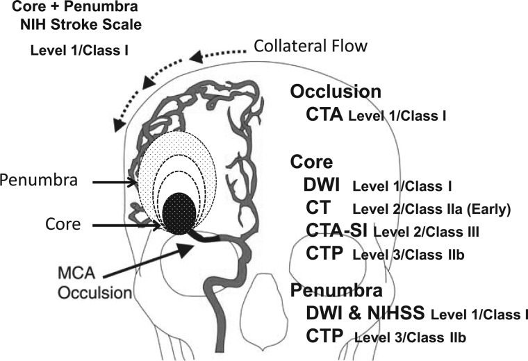
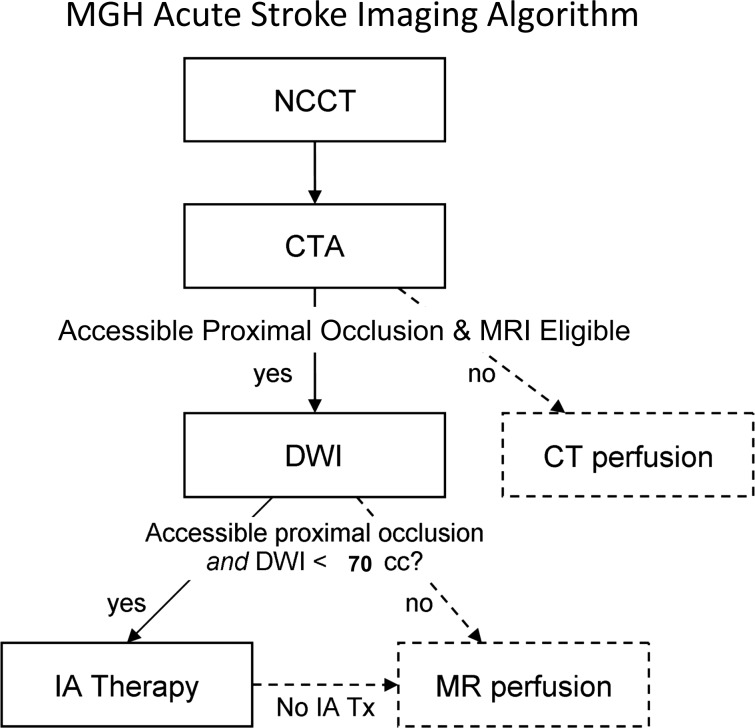
The Massachusetts General Hospital Neuroradiology Division employed an experience and evidence based approach to develop a neuroimaging algorithm to best select patients with severe ischemic strokes caused by anterior circulation occlusions (ACOs) for intravenous tissue plasminogen activator and endovascular treatment. Methods found to be of value included the National Institutes of Health Stroke Scale (NIHSS), non-contrast CT, CT angiography (CTA) and diffusion MRI. Perfusion imaging by CT and MRI were found to be unnecessary for safe and effective triage of patients with severe ACOs. An algorithm was adopted that includes: non-contrast CT to identify hemorrhage and large hypodensity followed by CTA to identify the ACO; diffusion MRI to estimate the core infarct; and NIHSS in conjunction with diffusion data to estimate the clinical penumbra.


Figures

Figure 1
Figure 1
Measurements of physiological components of middle cerebral artery (MCA) occlusion. Depicted is a representation of a right MCA occlusion with a growing infarct core coupled to a shrinking penumbra at a rate determined by collateral flow. The value of each method used to measure each component of stroke physiology is shown, as judged according to criteria stated in the methodology and in tables 1 and 2. CTA, CT angiography; CTA-SI, CT angiography-source images; CTP, CT perfusion; DWI, diffusion weighted imaging; NIHSS, National Institutes of Health Stroke Scale.
Figure 2
Figure 2
Relationship of final infarct size to clinical outcomes in patients with major anterior circulation occlusions (ACO) treated endovascularly. The bar graphs depict the proportion of good outcomes defined as a modified Rankin Scale score of 0–2 at 3 months (blue bars) by final infarct volume strata. The data are from 107 patients with ACO. The figure was derived from Yoo et al.
Figure 3
Figure 3
Changes in hemodynamic parameters that may occur in major anterior circulation occlusions. The change in cerebral perfusion pressure (CPP) may or may not be fully compensated by the collateral circulation. In each of the four scenarios, arrows indicate possible increase or decrease in cerebral blood volume (CBV), cerebral blood flow (CBF), mean transit time (MTT) and Tmax. This is a derivation of the more detailed graph shown in the online supplementary appendix figure A1.
Figure 4
Figure 4
Massachusetts General Hospital acute stroke imaging algorithm for triage of patients with severe ischemic strokes caused by anterior circulation occlusions. All patients undergo non-contrast CT (NCCT) followed by CT angiography (CTA). If the patient has severe neurological deficits (National Institutes of Health Stroke Scale score ≥10), no large hypodensity and no hemorrhage on NCCT, and an occlusion is identified of the distal internal carotid artery and/or proximal middle cerebral artery that is accessible by microcatheter, then the patient is immediately evaluated by diffusion MRI if there are no contraindications to MRI. If the diffusion weighted imaging (DWI) lesion is small, defined as <70 ml, then the patient is immediately triaged to endovascular therapy if the patient is otherwise eligible for such treatment. If the patient is not eligible for MRI, he/she may undergo a CT perfusion study for possible guidance for therapy or for prognostic information. Also, if the patient is not eligible for endovascular therapy, CT or MR perfusion may be performed for similar reasons. Only the first step in this algorithm (NCCT) and the time from stroke onset are needed for the decision to treat with intravenous tissue plasminogen activator. IA, intra-arterial.

References

    1. Baron JC. Mapping the ischaemic penumbra with PET: implications for acute stroke treatment. Cerebrovasc Dis 1999;9:193–201. - PubMed
    1. Jauch EC, Saver JL, Adams HP, Jr, et al. Guidelines for the early management of patients with acute ischemic stroke: a guideline for healthcare professionals from the American Heart Association/American Stroke Association. Stroke 2013;44:870–947. - PubMed
    1. Smith WS, Lev MH, English JD, et al. Significance of large vessel intracranial occlusion causing acute ischemic stroke and TIA. Stroke 2009;40:3834–40. - PMC - PubMed
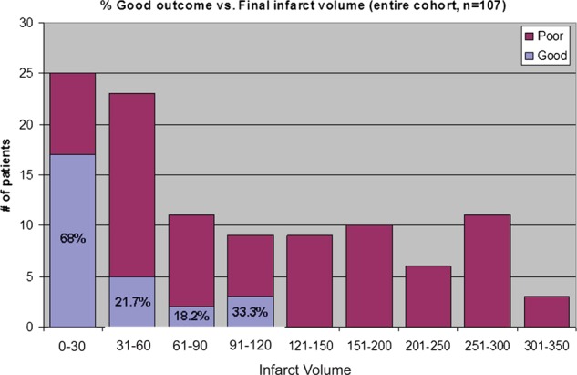
    1. Yoo AJ, Chaudhry ZA, Nogueira RG, et al. Infarct volume is a pivotal biomarker after intra-arterial stroke therapy. Stroke 2012;43:1323–30. - PubMed
    1. Zaidi SF, Aghaebrahim A, Urra X, et al. Final infarct volume is a stronger predictor of outcome than recanalization in patients with proximal middle cerebral artery occlusion treated with endovascular therapy. Stroke 2012;43:3238–44. - PubMed

MeSH terms

Substances