. 2013 Feb;38(1):2-10.
doi: 10.5395/rde.2013.38.1.2. Epub 2013 Feb 26.

In-depth morphological study of mesiobuccal root canal systems in maxillary first molars: review

Affiliations

In-depth morphological study of mesiobuccal root canal systems in maxillary first molars: review

Seok-Woo Chang et al. Restor Dent Endod. 2013 Feb.

Abstract

A common failure in endodontic treatment of the permanent maxillary first molars is likely to be caused by an inability to locate, clean, and obturate the second mesiobuccal (MB) canals. Because of the importance of knowledge on these additional canals, there have been numerous studies which investigated the maxillary first molar MB root canal morphology using in vivo and laboratory methods. In this article, the protocols, advantages and disadvantages of various methodologies for in-depth study of maxillary first molar MB root canal morphology were discussed. Furthermore, newly identified configuration types for the establishment of new classification system were suggested based on two image reformatting techniques of micro-computed tomography, which can be useful as a further 'Gold Standard' method for in-depth morphological study of complex root canal systems.

Keywords: Clearing technique; Mesiobuccal root; Micro-computed tomography; Minimum-intensity projection; Three-dimensional volume rendering.


Conflict of interest statement

No potential conflict of interest relevant to this article was reported.

Figures

Figure 1
Figure 1
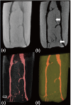
Representative images by (a, c) clearing technique; (b, d) Three-dimensional (3D) volume rendering technique of micro-computed tomography (MCT) showed restricted penetration of dye solution into a root canal system (a, c, white arrows with black outline).
Figure 2
Figure 2
Original cone-beam computed tomography (CBCT) images of a maxillary first molar with two canals in the mesiobuccal (MB) root as viewed coronal, sagittal, axial direction by using OnDemand3D software. (a) Coronal view with the MB root exhibiting Type III canal configuration; (b) Sagittal view; (c) Axial view (arrow indicates the existence of two canals in MB root); (d) Three-dimensional (3D) reconstruction image.
Figure 3
Figure 3
Canal images with different reformatting methods. (a) Simulation of conventional ray projection image; (b) Curved thin slab-minimum intensity projection (TS-MinIP) image; (c) Volume rendered image with segmentation of canal structure; (d) Surface rendered image. TS-MinIP image shows the most clear view of fine canal structures (b, white arrow), while an accessory canal hiding behind main root canal is shown only in three-dimensional (3D) volume rendered image (c, black arrow with white outline).
Figure 4
Figure 4
The slab is defined between two planes in (a), and (b) is a rotated image of (a) which shows root surface concavity included in the slab. (c) is a resultant thin slab-minimum intensity projection (TS-MinIP) image with pseudo-canal. If the slab extends beyond the extent of the root due to root concavity or severe curvature (b, white arrow), the pixel value outside the root is recognized as the minimum value and thus can result in a pseudo-canal image (c, black arrow with white outline).
Figure 5
Figure 5
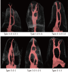
Representative images obtained by three-dimensional rendering which shows six non-classifiable configuration types when the modified Vertucci's classification is applied (Reprinted with the permission of the Clin Oral Investig. Originally published as: Clin Oral Investig 2012, 10.1007/s00784-012-0725-1.).
Figure 6
Figure 6
Three-dimensional reconstructed images of 3 non-classifiable configuration types when modified Vertucci's classification was applied. The configuration types were not reported in maxillary first molar MB roots in the literature (Reprinted with the permission of the Clin Oral Investig. Originally published as: Clin Oral Investig 2012, 10.1007/s00784-012-0725-1.).

Similar articles

Cited by

References

    1. Kim Y, Chang SW, Lee JK, Chen IP, Kaufman B, Jiang J, Cha BY, Zhu Q, Safavi KE, Kum KY. A micro-computed tomography study of canal configuration of multiplecanalled mesiobuccal root of maxillary first molar. Clin Oral Investig. 2012 Oct; [Epub ahead of print]. doi: 10.1007/s00784-012-0852-8. - DOI - PubMed
    1. Versiani MA, Pécora JD, de Sousa-Neto MD. Root and root canal morphology of four-rooted maxillary second molars: a micro-computed tomography study. J Endod. 2012;38:977–982. - PubMed
    1. Du Y, Soo I, Zhang CF. A case report of six canals in a maxillary first molar. Chin J Dent Res. 2011;14:151–153. - PubMed
    1. Badole GP, Bahadure RN, Warhadpande MM, Kubde R. A rare root canal configuration of maxillary second molar: a case report. Case Rep Dent. 2012;2012:767582. doi: 10.1155/2012/767582. Epub 2012 Jul 8. - DOI - PMC - PubMed
    1. Spagnuolo G, Ametrano G, D'Antò V, Formisano A, Simeone M, Riccitiello F, Amato M, Rengo S. Microcomputed tomography analysis of mesiobuccal orifices and major apical foramen in first maxillary molars. Open Dent J. 2012;6:118–125. - PMC - PubMed

LinkOut - more resources