Review
. 2013 Dec;34(12):2233-40.
doi: 10.3174/ajnr.A3415. Epub 2013 Mar 14.

Illustrated review of the embryology and development of the facial region, part 1: Early face and lateral nasal cavities

Affiliations
Review

Illustrated review of the embryology and development of the facial region, part 1: Early face and lateral nasal cavities

P M Som et al. AJNR Am J Neuroradiol. 2013 Dec.

Abstract

The early embryological development of the face has been reviewed. One repeating theme to note is the serial closing and then the re-opening of a space. This is seen in the separation of the nasal and oral cavities, the nostrils, and in part 2 the developing eyelids fusing and then re-opening. Part 2 will discuss the further facial development as well as the changes in facial bone appearance after birth.


Figures

Fig 1.
Fig 1.
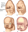
Anterior oblique drawing (A) of a 5-week embryo showing the appearance of the nasal (olfactory) placode. B, Drawing shows the development of the medial and lateral nasal processes forming a downward-facing “horseshoe” around the sinking nasal placode, which forms the nasal pit.
Fig 2.
Fig 2.
Anterior oblique drawing of an embryo in the late fourth week (A) shows the stomodeum with the oropharyngeal membrane surrounded by the further development of the frontonasal process and the maxillary and mandibular processes. B, Drawing shows the breakdown of the oropharyngeal membrane by the fifth week. The early appearance of the lens and otic placodes is also seen.
Fig 3.
Fig 3.
Drawing (A) of a ventral view of a 3- to 4-week embryo shows the anterior neuropore and the early formation of the maxillary processes. Drawing (B) in the late fourth week shows closure of the anterior neuropore and the location of the future frontonasal process. (Modified with permission from Netter's Atlas of Human Embryology. Edited by Cochard, L.R., PhD. 2002. Icon Learning Systems, Teterboro, New Jersey, Figures 9.5. Netter Illustrations from www.netterimages.com, © Elsevier Inc, All rights reserved).
Fig 4.
Fig 4.
Anterior oblique drawing of a 5-week embryo (A) shows the further growth of the medial and lateral nasal processes and the development of the nasal sac. The bucconasal groove is shown. B, Anterior oblique drawing of a 6-week embryo shows closure of bucconasal groove completing the floor of the nasal cavity and progressive flattening of the nasal sac openings, mainly as a result of ventrolateral growth of the medial nasal processes. The nasal sacs are also pushed toward the midline as the maxillary processes grow. Anterior oblique drawing of a 7-week embryo (C) and a 10-week (D) embryo shows the progressive medial movement of the nasal sacs and the resulting progressive pushing upwards of the frontonasal process.
Fig 5.
Fig 5.
Frontal drawings of 4- to 5-week (A) and 5- to 6-week (B) embryos illustrate the progressive displacement of the nasal sacs toward the midline as a result of medial growth of the maxillary processes. Frontal view of a 6- to 7-week embryo (C) shows the nasolacrimal groove. Closure of this groove establishes continuity between the side of the nose formed by the lateral nasal process and the cheek formed by the maxillary process. (Modified with permission from Levine HL, Clemente MP, eds. Chapter 1, Surgical Anatomy of the Paranasal Sinus. China: 2005. Sinus Surgery Endoscopic and Microscopic Approaches. Figures 1–2. Thieme Medical Publishers Inc., Georg Thieme Verlag Stuttgart).
Fig 6.
Fig 6.
Drawing from below of a 6-week fetus showing the emergence of the intermaxillary segment below the medial nasal processes. This segment may actually arise from the globular processes of His. Also shown is the maxillary process and, arising from its medial surface, the lateral palatine process, which will form the secondary palate. (Modified with permission from Levine HL, Clemente MP, eds. Chapter 1, Surgical Anatomy of the Paranasal Sinus. China: 2005. Sinus Surgery Endoscopic and Microscopic Approaches. Figures 1–3. Thieme Medical Publishers Inc., Georg Thieme Verlag Stuttgart).
Fig 7.
Fig 7.
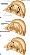
Drawings of the inferior view of an embryo from the sixth (A) to seventh weeks (C) show the bucconasal groove starting to close because of medial growth of the maxillary processes (A). B, There is fusion along most of the course of the bucconasal groove and membranous closure posteriorly by the bucconasal membrane. C, The oronasal membrane (thinned bucconasal membrane) has ruptured, creating an opening for communication between the primitive nasal and oral cavities (posterior choana).
Fig 8.
Fig 8.
Drawings from below of a 7-week embryo (A) and a 13- to 15-week fetus (B) show epithelial plugs closing the nostrils (A) and then dissolving (B). If a plug does not dissolve, there will be atresia of the nostril. Lateral drawing of the anterior nose (C) shows the nasal vestibule and limen nasi. These areas mark the location of the nasal epithelial plugs.
Fig 9.
Fig 9.
Frontal drawings of the upper lip region in embryos at 5 (A), 6 (B) and 7 (C) weeks show the progressive medial growth of the maxillary processes toward the midline. However, the actual midline of the upper lip (the philtrum) is formed by the medial nasal processes. (Modified with permission from Levine HL, Clemente MP, eds. Chapter 1, Surgical Anatomy of the Paranasal Sinus. China: 2005. Sinus Surgery Endoscopic and Microscopic Approaches. Figures 1–3. Thieme Medical Publishers Inc., Georg Thieme Verlag Stuttgart).
Fig 10.
Fig 10.
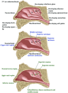
Lateral drawings of the developing lateral nasal wall showing the appearance of the nasal turbinals and their eventual development into the nasal turbinates and meati.

References

    1. Kim CH, Park HW, Kim K, et al. . Early development of the nose in human embryos: a stereomicroscopic and histologic analysis. Laryngoscope 2004;114:1791–800 - PubMed
    1. Warbrick JG. The early development of the nasal cavity and upper lip in the human embryo. J Anat 1960;94:351–62 - PMC - PubMed
    1. Osumi-Yamashita N. Retinoic acid and mammalian craniofacial morphogenesis. J Biosci 1996;21:313–27
    1. Streit A. The preplacodal region: an ectodermal domain with multipotential progenitors that contribute to sense organs and cranial sensory ganglia. Int J Dev Biol 2007;51:447–61 - PubMed
    1. Bossy J. Development of olfactory and related structures in staged human embryos. Anat Embryol (Berl) 1980;161:225–36 - PubMed

LinkOut - more resources