

Dermoscopic patterns of melanoma metastases: interobserver consistency and accuracy for metastasis recognition
- PMID: 23495915
- PMCID: PMC3707959
- DOI: 10.1111/bjd.12314
Dermoscopic patterns of melanoma metastases: interobserver consistency and accuracy for metastasis recognition
Abstract
Background: Cutaneous metastases of malignant melanoma (CMMM) can be confused with other skin lesions. Dermoscopy could be helpful in the differential diagnosis.
Objectives: To describe distinctive dermoscopic patterns that are reproducible and accurate in the identification of CMMM.
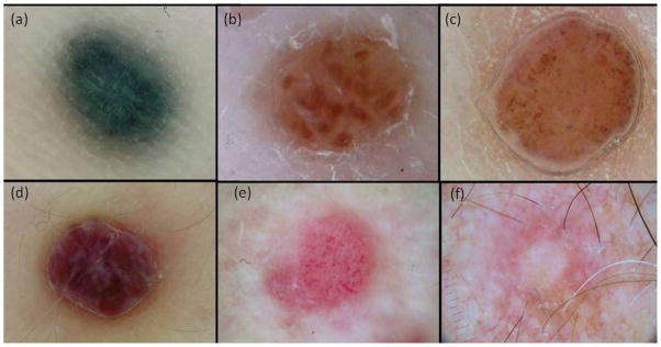
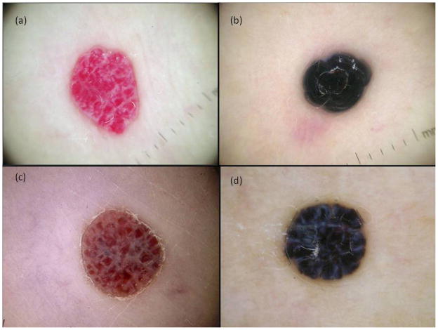
Methods: A retrospective study of 146 dermoscopic images of CMMM from 42 patients attending a melanoma unit between 2002 and 2009 was performed. Firstly, two investigators established six dermoscopic patterns for CMMM. The correlation of 73 dermoscopic images with their distinctive patterns was assessed by four independent dermatologists to evaluate the reproducibility in the identification of the patterns. Finally, 163 dermoscopic images, including CMMM and nonmetastatic lesions, were evaluated by the same four dermatologists to calculate the accuracy of the patterns in the recognition of CMMM.
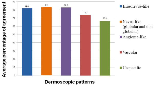
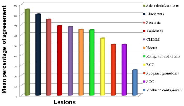
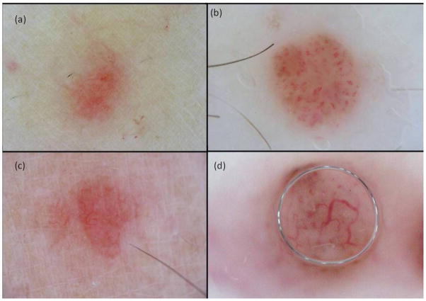
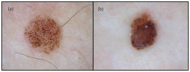
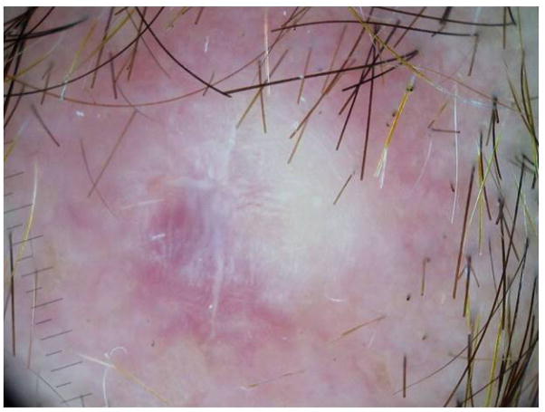
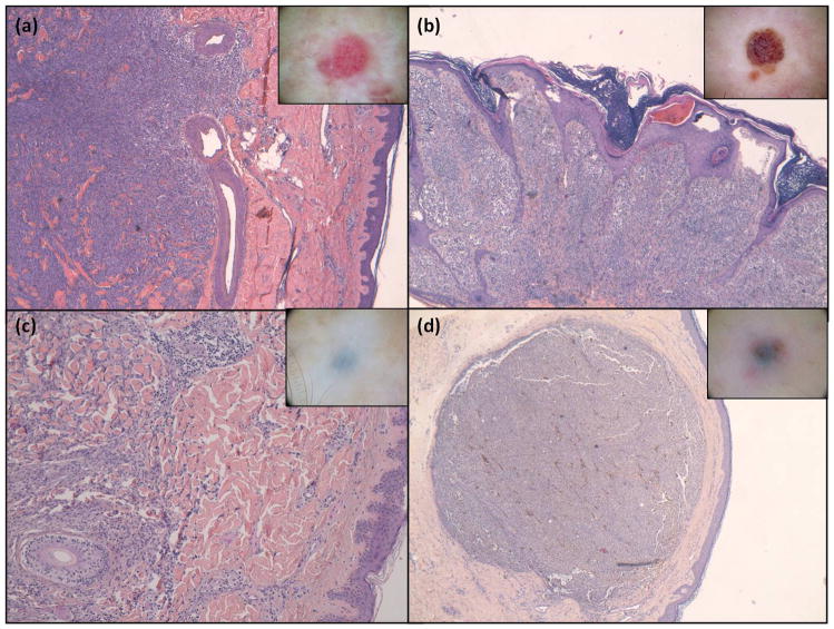
Results: Five CMMM dermoscopic patterns had a good interobserver agreement (blue naevus-like, naevus-like, angioma-like, vascular and unspecific). When CMMM were classified according to these patterns, correlation between the investigators and the four dermatologists ranged from κ = 0.56 to κ = 0.7. In total, 71 CMMM, 16 angiomas, 22 blue naevi, 15 malignant melanomas, 11 seborrhoeic keratoses, 15 melanocytic naevi with a globular pattern and 13 pink lesions with a vascular pattern were evaluated according to the previously described CMMM dermoscopy patterns, showing an overall sensitivity of 67.9% (range 54.9-76%) and a specificity of 79.9% (range 68.5-93.5%) for the diagnosis of CMMM.
Conclusions: Five dermoscopic patterns of CMMM with good interobserver agreement obtained a high sensitivity and specificity in the diagnosis of metastasis, with the accuracy varying according to the experience of the observer.
© 2013 The Authors BJD © 2013 British Association of Dermatologists.
Figures
Similar articles
-
Dermoscopy of cutaneous melanoma metastases: A color-based pattern classification.J Dermatol. 2019 Jul;46(7):564-569. doi: 10.1111/1346-8138.14926. Epub 2019 May 23. J Dermatol. 2019. PMID: 31120139
-
High-resolution ultrasonography assists the differential diagnosis of blue naevi and cutaneous metastases of melanoma.Br J Dermatol. 2010 Sep;163(3):550-6. doi: 10.1111/j.1365-2133.2010.09903.x. Epub 2010 Jul 26. Br J Dermatol. 2010. PMID: 20545694
-
Clinical and Histopathologic Characteristics of Melanocytic Lesions on the Volar Skin Without Typical Dermoscopic Patterns.JAMA Dermatol. 2019 May 1;155(5):578-584. doi: 10.1001/jamadermatol.2018.5926. JAMA Dermatol. 2019. PMID: 30865233 Free PMC article.
-
Surveillance of patients at high risk for cutaneous malignant melanoma using digital dermoscopy.Br J Dermatol. 2005 Jan;152(1):87-92. doi: 10.1111/j.1365-2133.2005.06370.x. Br J Dermatol. 2005. PMID: 15656806 Review.
-
Dermoscopic patterns of cutaneous melanoma metastases.Int J Dermatol. 2014 Apr;53(4):404-12. doi: 10.1111/ijd.12346. Epub 2013 Dec 10. Int J Dermatol. 2014. PMID: 24320196 Review.
Cited by
-
Dermatoscopic patterns of cutaneous metastases: A multicentre cross-sectional study of the International Dermoscopy Society.J Eur Acad Dermatol Venereol. 2024 Jul;38(7):1432-1438. doi: 10.1111/jdv.19962. Epub 2024 Mar 14. J Eur Acad Dermatol Venereol. 2024. PMID: 38483241 Free PMC article.
-
Dermoscopy of skin metastases in advanced cancer-systemic (visceral, hematologic) and cutaneous.Front Med (Lausanne). 2024 Jul 30;11:1445811. doi: 10.3389/fmed.2024.1445811. eCollection 2024. Front Med (Lausanne). 2024. PMID: 39139791 Free PMC article.
-
Dermoscopy of nodular skin metastases from the gastrointestinal primary cancer.Postepy Dermatol Alergol. 2015 Aug;32(4):312-6. doi: 10.5114/pdia.2015.48043. Epub 2015 Aug 12. Postepy Dermatol Alergol. 2015. PMID: 26366159 Free PMC article. No abstract available.
-
Skin Cancer Diagnosis by Lesion, Physician, and Examination Type: A Systematic Review and Meta-Analysis.JAMA Dermatol. 2025 Feb 1;161(2):135-146. doi: 10.1001/jamadermatol.2024.4382. JAMA Dermatol. 2025. PMID: 39535756
-
Can Multispectral Dermoscopy Help In Distinguishing Blue Color?Dermatol Pract Concept. 2023 Jan 1;13(1):e2023058. doi: 10.5826/dpc.1301a58. Dermatol Pract Concept. 2023. PMID: 36892379 Free PMC article.
References
-
- Savoia P, Fava P, Nardò T, et al. Skin metastases of malignant melanoma: a clinical and prognostic survey. Melanoma Res. 2009;19:321–6. - PubMed
-
- Meier F, Will S, Ellwanger U, et al. Metastatic pathways and time courses in the orderly progression of cutaneous melanoma. Br J Dermatol. 2002;147:62–70. - PubMed
-
- Bono R, Giampetruzzi AR, Concolino F, et al. Dermoscopic patterns of cutaneous melanoma metastases. Melanoma Res. 2004;14:367–73. - PubMed
-
- Brauer JA, Wriston CC, Troxel AB, et al. Characteristics associated with early and late melanoma metastases. Cancer. 2010;116:415–23. - PubMed
Publication types
MeSH terms
Grants and funding
LinkOut - more resources
Full Text Sources
Other Literature Sources
Medical
