

Intravitreal bevacizumab (Avastin) and panretinal photocoagulation in the treatment of high-risk proliferative diabetic retinopathy
- PMID: 23495932
- PMCID: PMC3708621
- DOI: 10.1089/jop.2012.0202
Intravitreal bevacizumab (Avastin) and panretinal photocoagulation in the treatment of high-risk proliferative diabetic retinopathy
Abstract
Purpose: To report the short-term efficacy and safety of intravitreal bevacizumab (Avastin) injection with panretinal laser photocoagulation (PRP) in patients with high-risk proliferative diabetic retinopathy (PDR) according to the Early Treatment Diabetic Retinopathy Study criteria.
Methods: A prospective, interventional case series study was conducted in 17 patients (20 eyes) with high-risk PDR, who were treated with intravitreal bevacizumab (2.5 mg) followed by PRP when the peripheral vitreous became clear or 2 weeks after injection. Patients underwent complete ophthalmic evaluation, including Snellen visual acuity and fluorescein angiography at baseline, 1, 3, and 6 months after bevacizumab injection. Main outcome measures included the serial changes in visual acuity, vitreous clear-up time, and neovascularization on the disc (NVD) regression time.
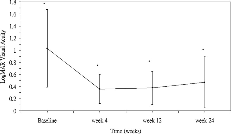
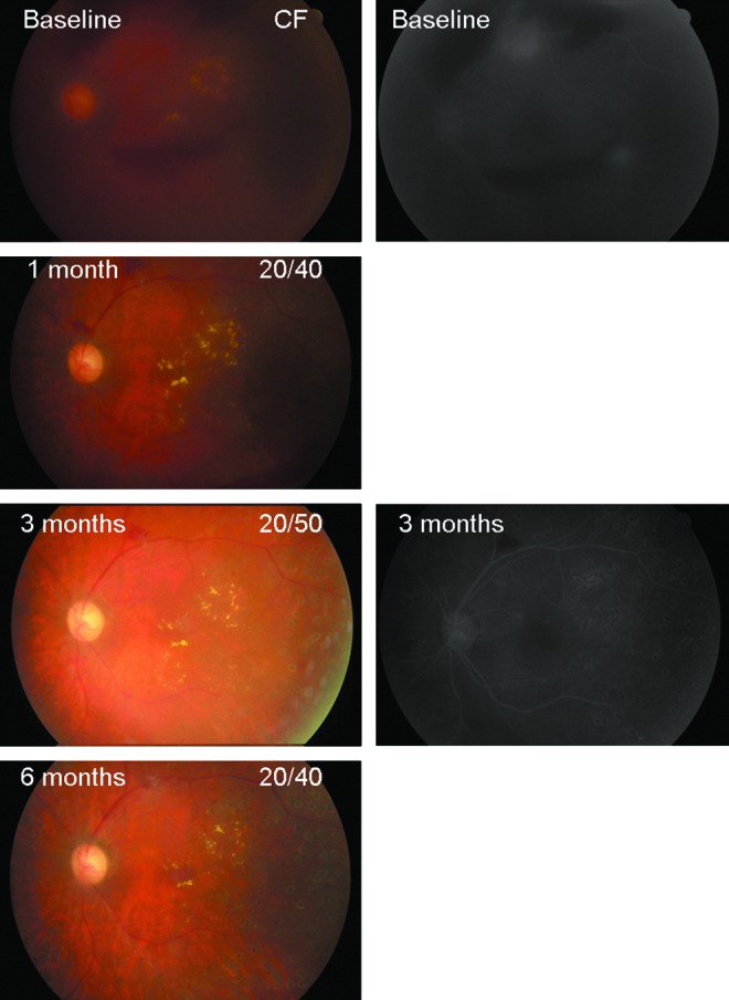
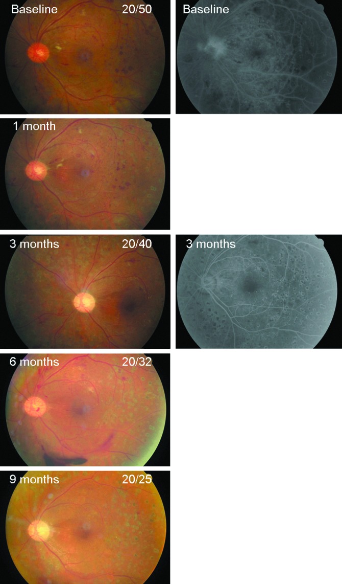
Results: All patients had obvious reduction in angiographic leakage and involution of retinal neovascularization (NV) at the 1- and 3-month follow-up. The mean follow-up time was 7.5 months. The vitreous hemorrhage (VH) showed a partial resolution as early as 1 week, and complete regression at 3 months. The mean vitreous clear-up time after intravitreal Avastin was 8.5±2.2 weeks. The mean time interval from intravitreal Avastin to NVD regression was 10.8±3.4 weeks. Mean logarithm of the minimum angle resolution visual acuity improved from 1.03 at baseline to 0.36 at 1-month, 0.38 at 3-month, and 0.48 at the 6-month follow-up (P<0.01). Three eyes (18%) required vitrectomy surgery during follow-up. The indication for vitrectomy was dense, persistent VH in 2 eyes, and focal tractional retinal detachment (TRD) in 1 eye. Recurrent retinal NV with minor preretinal hemorrhage was observed in 6 eyes (30%) 3 months after the first injection, and resolved after repeated bevacizumab injections. Patients received an average of 1.4 injections (range: 1-2). Seven eyes (35%) underwent 2 injections. One eye (5%) had ocular complication of PDR progression to TRD. No systemic adverse events were observed following injections.
Conclusions: Short-term results suggest combined intravitreal bevacizumab and PRP achieved rapid clearance of VH, regression of retinal NV, and visual improvement in the treatment of high-risk PDR. Long-term study is warranted to assess the long-term efficacy and safety.
Figures
Comment in
-
Intravitreal anti-VEGF treatment as adjunctive treatment in the management of diabetic retinopathy.J Ocul Pharmacol Ther. 2014 May;30(4):303. doi: 10.1089/jop.2013.0217. Epub 2013 Dec 14. J Ocul Pharmacol Ther. 2014. PMID: 24329129 No abstract available.
-
Response to Carifi et al.'s letter.J Ocul Pharmacol Ther. 2014 May;30(4):304-5. doi: 10.1089/jop.2013.0217.rs. Epub 2014 Jan 2. J Ocul Pharmacol Ther. 2014. PMID: 24383445 No abstract available.
References
-
- Chew E.Y. Major clinical trials of vitreoretinal diseases. In: Regillo C.D., editor; Brown G.C., editor; Flyn H.W., editor. Vitreoretinal Disease, the Essentials. New York: Theme Medical Publishers, Inc.; 1999. pp. 667–677.
-
- Early Treatment Diabetic Retinopathy Study Research Group. Fundus photographic risk factors for progression of diabetic retinopathy. Ophthalmology. 1991;98:823–833. ETDRS Report No. 12. - PubMed
-
- Adamis A.P. Miller J.W. Bernal M.T., et al. Increased vascular endothelial growth factor levels in the vitreous of eyes with proliferative diabetic retinopathy. Am. J. Ophthalmol. 1994;118:445–450. - PubMed
-
- Aiello L.P. Avery R.L. Arrigg P.G, et al. Vascular endothelial growth factor in ocular fluid of patients with diabetic retinopathy and other retinal disorders. N. Engl. J. Med. 1994;331:1480–1487. - PubMed
Publication types
MeSH terms
Substances
LinkOut - more resources
Full Text Sources
Other Literature Sources
Medical
Research Materials
