Case Reports
. 2013 Mar 16:10:39.
doi: 10.1186/1742-2094-10-39.

Corticosteroid therapy in a patient with cerebral amyloid angiopathy-related inflammation

Affiliations
Case Reports

Corticosteroid therapy in a patient with cerebral amyloid angiopathy-related inflammation

Akio Kimura et al. J Neuroinflammation. .

Abstract

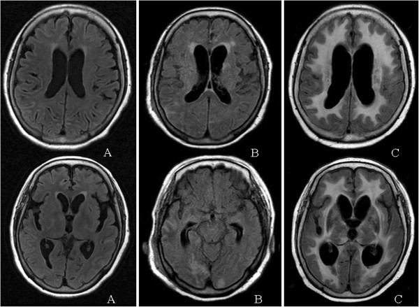
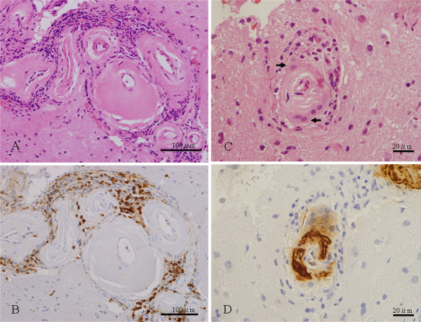
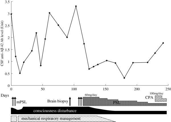
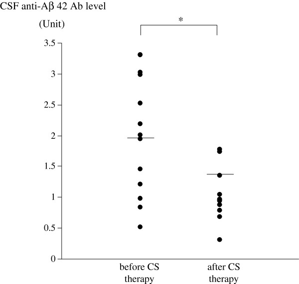
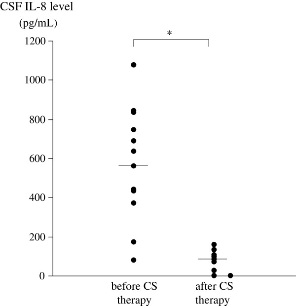
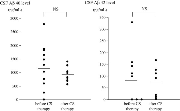
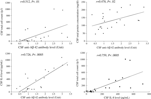
We studied longitudinal changes of the levels of anti-amyloid β (anti-Aβ) antibody, amyloid β (Aβ) protein, and interleukin 8 (IL-8) in cerebrospinal fluid (CSF) of a patient with cerebral amyloid angiopathy-related inflammation (CAA-ri) in whom steroid treatment resulted in clinical improvement. The diagnosis of CAA-ri was established with brain biopsy. Levels of anti-Aβ 42 antibody, Aβ 40, Aβ 42 and IL-8 in CSF were measured in the CAA-ri patient at 23 time points in the 8-month clinical course. These CSF samples were divided into 2 groups: those obtained before (n = 12) and those after (n = 11) oral corticosteroid therapy was started. We compared these levels between CSF samples obtained before and after therapy. The mean levels of anti-Aβ 42 antibody and IL-8 were significantly higher in CSF samples of the CAA-ri patient before oral corticosteroid therapy than those after therapy. A positive correlation was noted between levels of anti-Aβ 42 antibodies and IL-8 in CSF of this patient. There were no significant differences of mean levels of Aβ 40 and Aβ 42 between CSF samples obtained before and after oral corticosteroid therapy. It was possible that the autoinflammatory process with anti-Aβ 42 antibodies and IL-8 may have been involved in the pathogenesis of CAA-ri, and that corticosteroid therapy directly affected levels of anti-Aβ 42 antibody and IL-8. In summary, CAA-ri encephalopathy is a relapsing or progressive disorder and may be treatable by adequate immunosuppressive therapy. The anti-Aβ 42 antibody in CSF is a useful biological marker for therapeutic monitoring of CAA-ri.


Figures

Figure 1
Figure 1
Axial fluid-attenuated inversion recovery (FLAIR) brain MRI scans of the patient with CAA-ri. (A) At time of admission, there was increased signal intensity in the leptomeninges and sulci; (B) At 49 days after admission, there were more extensive hyperintensity lesions in the leptomeninges and sulci, and there was asymmetric subcortical white matter of the occipital lobes; (C) At 195 days after admission, the hyperintensity lesions extended diffusely through the white matter.
Figure 2
Figure 2
Neuropathological findings of the biopsy specimen from the right parietal lobe of the patient with CAA-ri. (A) Multifocal lymphocytic infiltrates around the leptomeningeal-parenchymal blood vessels, with hyaline thickening and splitting of the walls (double-barrel formation) (hematoxylin-eosin); (B) The perivascular lymphocytic infiltrates consisting primarily of UCHL1 + T cells (serial section of [A]; UCHL1 immunostain); (C) An affected blood vessel, showing perivascular multinucleated giant cells (arrows) (hematoxylin-eosin); (D) Dense deposition of Aβ protein in the affected blood vessel wall (serial section of [C]; Aβ immunostain).
Figure 3
Figure 3
Clinical course and longitudinal changes in levels of anti-Aβ 42 antibody in CSF of the patient with CAA-ri. CPA, cyclophosphamide; Days: time from admission; mPSL, methylprednisolone; PSL, prednisolone.
Figure 4
Figure 4
Levels of anti-Aβ 42 antibody in CSF of the patient with CAA-ri before and after oral corticosteroid therapy. Mean values are indicated by horizontal lines. *P <0.01.
Figure 5
Figure 5
Levels of IL-8 in CSF of the patient with CAA-ri before and after oral corticosteroid therapy. Mean values are indicated by a horizontal line. *P <0.0005.
Figure 6
Figure 6
Levels of Aβ 40 and Aβ 42 in CSF of the patient with CAA-ri before and after oral corticosteroid therapy. Mean values are indicated by a horizontal line. NS, not significant (P >0.05).
Figure 7
Figure 7
Correlation between the anti-Aβ 42 antibody level, IL-8 level, total cell count and total protein concentration in the CSF samples of the patient with CAA-ri.

References

    1. Eng JA, Frosch MP, Choi K, Rebeck GW, Greenberg SM. Clinical manifestations of cerebral amyloid angiopathy-related inflammation. Ann Neurol. 2004;55:250–256. doi: 10.1002/ana.10810. - DOI - PubMed
    1. Oh U, Gupta R, Krakauer JW, Khandji AG, Chin SS, Elkind MS. Reversible leukoencephalopathy associated with cerebral amyloid angiopathy. Neurology. 2004;62:494–497. doi: 10.1212/01.WNL.0000106951.94624.DF. - DOI - PubMed
    1. Scolding NJ, Joseph F, Kirby PA, Mazanti I, Gray F, Mikol J, Ellison D, Hilton DA, Williams TL, MacKenzie JM, Xuereb JH, Love S. Abeta-related angiitis: primary angiitis of the central nervous system associated with cerebral amyloid angiopathy. Brain. 2005;128:500–515. doi: 10.1093/brain/awh379. - DOI - PubMed
    1. Kloppenborg RP, Richard E, Sprengers ME, Troost D, Eikelenboom P, Nederkoorn PJ. Steroid responsive encephalopathy in cerebral amyloid angiopathy: a case report and review of evidence for immunosuppressive treatment. J Neuroinflammation. 2010;7:18. doi: 10.1186/1742-2094-7-18. - DOI - PMC - PubMed
    1. Sakaguchi H, Ueda A, Kosaka T, Yamashita S, Kimura E, Yamashita T, Maeda Y, Hirano T, Uchino M. Cerebral amyloid angiopathy-related inflammation presenting with steroid-responsive higher brain dysfunction: case report and review of the literature. J Neuroinflammation. 2011;8:116. doi: 10.1186/1742-2094-8-116. - DOI - PMC - PubMed

Publication types

MeSH terms