. 2013 Mar 15:13:43.
doi: 10.1186/1471-2229-13-43.

Homo-dimerization and ligand binding by the leucine-rich repeat domain at RHG1/RFS2 underlying resistance to two soybean pathogens

Affiliations

Homo-dimerization and ligand binding by the leucine-rich repeat domain at RHG1/RFS2 underlying resistance to two soybean pathogens

Ahmed J Afzal et al. BMC Plant Biol. .

Abstract

Background: The protein encoded by GmRLK18-1 (Glyma_18_02680 on chromosome 18) was a receptor like kinase (RLK) encoded within the soybean (Glycine max L. Merr.) Rhg1/Rfs2 locus. The locus underlies resistance to the soybean cyst nematode (SCN) Heterodera glycines (I.) and causal agent of sudden death syndrome (SDS) Fusarium virguliforme (Aoki). Previously the leucine rich repeat (LRR) domain was expressed in Escherichia coli.

Results: The aims here were to evaluate the LRRs ability to; homo-dimerize; bind larger proteins; and bind to small peptides. Western analysis suggested homo-dimers could form after protein extraction from roots. The purified LRR domain, from residue 131-485, was seen to form a mixture of monomers and homo-dimers in vitro. Cross-linking experiments in vitro showed the H274N region was close (<11.1 A) to the highly conserved cysteine residue C196 on the second homo-dimer subunit. Binding constants of 20-142 nM for peptides found in plant and nematode secretions were found. Effects on plant phenotypes including wilting, stem bending and resistance to infection by SCN were observed when roots were treated with 50 pM of the peptides. Far-Western analyses followed by MS showed methionine synthase and cyclophilin bound strongly to the LRR domain. A second LRR from GmRLK08-1 (Glyma_08_g11350) did not show these strong interactions.

Conclusions: The LRR domain of the GmRLK18-1 protein formed both a monomer and a homo-dimer. The LRR domain bound avidly to 4 different CLE peptides, a cyclophilin and a methionine synthase. The CLE peptides GmTGIF, GmCLE34, GmCLE3 and HgCLE were previously reported to be involved in root growth inhibition but here GmTGIF and HgCLE were shown to alter stem morphology and resistance to SCN. One of several models from homology and ab-initio modeling was partially validated by cross-linking. The effect of the 3 amino acid replacements present among RLK allotypes, A87V, Q115K and H274N were predicted to alter domain stability and function. Therefore, the LRR domain of GmRLK18-1 might underlie both root development and disease resistance in soybean and provide an avenue to develop new variants and ligands that might promote reduced losses to SCN.


Figures

Figure 1
Figure 1
Sequences of the GmRLK18-1 protein and the LRR domains expressed in E. coli. The whole RLK protein theoretical pI was 8.42 and the molecular weight was 92,388.98 Da. B. LRR domain fragment expressed in E. coli. Boxed was the peptide used to raise a specific antibody. In bold are the cysteine residue labeled by cross linking and the histidine residue polymorphic in resistant and susceptible plants. Boxed red is the trypsin fragment in contact with the cysteine when the homo-dimer forms; note it contains the histidine residue. The protein predicted pI was 9.54 and molecular weight 38,404.55 Da. Also shown was the amino acid sequence of the LRR domain of GmRLK08-1 near Rhg4 that was expressed in E. coli and used for ligand binding assays. The proteins predicted pI was 5.2 and molecular weight 38,086.11 Da. The LRR domain showed 45% similarity with that of GmRLK18-1.
Figure 2
Figure 2
Evidence for dimerization by the GmRLK18-1 LRR-domain. Panel (A) 12 % (w/v) non-denatured PAGE of; lane 1 BenchmarkTM prestained protein ladder; lane 2, purified GmRLK18-1-LRR. Refolded GmRLK18-1-LRR showed presence of monomer as well as homodimer. Loading on SDS PAGE under reduced conditions showed only a single band [6]. Panel (B); A 12 % (w/v) non-denatured PAGE of; lane 1 BenchmarkTM prestained protein ladder; lane 2, GmRLK18-1 proteins detected by the anti-GmRLK18-1 antibody. Proteins were isolated from roots and refolded GmRLK18-1 showed presence of monomers as well as complexes in the correct position to be homo-dimers.
Figure 3
Figure 3
Circular dichroism of GmRLK18-1-LRR in 20 mM phosphate buffer pH 6.0. The CD data was processed using an integrated software package termed CDTOOL. The CD profile of GmRLK18-1 LRR is intermediary to LRRs with complete secondary structure (PGIP from Phaseolus vulgaris) and spectra generated from intrinsically unstructured proteins.
Figure 4
Figure 4
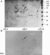
Far-Western analysis of soybean root proteins at 24 dap (10 dai) probed with the LRR domain of GmRLK18-1. Panel (A): Shown is a portion of a 2D gel (14.4-21.5 KDa; 7.5-10.0 pI) from 34–23 (resistant) SCN inoculated total root proteins with spots visualized with silver staining. Panel (B): Proteins transferred to a membrane and probed with purified GmRLK18-1 LRR domain and 6X his-RHG1. Anti-His-HRP was used as the secondary probe. The single spot identified (arrowed) was excised from the duplicate gel and analyzed by Q-TOF (MS-MS) to identify a cyclophilin as a GmRLK18-1 LRR domain interacting partner.
Figure 5
Figure 5
Far-Western analysis of soybean root proteins at 42 dap (28 dai) probed with the LRR domain of GmRLK18-1. Panel (A): Shown is a whole 2D gel (6.5-116.0 KDa; 3.0-10.0 pI) from 34–23 (resistant) SCN inoculated total root proteins with spots visualized with silver staining. Panel (B): Proteins transferred to a membrane and probed with purified GmRLK18-1 LRR domain and 6X his-RHG1. Anti-His-HRP was used as the secondary probe. The single spot identified (arrowed) was excised from the duplicate gel and analyzed by Q-TOF (MS-MS) to identify methionine synthase (GI: 33325957) at 84.2 KDa and pI 5.93 as a GmRLK18-1 LRR domain interacting partner. The other 3 proteins were of higher abundance and so not likely to be specific interactions.
Figure 6
Figure 6
Predicted structures of the GmRLK18-1 LRR monomer. Panels (A-D) The β sheet regions are shown in yellow and helical regions in red. The modeling results suggest LRRs of GmRLK18-1 at Rhg1/Rfs2 (panel A) and GmRLK8-1 at Rhg4 adopt horse shoe type architectures. In GmRLK18-1, the N and C terminal helices are longer and the shorter helices are evenly spaced in the repeats. Also seen are 4 pockets in the protein where helices do not form and the unstructured regions are the two nearest the C terminus. In GmRLK18-1 prediction, the N and C terminal helices are longer and the shorter helices are evenly spaced in the repeats whereas in the GmRLK8-1 prediction, the helices are unevenly spaced and are present only at the N or C terminal. Panel C shows the predicted GmRLK18-1 structure looking at the concave surface and panel D was looking at the convex surface. Panel (E) GmRLK18-1 was modeled as a crystal homo-dimer based on the RI template. The homo-dimer interface was held together by anti parallel β sheets involving many residues from each monomeric chain. Chains were offset by about 90 residues. Circled in white is the cysteine less than 11 A from the partnering homo-dimer chain as detected by cross-linking, circled in yellow is the cysteine not near the dimer interface. (E) The predicted structure by SWISS-PROT [49] for only the LRR domain from amino acid 141–435 of GmRLK18-1 that was expressed in E. coli. The N-terminus lacked the signaling peptide. The C terminus was 61 amino acids (−61) short of the start of the trans-membrane domain.

References

    1. Jones JDG, Dangl JL. The plant immune system. Nature. 2006;444:323–329. doi: 10.1038/nature05286. - DOI - PubMed
    1. Afzal AJ, Wood AJ, Lightfoot DA. Plant receptor-like serine threonine kinases: Roles in signaling plant defense. Molec Plant Microb Interact. 2008;21:507–517. doi: 10.1094/MPMI-21-5-0507. - DOI - PubMed
    1. Wrather JA, Koenning SR, Anderson TR. Effect of diseases on soybean yields in the United States and Ontario (1999 to 2002) Plant Health Progr. 2003. - DOI
    1. Palmateer AJ, Schmidt ME, Stetina SR, Russin JS. Temperature effects on race determination in Heterodera glycines. J Nematol. 2000;32:349–355. - PMC - PubMed
    1. Ruben E, Aziz J, Afzal J, Njiti VN, Triwitayakorn K, Iqbal MJ, Yaegashi S, Arelli PR, Town CD, Ishihara H, Meksem K, Lightfoot DA. Genomic analysis of the ‘Peking’ rhg1 locus: Candidate genes that underlie soybean resistance to the cyst nematode. Mol Genet Genome. 2006;276:320–330. - PubMed

Publication types