. 2013 Mar 12:12:24.
doi: 10.1186/1475-2859-12-24.

Optimizing heterologous protein production in the periplasm of E. coli by regulating gene expression levels

Affiliations

Optimizing heterologous protein production in the periplasm of E. coli by regulating gene expression levels

Susan Schlegel et al. Microb Cell Fact. .

Abstract

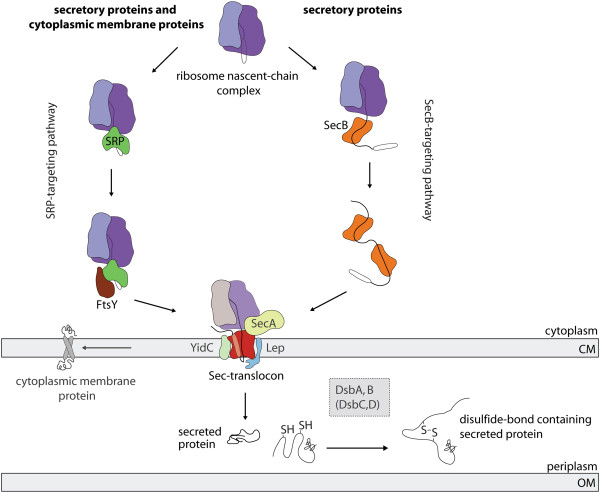
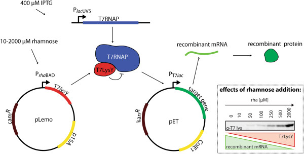
Background: In Escherichia coli many heterologous proteins are produced in the periplasm. To direct these proteins to the periplasm, they are equipped with an N-terminal signal sequence so that they can traverse the cytoplasmic membrane via the protein-conducting Sec-translocon. For poorly understood reasons, the production of heterologous secretory proteins is often toxic to the cell thereby limiting yields. To gain insight into the mechanism(s) that underlie this toxicity we produced two secretory heterologous proteins, super folder green fluorescent protein and a single-chain variable antibody fragment, in the Lemo21(DE3) strain. In this strain, the expression intensity of the gene encoding the target protein can be precisely controlled.

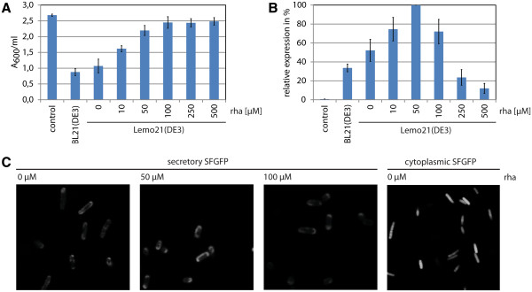
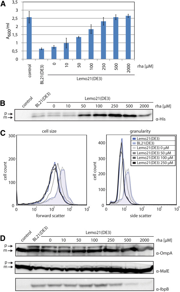
Results: Both SFGFP and the single-chain variable antibody fragment were equipped with a DsbA-derived signal sequence. Producing these proteins following different gene expression levels in Lemo21(DE3) allowed us to identify the optimal expression level for each target gene. Too high gene expression levels resulted in saturation of the Sec-translocon capacity as shown by hampered translocation of endogenous secretory proteins and a protein misfolding/aggregation problem in the cytoplasm. At the optimal gene expression levels, the negative effects of the production of the heterologous secretory proteins were minimized and yields in the periplasm were optimized.

Conclusions: Saturating the Sec-translocon capacity can be a major bottleneck hampering heterologous protein production in the periplasm. This bottleneck can be alleviated by harmonizing expression levels of the genes encoding the heterologous secretory proteins with the Sec-translocon capacity. Mechanistic insight into the production of proteins in the periplasm is key to optimizing yields in this compartment.


Figures

Figure 1
Figure 1
The biogenesis of Sec-translocon dependent secretory and cytoplasmic membrane proteins in E. coli. In E. coli, most secretory and cytoplasmic membrane proteins require the Sec-translocon for their biogenesis. The Sec-translocon is a protein conducting channel in the cytoplasmic membrane (CM), which mediates the vectorial transfer of secretory proteins across and the biogenesis of membrane proteins in the cytoplasmic membrane [7]. Secretory proteins are equipped with a cleavable N-terminal signal sequence. The signal sequence determines whether a secretory protein is targeted to the Sec-translocon via the post-translational SecB-targeting pathway or the co-translational signal recognition particle (SRP)-targeting pathway, which is comprised of the SRP and its receptor FtsY. Upon translocation, the signal sequence is cleaved off by leader peptidase (Lep) and the secretory protein is released into the periplasm. In this compartment, the Dsb-system can catalyze the formation of disulfide bonds. The disulfide oxidoreductase DsbA catalyzes the de-novo formation of disulfide bonds in polypeptide chains. The disulfide bond formation protein B (DsbB) is essential to maintain DsbA in an oxidized state. Incorrectly formed disulfide bonds can be corrected by DsbC/D. For a more detailed description of disulfide bond formation in the periplasm of E. coli see [3,5]. Cytoplasmic membrane proteins are targeted to the Sec-translocon via the SRP-targeting pathway. SecA = peripheral membrane ATPase associated with the Sec-translocon [18], OM = outer membrane, YidC = cytoplasmic membrane protein translocase/insertase [18].
Figure 2
Figure 2
Regulating target gene expression levels using the Lemo21(DE3) strain. Lemo21(DE3) is a BL21(DE3) derivative harboring the pLemo plasmid. In Lemo21(DE3), expression of the gene encoding the target protein is driven by T7 RNAP. The gene encoding T7 RNAP is located on the chromosome. Its expression is governed by the not well-titratable and very strong, IPTG inducible lacUV5 promoter. The activity of T7 RNAP can be modulated by expression of the gene encoding the natural inhibitor of the T7 RNAP, T7 lysozyme, from pLemo. The pLemo plasmid has a p15A ori and a chloramphenicol resistance marker. Expression of the gene encoding the T7 lysozyme is governed by the well-titratable rhamnose promoter. The gene encoding the target protein is located on a pET-vector. pET-vectors have a ColE1 ori and the version used in this study has a kanamycin resistance marker. The expression of the gene encoding the target protein from the pET-vector is governed by the T7lac promoter. The expression levels of the gene encoding the target protein can be increasingly dampened by the addition of increasing amounts of rhamnose to the culture. The more rhamnose is added the more T7 lysozyme is synthesized (see immuno-blot of T7 lysozyme on the right). As a consequence, T7 RNAP is increasingly inhibited and the expression levels of the target gene decrease (see inset).
Figure 3
Figure 3
Production of secretory SFGFP following varying gene expression levels. Lemo21(DE3) cells harboring a pET-vector with the gene encoding secretory SFGFP were cultured in LB medium at 30°C. The expression of secretory SFGFP was induced with 400 μM IPTG for 4 h. Rhamnose was present as indicated. Lemo21(DE3) harboring an empty expression vector (control) and BL21(DE3) producing secretory SFGFP were included as controls. A The effect of the production of secretory SFGFP following varying gene expression levels on biomass formation was monitored by measuring the A600. B The effect of the production of secretory SFGFP following varying gene expression levels on protein yields was monitored as fluorescence per ml of culture. The highest level of fluorescence was set to 100%; the other values were adjusted accordingly. C The localization of secretory SFGFP in Lemo21(DE3) cells cultured in the absence and presence of increasing concentrations of rhamnose was monitored directly in whole cells using fluorescence microscopy. Lemo21(DE3) cells producing cytoplasmic SFGFP (i.e., SFGFP not equipped with a signal sequence) in the absence of rhamnose were included as control.
Figure 4
Figure 4
Consequences of varying expression levels of the gene encoding secretory SFGFP. The expression of the gene encoding secretory SFGFP was induced with IPTG in Lemo21(DE3) in the absence and presence of increasing concentrations of rhamnose. Lemo21(DE3) harboring an empty expression vector (control) and BL21(DE3) harboring the secretory SFGFP expression vector were included as controls. 4 h after induction of the expression of the gene encoding secretory SFGFP cells were analysed by flow cytometry and immuno-blotting. A Cell size (forward scatter) and granularity (side scatter) were monitored in cells producing secretory SFGFP. B SDS-PAGE/immuno-blotting using antisera against IbpB, OmpA and MalE. For the immuno-blotting analysis, the precursor (p) and the mature form (m) of OmpA and MalE are indicated.
Figure 5
Figure 5
Production of the secretory scFv BL1 following varying gene expression levels. Expression of the gene encoding secretory BL1 was induced with IPTG in Lemo21(DE3) cells grown in the absence and presence of increasing concentrations of rhamnose. BL21(DE3) expressing the gene encoding secretory BL1 and Lemo21(DE3) harboring an empty expression vector were included as controls. A 4 h after induction, cell growth was monitored by measuring A600. B Levels and processing of secretory BL1 were monitored using SDS-PAGE followed by immuno-blotting using an α-His antibody. The precursor (p) and the mature (m) form of the protein are indicated. C Using flow cytometry, cell size (forward scatter) and granularity (side scatter) of cells producing secretory BL1 were monitored. D Levels of IbpB, OmpA and MalE were monitored using a combination of SDS-PAGE and immuno-blotting. For OmpA and MalE, the precursor (p) and the mature form (m) of the respective protein are indicated.
Figure 6
Figure 6
Characterization of secretory BL1 expressed at the optimal rhamnose concentration in Lemo21(DE3). Expression of the gene encoding secretory BL1 was induced with IPTG in Lemo21(DE3) in the presence of 500 μM of rhamnose (see Figure 5). 4 h after induction a whole cell lysate was prepared and the periplasmic fraction isolated. A Nitrocellulose membranes containing increasing amounts of β-galactosidase were incubated with the whole cell lysate (top panel) and whole cell lysate that had been incubated with β-mercaptoethanol (middle panel). Binding of BL1 to the β-galactosidase spotted on the nitrocellulose membranes was detected using an α-His antibody recognizing the C-terminal His-tag of BL1. A nitrocellulose membrane containing spots with increasing amounts of BSA and incubated with the same lysate used in the top panel was included as a control (bottom panel). B The periplasmic fraction was isolated as described in Methods. SEC was used to analyze the BL1 that was isolated from the periplasmic fraction by means of IMAC. Indicated fractions (inset) from the SEC were analyzed by SDS-PAGE followed by Coomassie staining (top panel inset) or immuno-blotting using an α-His antibody (middle panel inset). The fractions representing the indicated peak were pooled and the BL1 was tested for binding to β-galactosidase (bottom panel inset). β-galactosidase concentrations correspond to the setup described in A. The bottom panel of the inset shows only the first 4 concentrations.

References

    1. Makino T, Skretas G, Georgiou G. Strain engineering for improved expression of recombinant proteins in bacteria. Microb Cell Fact. 2011;10:32. doi: 10.1186/1475-2859-10-32. - DOI - PMC - PubMed
    1. Mergulhao FJ, Summers DK, Monteiro GA. Recombinant protein secretion in Escherichia coli. Biotechnol Adv. 2005;23(3):177–202. doi: 10.1016/j.biotechadv.2004.11.003. - DOI - PubMed
    1. Kadokura H, Beckwith J. Mechanisms of oxidative protein folding in the bacterial cell envelope. Antioxid Redox Signal. 2010;13(8):1231–1246. doi: 10.1089/ars.2010.3187. - DOI - PMC - PubMed
    1. de Marco A. Recent contributions in the field of the recombinant expression of disulfide bonded proteins in bacteria. Microb Cell Fact. 2012;11:129. doi: 10.1186/1475-2859-11-129. - DOI - PMC - PubMed
    1. Denoncin K, Collet JF. Disulfide bond formation in the bacterial periplasm: major achievements and challenges ahead. Antioxid Redox Signal. 2012. - DOI - PMC - PubMed

Publication types

MeSH terms