Clinical Trial
. 2013 Mar 9;14(1):33.
doi: 10.1186/1465-9921-14-33.

Smoking decreases the response of human lung macrophages to double-stranded RNA by reducing TLR3 expression

Affiliations
Clinical Trial

Smoking decreases the response of human lung macrophages to double-stranded RNA by reducing TLR3 expression

Jill C Todt et al. Respir Res. .

Abstract

Background: Cigarette smoking is associated with increased frequency and duration of viral respiratory infections, but the underlying mechanisms are incompletely defined. We investigated whether smoking reduces expression by human lung macrophages (Mø) of receptors for viral nucleic acids and, if so, the effect on CXCL10 production.

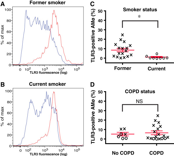
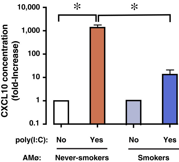
Methods: We collected alveolar macrophages (AMø) by bronchoalveolar lavage of radiographically-normal lungs of subjects undergoing bronchoscopies for solitary nodules (n = 16) and of volunteers who were current or former smokers (n = 7) or never-smokers (n = 13). We measured expression of mRNA transcripts for viral nucleic acid receptors by real-time PCR in those AMø and in the human Mø cell line THP-1 following phorbol myristate acetate/vitamin D3 differentiation and exposure to cigarette smoke extract, and determined TLR3 protein expression using flow cytometry and immunohistochemistry. We also used flow cytometry to examine TLR3 expression in total lung Mø from subjects undergoing clinically-indicated lung resections (n = 25). Of these, seven had normal FEV1 and FEV1/FVC ratio (three former smokers, four current smokers); the remaining 18 subjects (14 former smokers; four current smokers) had COPD of GOLD stages I-IV. We measured AMø production of CXCL10 in response to stimulation with the dsRNA analogue poly(I:C) using Luminex assay.

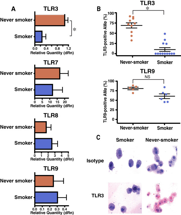
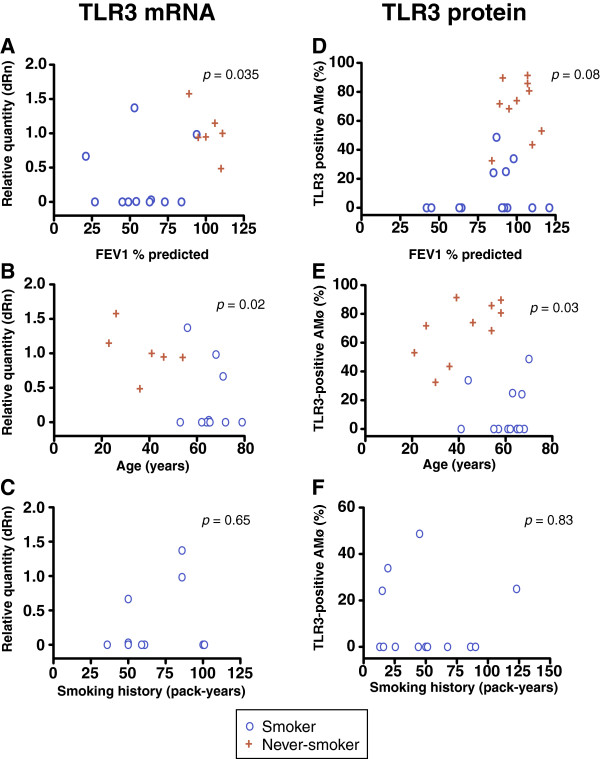
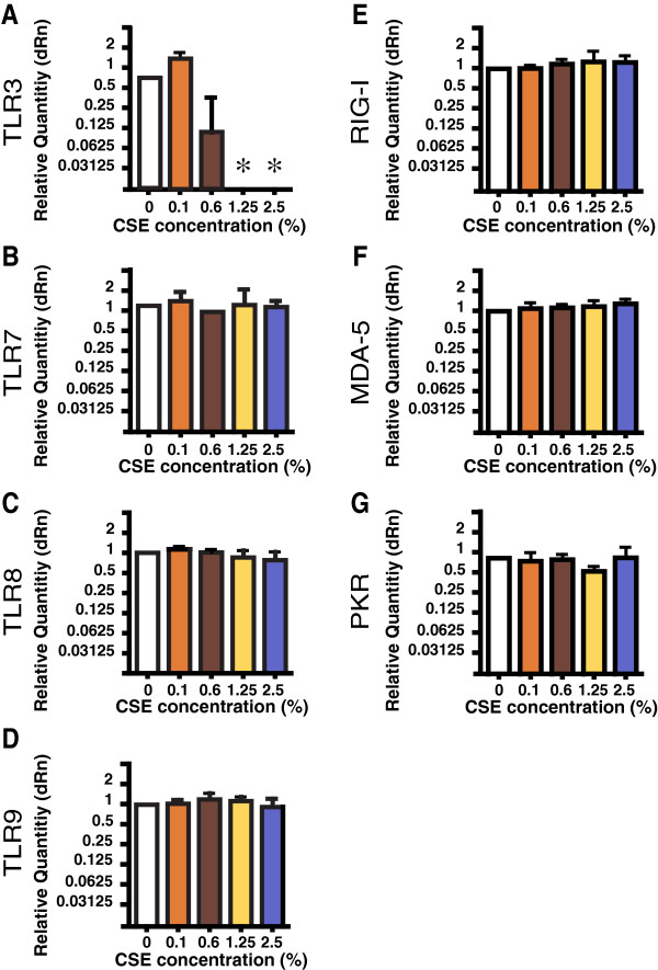
Results: Relative to AMø of never-smokers, AMø of smokers demonstrated reduced protein expression of TLR3 and decreased mRNA for TLR3 but not TLR7, TLR8, TLR9, RIG-I, MDA-5 or PKR. Identical changes in TLR3 gene expression were induced in differentiated THP-1 cells exposed to cigarette smoke-extract in vitro for 4 hours. Among total lung Mø, the percentage of TLR3-positive cells correlated inversely with active smoking but not with COPD diagnosis, FEV1% predicted, sex, age or pack-years. Compared to AMø of never-smokers, poly(I:C)-stimulated production of CXCL10 was significantly reduced in AMø of smokers.

Conclusions: Active smoking, independent of COPD stage or smoking duration, reduces both the percent of human lung Mø expressing TLR3, and dsRNA-induced CXCL10 production, without altering other endosomal or cytoplasmic receptors for microbial nucleic acids. This effect provides one possible mechanism for increased frequency and duration of viral lower respiratory tract infections in smokers.

Trial registration: ClinicalTrials.gov NCT00281190, NCT00281203 and NCT00281229.


Figures

Figure 1
Figure 1
AMø of current smokers show reduced expression of TLR3 mRNA transcripts and intracellular protein relative to AMø of never-smokers. A. mRNA transcripts. RNA was isolated from AMø, depleted of contaminating genomic DNA, reverse-transcribed and analyzed by quantitative real-time RT-PCR using Taqman chemistry and specific primer-probe sets, normalized to GAPDH transcripts. Data are expressed on the horizontal axis as mean ± SEM for relative quantity (dRn), calculated in comparison to a single never-smoker who was arbitrarily designated the reference sample. Never-smokers (n = 6), red bars; smokers (n = 11), blue bars. *, p < 0.05 by Mann–Whitney test. B. Intracellular TLR3 (top panel) and TLR9 (bottom panel) expression by flow cytometry. AMø were permeabilized, stained for TLR3 and TLR9 expression and analyzed by flow cytometry. Data are expressed as TLR-positive AMø as mean ± SEM, never-smokers (n = 10 for TLR3, n = 5 for TLR9), red circles; smokers (n = 13 for TLR3, n = 6 for TLR9), blue circles. *, p < 0.05 by Mann–Whitney test. C. Immunohistochemical staining. Cytospins from a representative smoker (right) and a never-smoker (left), stained for intracellular TLR3 expression using AEC (red product) and hematoxylin (original 20 X). Top row, isotype control staining, bottom row, specific TLR3 staining. Representative of three experiments with similar results.
Figure 2
Figure 2
AMø TLR3 expression does not correlate with lung function or total duration of smoking exposure. AMø were harvested, processed and analyzed as described in the legend to Figure 1. A-C. TLR3 mRNA transcripts by quantitative real-time PCR (smokers, n = 11; never-smokers, n = 6, except in panel C, which includes only current or former smokers). D-F. Flow cytometric analysis of TLR3 protein expression (smokers, n = 13; never-smokers, n = 10, except in panel F which includes only current or former smokers). A, D: FEV1 % predicted; B, E: participant age (years); C, F: smoking history (pack-years); note the difference in the vertical scale of panel F. Blue circles, smokers; red crosses, never-smokers. p values by Spearman correlation.
Figure 3
Figure 3
In current smokers, the frequency of lung Mø expressing TLR3 protein is reduced relative to lung Mø of former-smokers. To isolate total lung Mø, areas of human lung parenchyma, resected for clinical indications and remote from any evidence of nodules or infection, were processed by mechanical disaggregation without the use of enzymes. Lung Mø were permeabilized, stained for TLR3 expression and analyzed by flow cytometry. A, B. Representative histograms of intracellular TLR3 expression; A, former smoker; B, current smoker. C. Percentage of lung Mo expressing TLR3 relative to smoking status; x’s represent former smokers (n = 17); circles represent current smokers (n = 8). D. Percentage of TLR3-positive by lung Mø relative to COPD status; as in panel C, x’s represent former smokers (n = 17); circles represent current smokers (n = 8); *, p < 0.05; NS, non-significant. The Mann–Whitney test was used to calculate p values.
Figure 4
Figure 4
Exposure to CSE specifically and rapidly reduces TLR3 gene expression in a differentiated human Mø cell line. THP-1 cells differentiated using PMA & vitamin D3 for 48 hours were cultured in complete medium alone or with various concentration of CSE for an additional 4 hr, then analyzed by quantitative real-time RT-PCR. Data are mean ± SEM of four independent experiments, expressed as dRn (relative to differentiated THP-1 cells cultures in complete medium alone). A, TLR3; B, TLR7; C, TLR8; D, TLR9; E, RIG-1; F, MDA-5; G, PKR. Kruskal-Wallis one-way ANOVA was used to compare groups. *, p < 0.05, compared to cells that did not receive CSE exposure.
Figure 5
Figure 5
Production of CXCL10 by AMø of smokers in response to poly(I:C) stimulation is impaired. AMø from never-smokers (n = 4) (white and red columns) and smokers (n = 5) (light and dark blue columns) were cultured for 24 h in either complete medium alone or with poly(I:C). Supernatants were collected and CXCL10 protein was measured by Luminex assay; results are expressed as the fold-increase in CXCL10 over the unstimulated control condition; note the logarithmic scale of the vertical axis. Data are mean ± SEM. One-way ANOVA with Bonferroni’s Multiple Comparison post-hoc testing was used to compare differences among groups, *, p < 0.01.

References

    1. Johnston SL. Overview of virus-induced airway disease. Proc Am Thorac Soc. 2005;2:150–156. doi: 10.1513/pats.200502-018AW. - DOI - PubMed
    1. Proud D, Chow CW. Role of viral infections in asthma and chronic obstructive pulmonary disease. Am J Respir Cell Mol Biol. 2006;35:513–518. doi: 10.1165/rcmb.2006-0199TR. - DOI - PubMed
    1. Arcavi L, Benowitz NL. Cigarette smoking and infection. Arch Intern Med. 2004;164:2206–2216. doi: 10.1001/archinte.164.20.2206. - DOI - PubMed
    1. Aronson MD, Weiss ST, Ben RL, Komaroff AL. Association between cigarette smoking and acute respiratory tract illness in young adults. JAMA. 1982;248:181–183. doi: 10.1001/jama.1982.03330020025023. - DOI - PubMed
    1. Bensenor IM, Cook NR, Lee IM, Chown MJ, Hennekens CH, Buring JE, Manson JE. Active and passive smoking and risk of colds in women. Ann Epidemiol. 2001;11:225–231. doi: 10.1016/S1047-2797(00)00214-3. - DOI - PubMed

Publication types

MeSH terms

Associated data

LinkOut - more resources