. 2013 Mar 5;6(1):5.
doi: 10.1186/1756-8935-6-5.

A permissive chromatin structure is adopted prior to site-specific DNA demethylation of developmentally expressed genes involved in macronuclear differentiation

Affiliations

A permissive chromatin structure is adopted prior to site-specific DNA demethylation of developmentally expressed genes involved in macronuclear differentiation

Aneta Bulic et al. Epigenetics Chromatin. .

Abstract

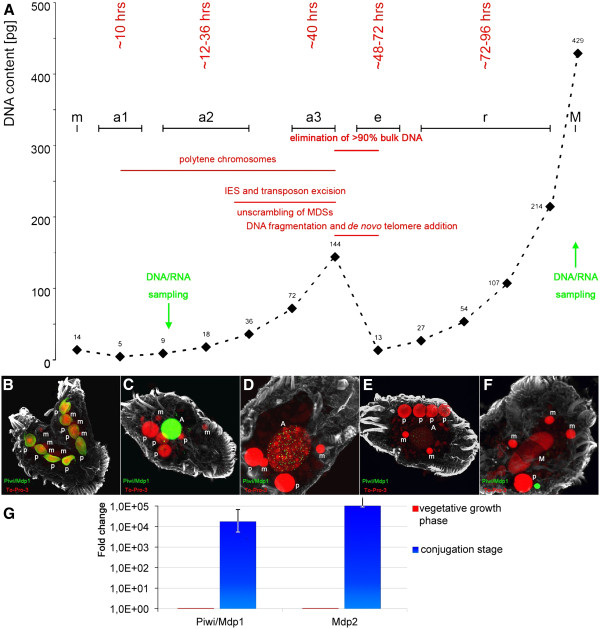
Background: DNA methylation and demethylation are important epigenetic regulatory mechanisms in eukaryotic cells and, so far, only partially understood. We exploit the minimalistic biological ciliate system to understand the crosstalk between DNA modification and chromatin structure. In the macronucleus of these cells, the DNA is fragmented into individual short DNA molecules, each representing a functional expression and replication unit. Therefore, long range epigenomic interaction can be excluded in this system.

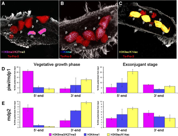
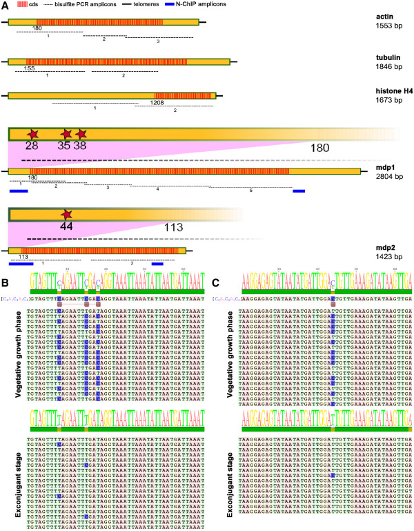
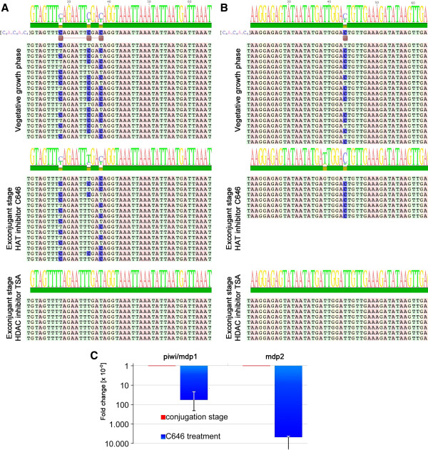
Results: In the stichotrichous ciliate Stylonychia lemnae, cytosine methylation occurs in a small subset of macronuclear nanochromosomes expressed only during sexual reproduction. Methylation pattern shows similarity to that observed in fungi and Drosophila. Cytosine methylation correlates with gene activity and chromatin structure. Upon gene activation, cytosines become demethylated and a redistribution of histone post-translational modifications (PTMs) takes place. Evidence is presented that the formation of a permissive chromatin structure in the vicinity of the 5meCs precedes cytosine methylation and is probably a necessary prerequisite for their demethylation. Shortly after demethylation of cytosines occurs, the parental macronucleus degenerates, a new macronucleus is formed from a micronuclear derivative and the specific methylation pattern is transmitted from the germline micronucleus to the new macronucleus.

Conclusions: We show that very few, or even only one, discrete methylated cytosines are required to assign regulatory functions at a specific locus. Furthermore, evidence is provided that a permissive chromatin structure is probably a necessary prerequisite for the demethylation of specific cytosines. Our results allow us to propose a mechanistic model for the biological function of cytosine methylation in the ciliate cell and its regulation during the cell cycle.


Figures

Figure 1
Figure 1
Macronuclear differentiation in the stichotrichous ciliate Stylonychia lemnae. (A) Schematic diagram of macronuclear differentiation. Time points at which DNA or RNA was isolated are indicated by an arrow. (B-F) Nuclear events during macronuclear differentiation and behavior of the developmentally expressed Mdp1 (Piwi) protein. (B) In the two conjugating cells, Piwi accumulates in the parental macronucleus (p). (C-D) During formation of polytene chromosomes, Piwi relocalizes from the parental macronucleus (p) to the developing macronucleus (a1-3). (E, F) During chromatin elimination in the anlage (e) and subsequent formation of the vegetative macronucleus (m), Piwi is no longer present. (G) Expression of the genes mdp1 and mdp2 during vegetative growth and at the onset of macronuclear differentiation. While no expression of these genes takes place during vegetative growth, they become transcribed approximately 30 hours after the onset of sexual reproduction. IES: sequences interrupting macronuclear-destined sequences in the micronuclear genome. MDS: macronuclear-destined sequences in the macronuclear genome.
Figure 2
Figure 2
Localization of different histone PTMs in parental macronuclei (p) and micronuclei (m) of exconjugant cells shortly after separation. (A-C) In situ staining using antibodies directed against H3K9me3/K27me3 (A, hot magenta), H3K4me (B, hot blue), H3K9ac/K14ac (C, yellow). DNA counterstaining with To-Pro-3 (red). (D, E) Distribution of these PTMs on the 5- and 3-subtelomeric regions of mdp1 as well as mdp2 nanochromosomes during vegetative growth and in exconjugant cells.
Figure 3
Figure 3
Macronuclear nanochromosomes studied and DNA methylation pattern of developmentally expressed genes during vegetative growth and sexual reproduction. (A) Actin, tubulin and histone H4 are constitutively expressed genes; mdp1 and mdp2 are only expressed during sexual reproduction in exconjugant cells. Red shaded areas show the open reading frames of the nanochromosomes. Cytosines are methylated at positions 28, 35 and 38 in mdp1 and at position 44 in mdp2 (red stars). Black dashed lines: amplicons analyzed for cytosine methylation; blue lines: amplicons used for ChIP analyses. (B, C) DNA methylation pattern in mdp1 (B) and mdp2 (C) during vegetative growth (mdp1/mdp2 repressed) and in exconjugant cells (mdp1/mdp2 expressed).
Figure 4
Figure 4
Inhibition of histone acetyltransferase and histone deacetylase. (A, B) Effect of histone acetyl transferase inhibitor C646 and histone deacetylase inhibitor TSA on the DNA methylation pattern of mdp1 (A) and mdp2 (B). (C) The effect of histone deacetylase inhibitor C646 on the expression of these nanochromosomes during sexual reproduction was analyzed by qPCR. In both cases a dramatic decline in expression is observed after treatment with C646.

References

    1. Bogdanovic O, Veenstra GJ. DNA methylation and methyl-CpG binding proteins: developmental requirements and function. Chromosoma. 2009;118:549–565. doi: 10.1007/s00412-009-0221-9. - DOI - PMC - PubMed
    1. Bannister AJ, Kouzarides T. Regulation of chromatin by histone modifications. Cell Res. 2011;21:381–395. doi: 10.1038/cr.2011.22. - DOI - PMC - PubMed
    1. Ray-Gallet D, Almouzni G. Nucleosome dynamics and histone variants. Essays Biochem. 2010;48:75–87. doi: 10.1042/bse0480075. - DOI - PubMed
    1. Berdasco M, Esteller M. Aberrant epigenetic landscape in cancer: how cellular identity goes awry. Dev Cell. 2010;19:698–711. doi: 10.1016/j.devcel.2010.10.005. - DOI - PubMed
    1. Doi A, Park IH, Wen B, Murakami P, Aryee MJ, Irizarry R, Herb B, Ladd-Acosta C, Rho J, Loewer S. Differential methylation of tissue- and cancer-specific CpG island shores distinguishes human induced pluripotent stem cells, embryonic stem cells and fibroblasts. Nat Genet. 2009;41:1350–1353. doi: 10.1038/ng.471. - DOI - PMC - PubMed

LinkOut - more resources