. 2013 Mar 15:8:41.
doi: 10.1186/1750-1172-8-41.

Autosomal recessive spastic ataxia of Charlevoix Saguenay (ARSACS): expanding the genetic, clinical and imaging spectrum

Affiliations

Autosomal recessive spastic ataxia of Charlevoix Saguenay (ARSACS): expanding the genetic, clinical and imaging spectrum

Matthis Synofzik et al. Orphanet J Rare Dis. .

Abstract

Background: Mutations in SACS, leading to autosomal-recessive spastic ataxia of Charlevoix-Saguenay (ARSACS), have been identified as a frequent cause of recessive early-onset ataxia around the world. Here we aimed to enlarge the spectrum of SACS mutations outside Quebec, to establish the pathogenicity of novel variants, and to expand the clinical and imaging phenotype.

Methods: Sequencing of SACS in 22 patients with unexplained early-onset ataxia, assessment of novel SACS variants in 3.500 European control chromosomes and extensive phenotypic investigations of all SACS carriers.

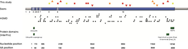
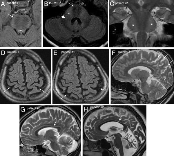
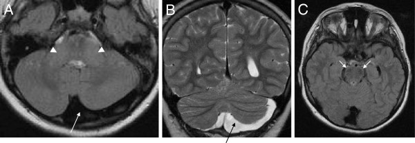
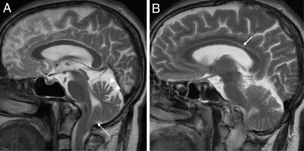
Results: We identified 11 index patients harbouring 17 novel SACS variants. 9/11 patients harboured two variants of at least probable pathogenicity which were not observed in controls and, in case of missense mutations, were located in highly conserved domains. These 9 patients accounted for at least 11% (9/83) in our series of unexplained early onset ataxia subjects. While most patients (7/9) showed the classical ARSACS triad, the presenting phenotype reached from pure neuropathy (leading to the initial diagnosis of Charcot-Marie-Tooth disease) in one subject to the absence of any signs of neuropathy in another. In contrast to its name "spastic ataxia", neither spasticity (absent in 2/9=22%) nor extensor plantar response (absent in 3/9=33%) nor cerebellar ataxia (absent in 1/9=11%) were obligate features. Autonomic features included urine urge incontinence and erectile dysfunction. Apart from the well-established MRI finding of pontine hypointensities, all patients (100%) showed hyperintensities of the lateral pons merging into the (thickened) middle cerebellar peduncles. In addition, 63% exhibited bilateral parietal cerebral atrophy, and 63% a short circumscribed thinning of the posterior midbody of the corpus callosum. In 2 further patients with differences in important clinical features, VUS class 3 variants (c.1373C>T [p.Thr458Ile] and c.2983 G>T [p.Val995Phe]) were identified. These variants were, however, also observed in controls, thus questioning their pathogenic relevance.

Conclusions: We here demonstrate that each feature of the classical ARSACS triad (cerebellar ataxia, spasticity and peripheral neuropathy) might be missing in ARSACS. Nevertheless, characteristic MRI features - which also extend to supratentorial regions and involve the cerebral cortex - will help to establish the diagnosis in most cases.


Figures

Figure 1
Figure 1
SACS mutations. Graphical overview of mutations found in this and other studies (Human Gene Mutation database, except gross deletions). Different mutation types are marked with different symbols: missense mutations = squares; insertions/duplications = triangles with downward orientation; deletions = triangles with upward orientation. Sequence variations identified in this study are coloured, with the colour indicating the pathogenicity class: red = pathogenic, orange = likely pathogenic, yellow = uncertain. Known protein domains of the sacsin protein are highlighted in green. AA = amino acid; HGMD = Human Gene Mutation Database.
Figure 2
Figure 2
Characteristic MRI findings in ARSACS. Axial FLAIR images of upper pons (A) and middle pons (B) show hypointense strips in the central pons representing the cortico-spinal tract (white arrows): Moreover, on the level of the mid-pons (B), they reveal diffuse slight hyperintensity of the lateral pons when merging into the middle cerebellar peduncles (stars) and thickened middle cerebellar peduncles (white arrow head) in patient #1. Also on coronal T2 images (C) the bilateral hyperintensity of the lateral pons can be seen (stars). Bilateral post-central and parietal atrophy is shown for the same patient in axial FLAIR (D, E) and sagittal T2 (F) images (white arrows). Thinning of the posterior mid-body of the corpus callosum on sagittal T2 images is shown for patient #5 (G) and patient #2 (H) (white arrows).
Figure 3
Figure 3
Developmental asymmetry of cerebellar hemispheres. Axial T1 (A) and coronal T2 (B) MRI images of patient #4 demonstrating developmental asymmetry of the cerebellar hemispheres (long arrows) without cerebellar atrophy. Apart from this finding, this patient shows the common infratentorial signs of ARSACS: thickening of the middle cerebellar peduncles (arrow head, A) and linear hypointensities of the mid-pons (best seen on axial FLAIR, arrows, C).
Figure 4
Figure 4
Developmental posterior fossa malformation resembling Chiari type I in the Thr458Ile+ p.Thr458Ile patient. (A) Midsagittal T2 MRI demonstrates herniation of cerebellar tonsils, resembling Chiari malformation type 1 (arrow). Also note the atrophy of the superior cerebellar vermis (arrow heads). (B) Para-midsagittal shows slight short-stretched thinning of the posterior mid-body of the corpus callosum (arrow).

References

    1. Vermeer S, van de Warrenburg BP, Kamsteeg EJ. In: GeneReviews [Internet] Pagon RA, Bird TC, Dolan CR, Stephens K, editor. Seattle (WA): University of Washington; 2012. ARSACS. http://www.ncbi.nlm.nih.gov/books/NBK1255/; accessed November 22nd, 2012.
    1. Gerwig M, Kruger S, Kreuz FR, Kreis S, Gizewski ER, Timmann D. Characteristic MRI and funduscopic findings help diagnose ARSACS outside Quebec. Neurology. 2010;75:2133. doi: 10.1212/WNL.0b013e318200d7f8. - DOI - PubMed
    1. Gazulla J, Benavente I, Vela AC, Marin MA, Pablo LE, Tessa A, Barrena MR, Santorelli FM, Nesti C, Modrego P. New findings in the ataxia of Charlevoix-Saguenay. J Neurol. 2012;259:869–878. doi: 10.1007/s00415-011-6269-5. - DOI - PubMed
    1. Shimazaki H, Takiyama Y, Sakoe K, Ando Y, Nakano I. A phenotype without spasticity in sacsin-related ataxia. Neurology. 2005;64:2129–2131. doi: 10.1212/01.WNL.0000166031.91514.B3. - DOI - PubMed
    1. Baets J, Deconinck T, Smets K, Goossens D, Van den Bergh P, Dahan K, Schmedding E, Santens P, Rasic VM, Van Damme P. Mutations in SACS cause atypical and late-onset forms of ARSACS. Neurology. 2010;75:1181–1188. doi: 10.1212/WNL.0b013e3181f4d86c. - DOI - PubMed

Publication types

MeSH terms

Supplementary concepts

LinkOut - more resources