Review
. 2013:86:1-36.
doi: 10.1016/B978-0-12-394315-6.00001-5.

Viruses and prions of Saccharomyces cerevisiae

Affiliations
Review

Viruses and prions of Saccharomyces cerevisiae

Reed B Wickner et al. Adv Virus Res. 2013.

Abstract

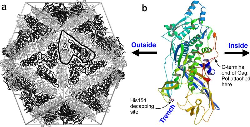
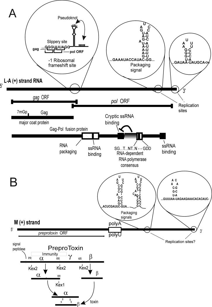
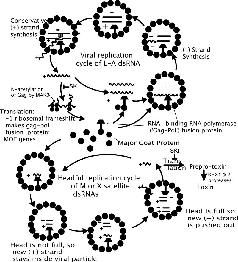
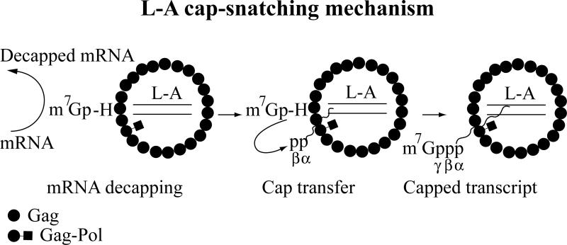
Saccharomyces cerevisiae has been a key experimental organism for the study of infectious diseases, including dsRNA viruses, ssRNA viruses, and prions. Studies of the mechanisms of virus and prion replication, virus structure, and structure of the amyloid filaments that are the basis of yeast prions have been at the forefront of such studies in these classes of infectious entities. Yeast has been particularly useful in defining the interactions of the infectious elements with cellular components: chromosomally encoded proteins necessary for blocking the propagation of the viruses and prions, and proteins involved in the expression of viral components. Here, we emphasize the L-A dsRNA virus and its killer-toxin-encoding satellites, the 20S and 23S ssRNA naked viruses, and the several infectious proteins (prions) of yeast.


Figures

Fig. 1
Fig. 1
L-A virion structure. a. “T=2” architecture with two non-identical forms of the Gag major coat protein (A and B). b. Structure of a single monomer. Note that the trench with His154, the site of cap attachment, is outside the particles, while the C-terminus of Gag, which is extended as Gag-Pol in one or two subunits per particle, is internal (modified from (Naitow et al. 2002)).
Fig. 2
Fig. 2
L-A and M dsRNA cis sites and encoded proteins. a. The L-A genome is shown with the sites of ribosomal frameshifting, the packaging site (and the overlapping internal replication enhancer), and the 3' replication site. The His154 cap attachment site is shown as 7mGp.
Fig. 3
Fig. 3
L-A and M replication cycles.
Fig. 4
Fig. 4
Schematic diagram of L-A cap-snatching mechanism. Gag of an L-A virion decaps mRNA and forms an intermediate with m7Gp through His-154. Then m7Gp is transferred to the diphosphorylated 5’ end of the viral transcript to form a 5’-5’ triphosphate linkage.
Fig. 5
Fig. 5
20S and 23S RNA viral genomes. A. Total RNA extracted from induced cells containing no viruses (lane 1), 20S RNA alone (lane 2), or 20S and 23S RNAs (lane 3). Ethidium bromide staining of an agarose gel is shown. 18S and 25S rRNAs are indicated. B. 20S and 23S RNA genomes and the encoded proteins, p91 and p104. P91 and p104 contain the consensus sequences (A-D) for RNA-dependent RNA polymerases, which are most closely related to those of RNA coliphages. 1-3 indicates amino acid stretches conserved between p91 and p104. C. Diagram of a launching plasmid. The complete sequence of the 20S or 23S RNA genome is inserted downstream of the constitutive PGK1 promoter (PGK1). The hepatitis delta virus ribozyme (R) is fused directly to the 3’ end of the viral genome. Thin lines indicate sequences derived from the vector.
Fig. 6
Fig. 6
Cis-acting signals. A. The 3’ terminal sequences of 20S and 23S RNAs with cis-acting signals. For comparison, the top half domain of tRNATyr is shown. B. The 5’ and 3’ terminal regions of the 20S RNA genome. The 5’ and 3’ cis sites are indicated by lightly shaded boxes. The initiation (Start) and termination (Stop) codons of p91 are also indicated.
Fig. 7
Fig. 7
Model for 20S RNA positive strand synthesis. The intermediate may contain two p91 molecules (filled circles). For simplicity, the interaction of p91 with the internal cis site of the positive strand is omitted.
Fig. 8
Fig. 8
Yeast prion parallel in-register beta sheet architecture and hypothesized mechanism of variant information propagation. Parallel in-register beta sheets are characterized by lines of identical amino acid side chains extending the length of the filaments. These yeast prion beta sheets are known to be folded along the filament long axis as shown. It is proposed that different prion variants have the folds in different locations. The parallel structure is maintained by the favorable interactions of identical side chains. The energetic advantage in these interactions force a new (unstructured) monomer joining the end of the filament to become in-register, thereby insuring that the folds/turns of this monomer are in the same locations as those of previous molecules in the filament. This insures inheritance of prion variant information (conformation) by templating, analogous to DNA templating of sequence information.

References

    1. Alberti S, Halfmann R, King O, Kapila A, Lindquist S. A systematic survey identifies prions and illuminates sequence features of prionogenic proteins. Cell. 2009;137:146–158. - PMC - PubMed
    1. Bai M, Zhou JM, Perrett S. The yeast prion protein Ure2 shows glutathione peroxidase activity in both native and fibrillar forms. J Biol Chem. 2004;279:50025–30. - PubMed
    1. Ball SG, Tirtiaux C, Wickner RB. Genetic control of L-A and L-BC dsRNA copy number in killer systems of Saccharomyces cerevisiae. Genetics. 1984;107:199–217. - PMC - PubMed
    1. Bateman DA, Wickner RB. [PSI+] prion transmission barriers protect Saccharomyces cerevisiae from infection: intraspecies ‘species barriers’. Genetics. 2012;190:569–579. - PMC - PubMed
    1. Baxa U, Cheng N, Winkler DC, Chiu TK, Davies DR, Sharma D, Inouye H, Kirschner DA, Wickner RB, Steven AC. Filaments of the Ure2p prion protein have a cross-beta core structure. J Struct Biol. 2005;150:170–9. - PubMed

Publication types

MeSH terms