

Review
doi: 10.1016/j.cell.2013.02.031.
Nuclear positioning
Affiliations
- PMID: 23498944
- PMCID: PMC3626264
- DOI: 10.1016/j.cell.2013.02.031
Item in Clipboard
Review
Nuclear positioning
Cell.
.
Abstract
The nucleus is the largest organelle and is commonly depicted in the center of the cell. Yet during cell division, migration, and differentiation, it frequently moves to an asymmetric position aligned with cell function. We consider the toolbox of proteins that move and anchor the nucleus within the cell and how forces generated by the cytoskeleton are coupled to the nucleus to move it. The significance of proper nuclear positioning is underscored by numerous diseases resulting from genetic alterations in the toolbox proteins. Finally, we discuss how nuclear position may influence cellular organization and signaling pathways.
Copyright © 2013 Elsevier Inc. All rights reserved.
Figures
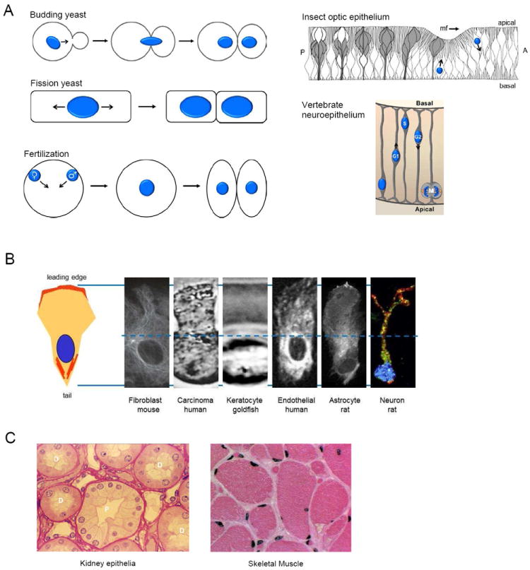
A. Schematics of nuclear positioning in dividing cells and developing epithelium. Arrows indicate movements of nuclei (blue). The nucleus is positioned relative to the plane of division in yeast and fertilized eggs. The diagram of insect optic epithelium represents a longitudinal section of a larval eye disc; two nuclei are shown. Nuclei anterior (A) to the morphogenetic furrow (mf), which moves anteriorly, move basally. Nuclei posterior (P) to the furrow move apically as cells are recruited into clusters comprising ommatidium (white cells, cones; gray cells, R-cells). Adapted from (Patterson et al., 2004; Tomlinson and Ready, 1986). The diagram of vertebrate neuroepithelium represents a longitudinal section of the developing cerebral cortex. Nuclei move basally during G1 and apically during G2. Mitosis (M) occurs near the apical surface. Adapted from (Buchman and Tsai, 2008). B. Rearward nuclear position is typical of migrating cells. Left, schematic of a migrating cell with protruding leading edge and contracting tail. Red, actin filaments. Right, montage of migrating cells with front-back dimensions normalized. Dotted line represents the midpoint between the front and back. Nuclei are positioned along the front-back axis but always rearward of the cell center. Images reproduced from: fibroblast (Gomes et al., 2005); breast carcinoma (McNiven, 2013); keratocyte (Barnhart et al., 2010); endothelial cell (Tsai and Meyer, 2012); astrocyte (Osmani et al., 2006); neuron (Godin et al., 2012). C. Nuclear positioning in mammalian tissues. Cross sections of kidney cortex and skeletal muscle stained with hematoxylin and eosin. Nuclei are positioned centrally in the distal (D) convoluted tubules and basally in proximal (P) convoluted tubules. Nuclei are positioned at the periphery of skeletal muscle fibers.

A. Schematic of an idealized LINC complex in nuclear envelope. The inner nuclear membrane (INM) SUNs bind within the perinuclear space to outer nuclear membrane (ONM) KASH proteins. KASH proteins bind directly or indirectly to cytoskeletal filaments including MTs, actin microfilaments and cytoplasmic intermediate filaments. In metazoans, SUNs bind to the nuclear lamina; in yeast and plants, other intranuclear proteins bind SUNs. A nuclear pore complex (NPC) is shown for reference. B. Side view of the structure of the SUN2-nesprin2 KASH complex. Trimeric SUN2 domains are represented by different shades of blue and the KASH peptide is in orange. The structure illustrates the orientation of the KASH peptide between adjacent SUN domains. Modified from Sosa et al., 2012 with permission. C. Schematic diagrams of KASH proteins from representative organisms and the cytoskeletal filaments to which they bind. Binding to actin filaments is mediated by CH domains and binding to cytoplasmic intermediate filaments by plectin. Binding to MTs is mediated by dynein and kinesins; direct binding to MTs has not been reported. The specific splice variants of nesprin-1 and nesprin-2 that interact with MT motors are unknown; for simplicity, a short variant of each is depicted. H.s. = Homo sapiens, M.m. = Mus musculus, D.m. = Drosophila melanogaster, C.e. = Caenorhabditis elegans, S.p. = Schizosaccharomyces pombe.
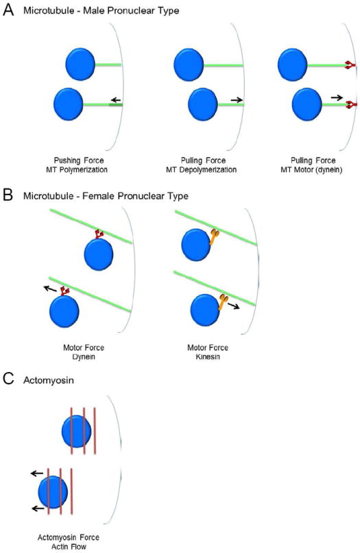
A. Schematic of male pronuclear type nuclear movement mediated by MTs (green). Forces (arrows) can be generated by MT polymerization, depolymerization or dynein motors (red) anchored in the cortex or cytoplasmic sites. B. Schematic of female pronuclear type nuclear movement mediated by MT dynein (red) and kinesin (orange) motors. Forces (arrows) are generated by motors that laterally connect nuclei to MTs. C. Schematic of actomyosin type nuclear movement. Force (arrows) is generated by the actomyosin-dependent flow of dorsal actin cables (red).
References
-
- Attali R, Warwar N, Israel A, Gurt I, McNally E, Puckelwartz M, Glick B, Nevo Y, Ben-Neriah Z, Melki J. Mutation of SYNE-1, encoding an essential component of the nuclear lamina, is responsible for autosomal recessive arthrogryposis. Hum Mol Genet. 2009;18:3462–3469. - PubMed
-
- Bione S, Maestrini E, Rivella S, Mancini M, Regis S, Romeo G, Toniolo D. Identification of a novel X-linked gene responsible for Emery-Dreifuss muscular dystrophy. Nat Genet. 1994;8:323–327. - PubMed
Publication types
MeSH terms
Grants and funding
LinkOut - more resources
Full Text Sources
Other Literature Sources
