. 2013 Mar 13;13(3):336-46.
doi: 10.1016/j.chom.2013.01.012.

Incoming RNA virus nucleocapsids containing a 5'-triphosphorylated genome activate RIG-I and antiviral signaling

Affiliations

Incoming RNA virus nucleocapsids containing a 5'-triphosphorylated genome activate RIG-I and antiviral signaling

Michaela Weber et al. Cell Host Microbe. .

Abstract

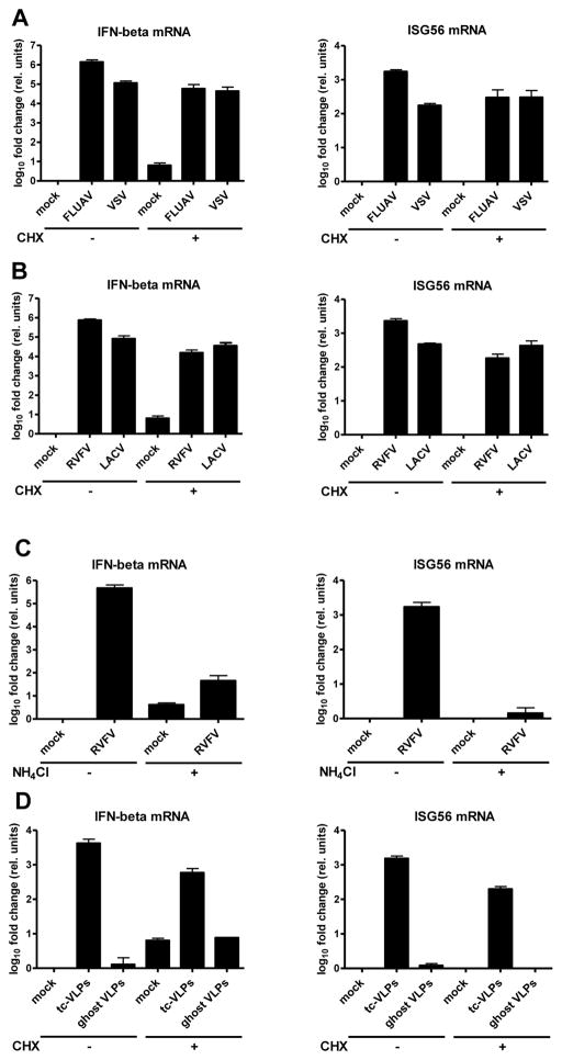
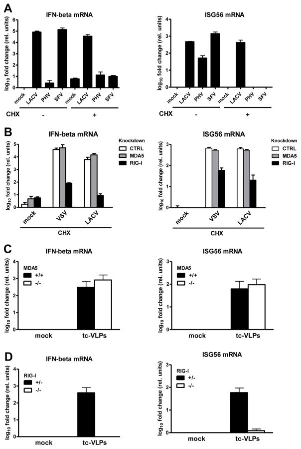
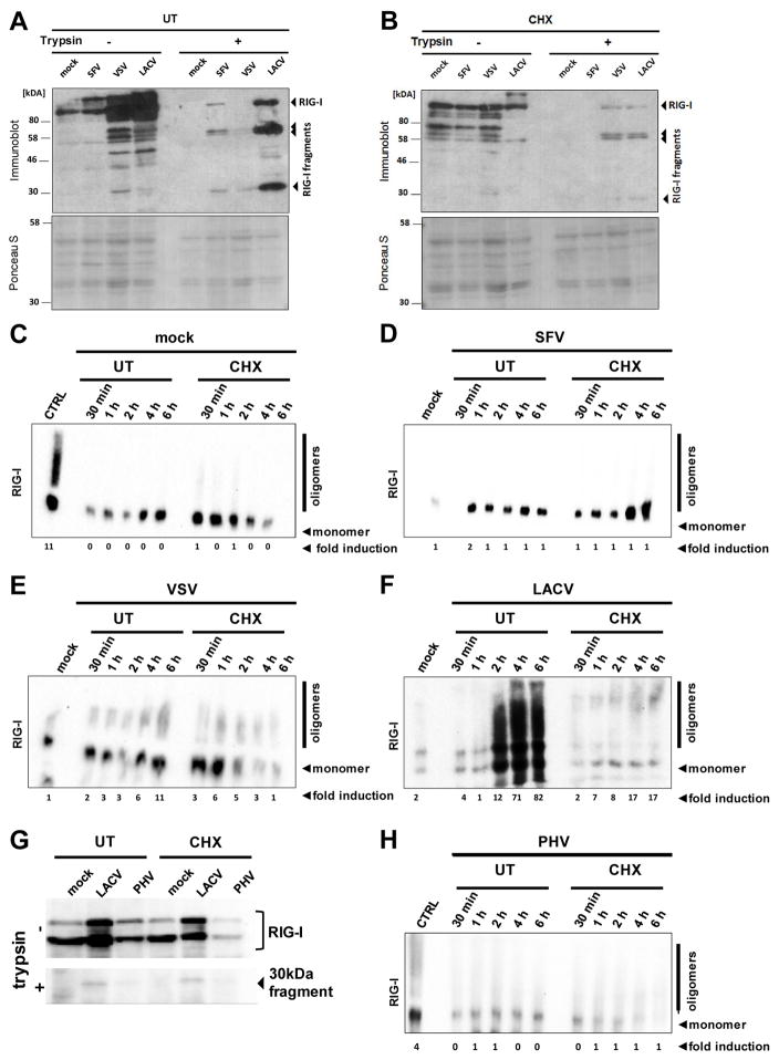
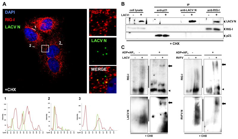
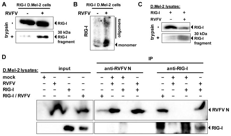
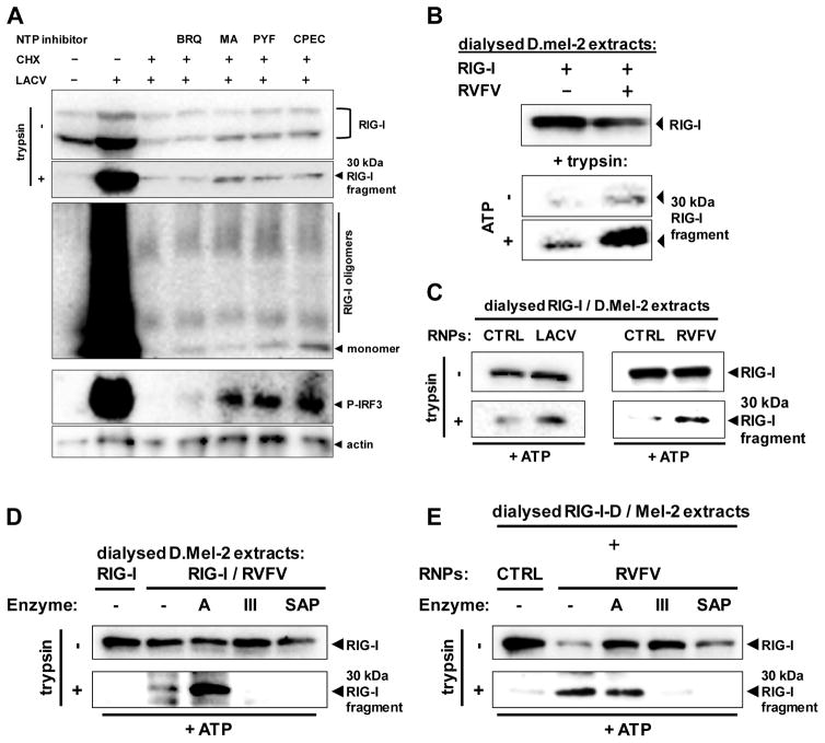
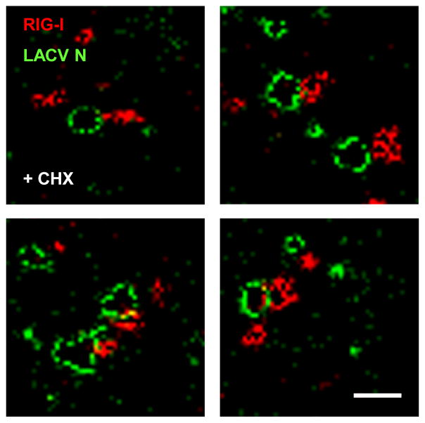
Host defense to RNA viruses depends on rapid intracellular recognition of viral RNA by two cytoplasmic RNA helicases: RIG-I and MDA5. RNA transfection experiments indicate that RIG-I responds to naked double-stranded RNAs (dsRNAs) with a triphosphorylated 5' (5'ppp) terminus. However, the identity of the RIG-I stimulating viral structures in an authentic infection context remains unresolved. We show that incoming viral nucleocapsids containing a 5'ppp dsRNA "panhandle" structure trigger antiviral signaling that commences with RIG-I, is mediated through the adaptor protein MAVS, and terminates with transcription factor IRF-3. Independent of mammalian cofactors or viral polymerase activity, RIG-I bound to viral nucleocapsids, underwent a conformational switch, and homo-oligomerized. Enzymatic probing and superresolution microscopy suggest that RIG-I interacts with the panhandle structure of the viral nucleocapsids. These results define cytoplasmic entry of nucleocapsids as the proximal RIG-I-sensitive step during infection and establish viral nucleocapsids with a 5'ppp dsRNA panhandle as a RIG-I activator.


Figures

Fig. 1
Fig. 1. IFN response to incoming negative-strand RNA viruses
Cells were treated with inhibitors, infected, incubated for 24 h, and then assayed for mRNA levels of IFN-β (left panels) and ISG56 (right panels) using real-time RT-PCR. Here and in all following figures, mean values and standard deviations from three independent experiments are shown. (A and B) IFN response in the absence of viral genome replication. Cells were treated for 1 h with 0 or 50 μg/ml CHX, and then infected either with FLUAVΔNS1 (FLUAV) and VSV (A), or with RVFVΔNSs::GFP (RVFV) and LACVdelNSs (LACV) (B). (C and D) Requirement for virus entry. (C) Cells were treated for 1 h with 0 or 50 mM NH4Cl, and then infected with RVFVΔNSs::GFP. (D) Infection with different types of VLPs. Cells were CHX treated and infected with tc-VLPs or the equivalent amount of ghost VLPs. See also Figs. S1 to S7.
Fig. 2
Fig. 2. Influence of the viral 5′ppp group and of RIG-I
(A) Cells were CHX treated, infected with LACVdelNSs, PHV or SFV, and assayed as described for Fig. 1. (B) Innate response to VSV or LACVdelNSs by cells treated with CHX and with siRNAs targeting MDA5 or RIG-I. (C) MEFs lacking MDA5 or (D) RIG-I, and the corresponding wt MEFs, were tested for their innate response to tc-VLPs. See also Figs. S8 to S10.
Fig. 3
Fig. 3. Activation of RIG-I
(A and B) Conformational switch. A549 cells were treated with 0 (A) or 50 μg/ml CHX (B), infected with the indicated viruses, and lysed 6 h later. Cell extracts were subjected to limited trypsin digestion (right panels), or left untreated (left panels). Upper panels show Western Blot analysis with an anti-RIG-I antibody, lower panels show Ponceau S staining as loading control. (C to F) Oligomerization. A549 cells were left untreated (UT) or treated with 50 μg/ml CHX. Then, cells were either mock infected (C) or infected with SFV (D), VSV (E), or LACVdelNSs (F). At the indicated time points, RIG-I was analyzed by native PAGE and Western blotting. As positive and negative controls, VSV-infected cells at 6 h post-infection (CTRL) and mock infected cells, respectively, were used. (G and H) PHV and RIG-I. A549 cells infected with LACVdelNSs or PHV were monitored for the RIG-I conformation at 6 h p.i. (G), or for RIG-I oligomerization over a time course (H). The ratio of oligomers to monomers (normalised to the actin signal and in relation to mock cells) is indicated below the blots (fold induction).
Fig. 4
Fig. 4. Interaction of RIG-I with LACV nucleocapsids
(A) Co-localization analysis. CHX-treated A549 cells were infected with LACVdelNSs and analyzed 5 h later by double immunofluorescence using antisera against LACV N (green channel) or RIG-I (red channel). Cell nuclei were counterstained with DAPI (blue channel). The square area of the inset is digitally magnified on the right hand side. Three fluorescence intensity profiles are shown on the bottom. (B) Co-immunoprecipitation. CHX-treated A549 cells were infected with LACVdelNSs (MOI 10), lysed 5 h later, and subjected to immunoprecipitation (IP) and Western blot analysis using antibodies against p21 (negative control), LACV N, and RIG-I. As input control, 10% of the cell lysate were analyzed in parallel (left lanes). (C) ADP–aluminium fluoride trapping. CHX-treated A549 cells were infected with LACVdelNSs (left panels) or RVFVΔNSs::REN (right panels). At 5 h post-infection lysates were incubated with ADP•AlF3 and analyzed by native PAGE and Western blot using antibodies against RIG-I (upper panels), or viral N (lower panels). Lines indicate oligomers, arrowheads monomers, and arrows point towards high molecular-weight complexes. See also Figs. S11 to S16.
Fig. 5
Fig. 5. Activation of RIG-I and binding to viral nucleocapsids in insect cells
D.Mel-2 cells expressing human RIG-I were infected for 72 h with RVFVΔNSs::REN (RVFV) and then tested for RIG-I conformation (A) and oligomerization (B). (C) In vitro activation of RIG-I. Lysates of RIG-I-expressing D.Mel-2 cells were mixed with lysates of naïve or RVFVΔNSs::REN-infected D.Mel-2 cells, and assayed for RIG-I conformation. (D) Co-immunoprecipitation. D.Mel-2 cells were either left naïve (mock), or only infected with RVFVΔNSs::REN (RVFV), only expressing RIG-I (RIG-I), or were both expressing RIG-I and superinfected with RVFVΔNSs::REN (RIG-I/RVFV). Combinations of lysates were subjected to IP and Western blot analysis using antibodies against RVFV N or RIG-I. As input control, 10% of the cell lysate were analyzed in parallel (left lanes).
Fig. 6
Fig. 6. Activation of RIG-I solely depends on the nucleocapsid-borne panhandle structure
(A) Effect of NTP withdrawal on activation of RIG-I and IRF-3. Pretreated A549 cells were infected with LACVdelNSs for 5 h. Pretreatment with CHX (50 μg/ml) was for 1h, and with BRQ (10 μM, stocks dissolved in DMSO), MA (10 μM, stocks dissolved in methanol), PYF (10 μM, stocks dissolved in DMSO), or CPEC (5 μM, stocks dissolved in DMSO) for 24 h. RIG-I conformation (upper two panels), RIG-I oligomerization (upper middle panel), and IRF-3 phosphorylation (lower middle panel) were monitored. Immunoblot for actin served as loading control. (B) RIG-I activation in dialysed samples. Lysates from D.Mel-2 cells expressing RIG-I (RIG-I) or infected with RVFVΔNSs::REN (RVFV) were dialysed against PBS, mixed with each other, and incubated with or without ATP. After 1 h incubation, mixes were subjected to the RIG-I conformational switch assay. (C) RIG-I activation by purified viral nucleocapsids (RNPs). Lysates from D.Mel-2 cells expressing RIG-I were dialysed against PBS and mixed with purified nucleocapsids from particles of LACV (left panels) or RVFV (right panels). Incubation with ATP and conformational switch assay were performed as described for (B). Equivalent fractions of gradient-purified supernatants from mock-infected cells were used as negative control (CTRL). (D and E) Structural requirements for viral nucleocapsids to activate RIG-I. Lysates from RVFV-infected D.Mel-2 cells (D) or purified RVFV nucleocapsids (E) were incubated with ATP and one of the indicated enzymes, namely RNase A (A), RNase III (III), or Shrimp Alkaline Phosphatase (SAP). After mixing and incubation with dialysed lysates from RIG-I-expressing D.Mel-2 cells, the RIG-I conformational switch assay was performed. Negative controls (CTRL) were performed as described for (C). See also Figs. S17 and S18.
Fig. 7
Fig. 7. Super-resolution immunofluorescence microscopy of RIG-I/LACV nucleocapsid complexes
CHX-treated A549 cells were infected with LACVdelNSs and analyzed 5 h later by GSDIM double immunofluorescence using antisera against LACV N (green channel) or RIG-I (red channel). Four example areas with nucleocapsids are shown. Scale bar 200 nm. See also Fig. S19.

Comment in

  • RIG-I goes beyond naked recognition.
    Bowzard JB, Ranjan P, Sambhara S. Bowzard JB, et al. Cell Host Microbe. 2013 Mar 13;13(3):247-9. doi: 10.1016/j.chom.2013.02.012. Cell Host Microbe. 2013. PMID: 23498950 Free PMC article.

References

    1. Baum A, Garcia-Sastre A. Differential recognition of viral RNA by RIG-I. Virulence. 2011;2:166–169. - PMC - PubMed
    1. Baum A, Sachidanandam R, Garcia-Sastre A. Preference of RIG-I for short viral RNA molecules in infected cells revealed by next-generation sequencing. Proc Natl Acad Sci U S A. 2010;107:16303–16308. - PMC - PubMed
    1. Binder M, Eberle F, Seitz S, Mucke N, Huber CM, Kiani N, Kaderali L, Lohmann V, Dalpke A, Bartenschlager R. Molecular Mechanism of Signal Perception and Integration by the Innate Immune Sensor Retinoic Acid-inducible Gene-I (RIG-I) J Biol Chem. 2011;286:27278–27287. - PMC - PubMed
    1. Blakqori G, Delhaye S, Habjan M, Blair CD, Sanchez-Vargas I, Olson KE, Attarzadeh-Yazdi G, Fragkoudis R, Kohl A, Kalinke U, et al. La Crosse bunyavirus nonstructural protein NSs serves to suppress the type I interferon system of mammalian hosts. J Virol. 2007;81:4991–4999. - PMC - PubMed
    1. Bunch TA, Grinblat Y, Goldstein LS. Characterization and use of the Drosophila metallothionein promoter in cultured Drosophila melanogaster cells. Nucleic Acids Res. 1988;16:1043–1061. - PMC - PubMed

Publication types

MeSH terms