. 2013 Apr 10;78(1):65-80.
doi: 10.1016/j.neuron.2013.02.029. Epub 2013 Mar 14.

VCP is essential for mitochondrial quality control by PINK1/Parkin and this function is impaired by VCP mutations

Affiliations

VCP is essential for mitochondrial quality control by PINK1/Parkin and this function is impaired by VCP mutations

Nam Chul Kim et al. Neuron. .

Erratum in

  • Neuron. 2013 Apr 24;78(2):403

Abstract

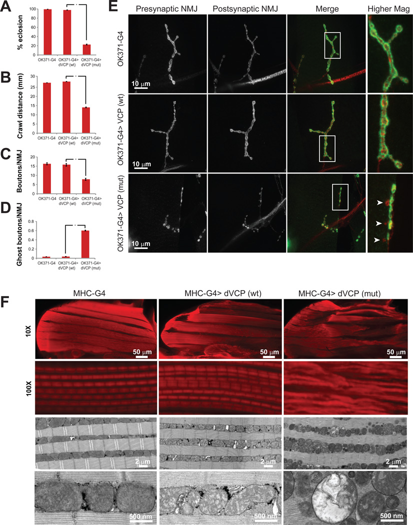
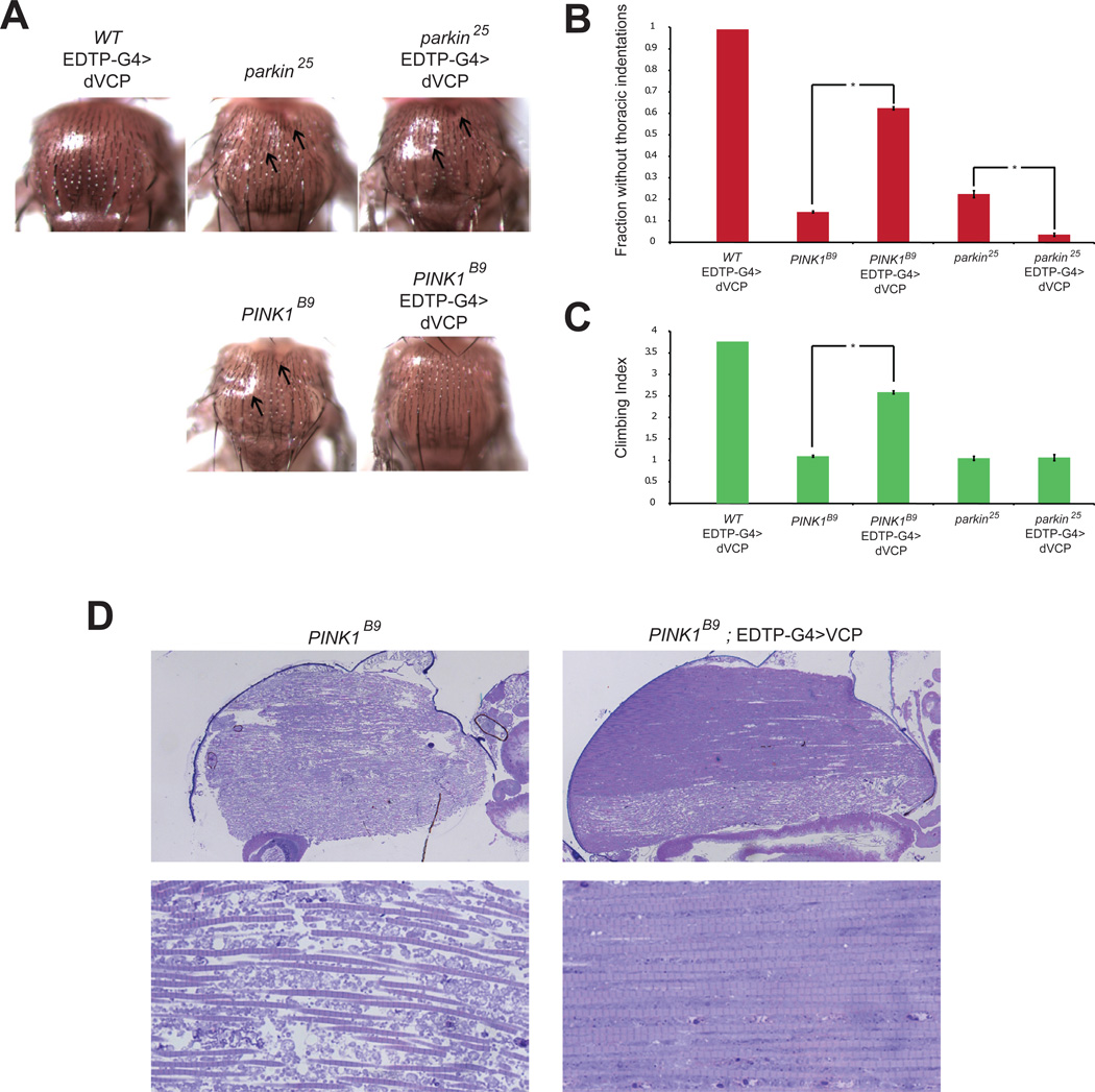
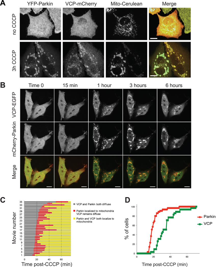
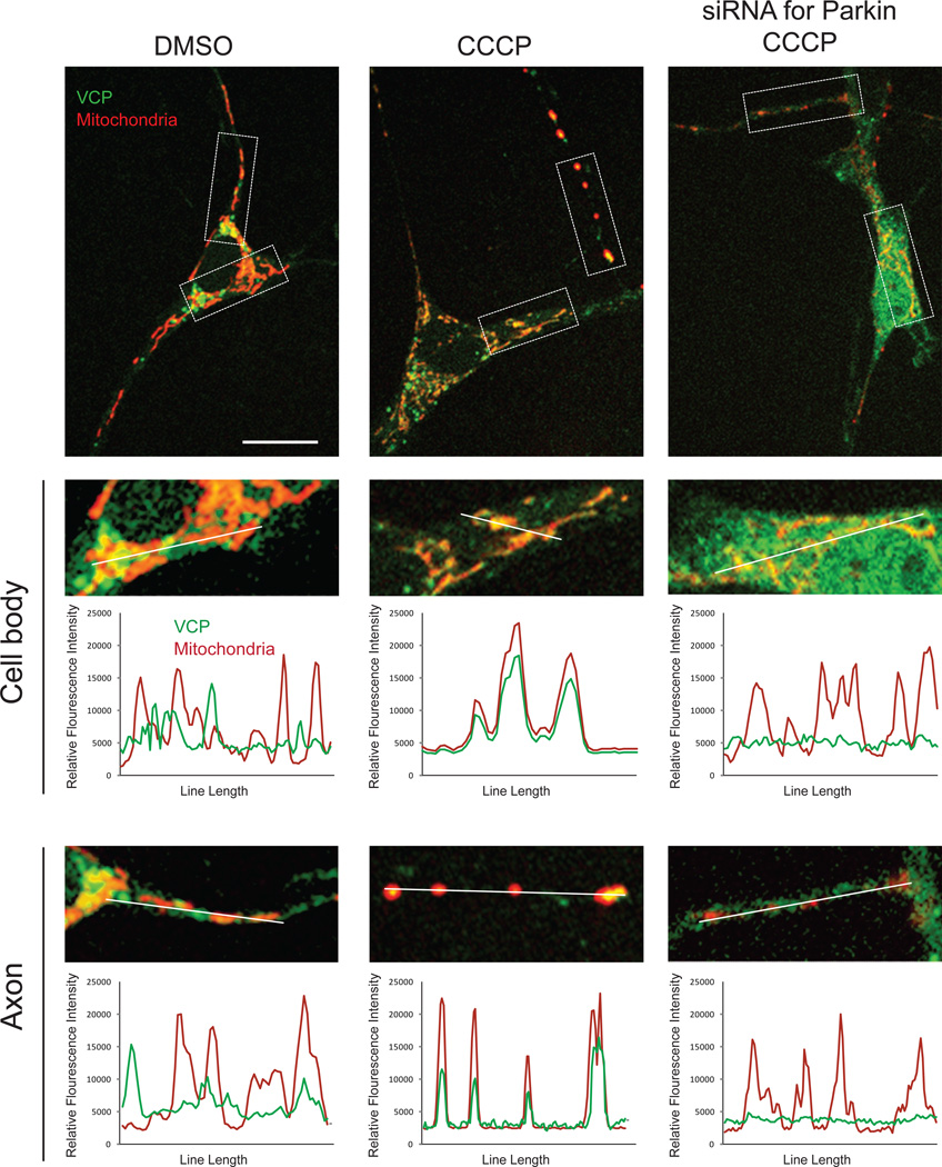
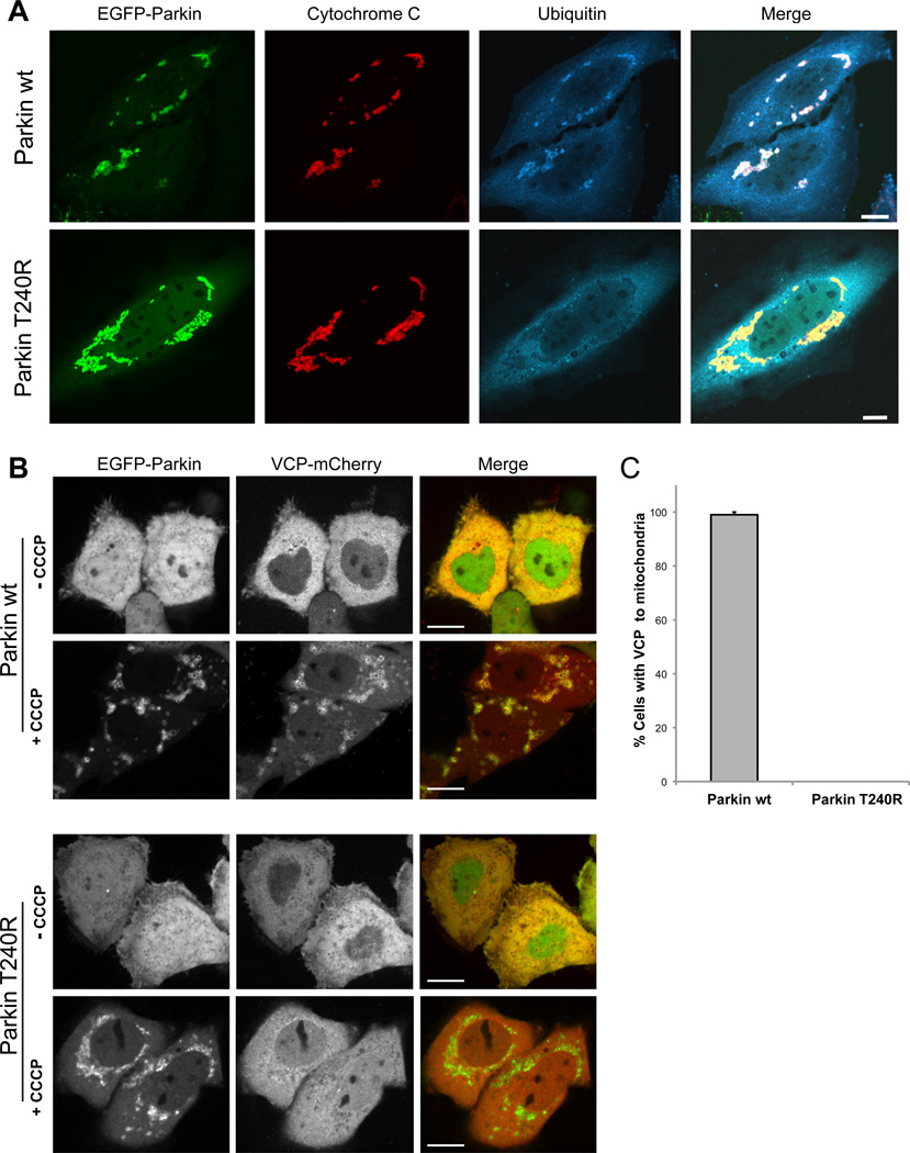
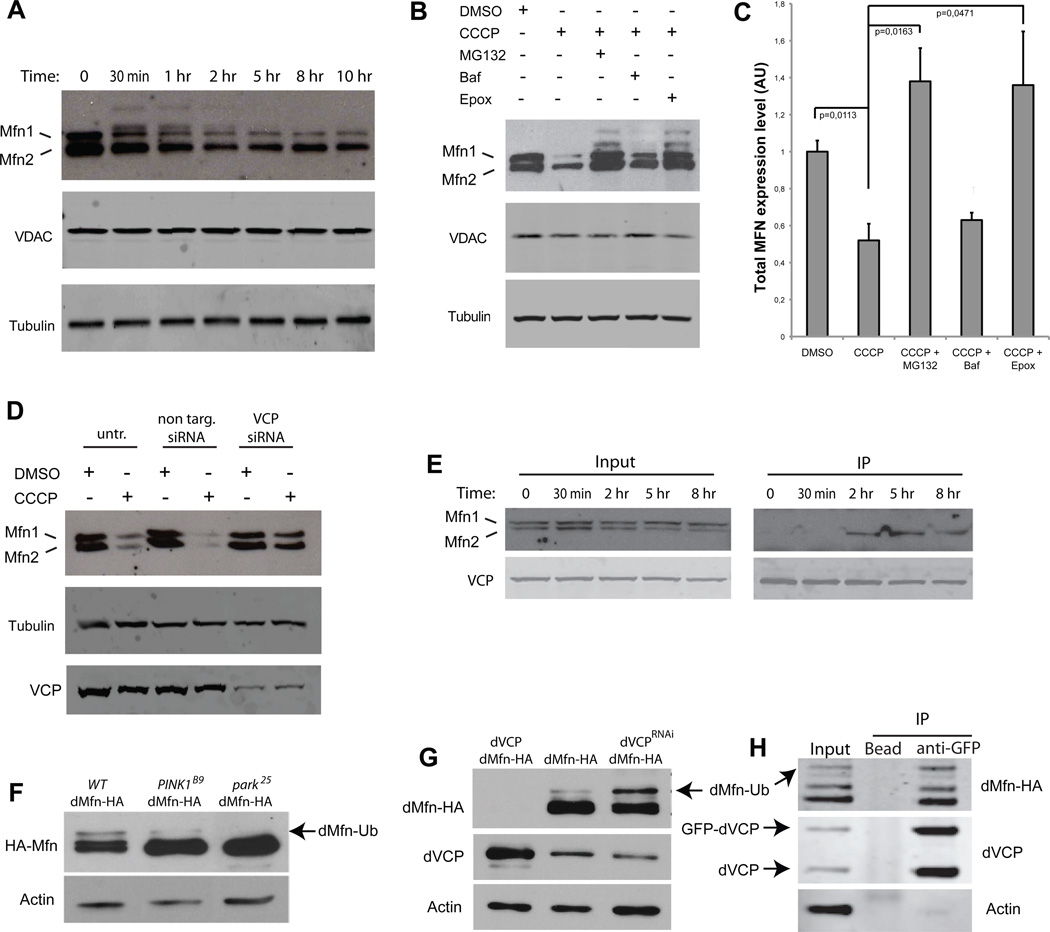
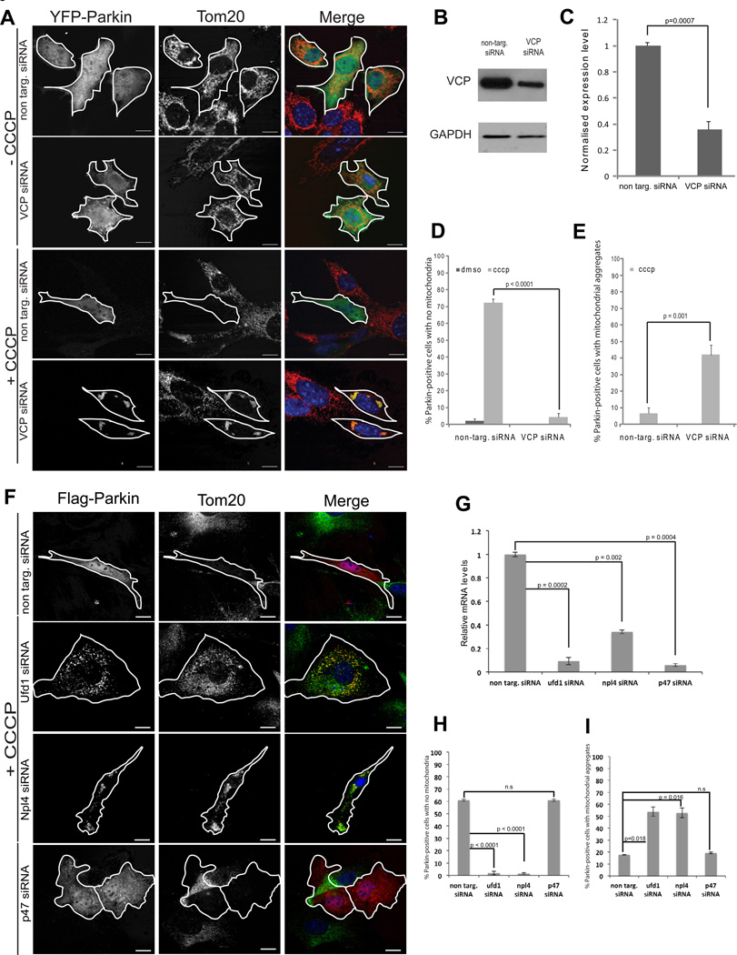
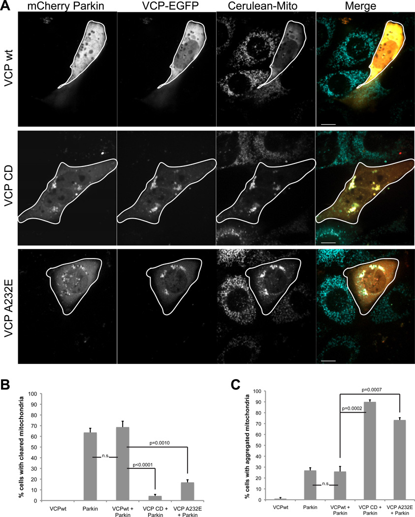
Mutations in VCP cause multisystem degeneration impacting the nervous system, muscle, and/or bone. Patients may present with ALS, Parkinsonism, frontotemporal dementia, myopathy, Paget's disease, or a combination of these. The disease mechanism is unknown. We developed a Drosophila model of VCP mutation-dependent degeneration. The phenotype is reminiscent of PINK1 and parkin mutants, including a pronounced mitochondrial defect. Indeed, VCP interacts genetically with the PINK1/parkin pathway in vivo. Paradoxically, VCP complements PINK1 deficiency but not parkin deficiency. The basis of this paradox is resolved by mechanistic studies in vitro showing that VCP recruitment to damaged mitochondria requires Parkin-mediated ubiquitination of mitochondrial targets. VCP recruitment coincides temporally with mitochondrial fission, and VCP is required for proteasome-dependent degradation of Mitofusins in vitro and in vivo. Further, VCP and its adaptor Npl4/Ufd1 are required for clearance of damaged mitochondria via the PINK1/Parkin pathway, and this is impaired by pathogenic mutations in VCP.


Figures

Figure 1
Figure 1. dVCP mutation-dependent motor neuron and muscle phenotypes in Drosophila
A. Expression of exogenous wild type dVCP in motor neurons with the driver OK371-GAL4 does not impact viability as assessed by rates of eclosion as adults. In contrast, expression of mutant dVCP (R152H) leads to a high rate of pupal lethality. The flies that do eclose die shortly thereafter. Error bars indicate standard error. B. Expression of mutant dVCP (but not wild type) in motor neurons results in a locomotor defect as assessed by monitoring crawling behavior of 3rd instar larvae. Error bars indicate standard error. C. Expression of mutant dVCP (but not wild type) in motor neurons results in abnormal NMJ morphology with reduced total bouton numbers. NMJs at muscle 4 were used for all analyses. Error bars indicate standard error. D. The abnormal NMJs in flies expressing mutant dVCP in motor neurons results in a frequent ‘ghost boutons’ in which presynaptic structure lacks appositional postsynaptic structure. Ghost boutons are very rarely observed in control animals or animals expressing wild type dVCP. Error bars indicate standard error. E. Representative images of NMJs from the genotypes indicated. The arrowheads point out ghost boutons in an animal expressing mutant dVCP. F. Hemithoraces stained with phalloidin from control flies (MHC-GAL4) and flies expressing wild type or mutant dVCP in muscle under control of MHC-GAL4 show a mutation-dependent myopathy at 10X, and a disruption of sarcomere architecture at 100X. TEM revealed profound abnormalities in mitochondrial morphology with numerous swollen mitochondria with disrupted cristae. See also Supplemental Figure 1 and 2 for further analysis.
Figure 2
Figure 2. dVCP overexpression suppresses degeneration associated with PINK1 deficiency but not Parkin deficiency
A. Micrographs of fly thoraces of the indicated genotypes showing thoracic indentations (arrows) caused by Parkin and PINK1 deficiency. dVCP over-expression rescues this phenotype in PINK1-deficient flies, but not in Parkin-deficient flies. B. Quantitation showing that over-expression of dVCP suppresses thoracic indentations in PINK1-deficient animals but actually enhances this phenotype in Parkin-deficient animals. Error bars indicate standard error. C. Quantitation showing that over-expression of dVCP suppresses the locomotor defect in PINK1-deficient animals but not in Parkin-deficient animals. Error bars indicate standard error. D. Micrographs of sections through fly thoraces showing that over-expression of dVCP suppresses the muscle and mitochondrial phenotypes in PINK1-deficient animals.
Figure 3
Figure 3. VCP recruitment to mitochondria follows Parkin
A. VCP-mCherry and Mito-Cerulean were expressed in YFP-Parkin stable HeLa cells and all 3 proteins were visualized at 0 and 3 hours after addition of CCCP. See also Supplemental Figure 3A for MEFs, Supplemental Figure 4 for Sy5y, Supplemental Figure 5 for C2C12. B. Time lapse imaging of VCP-EGFP and mCherry-Parkin following CCCP treatment in HeLa cells. These still images were extracted from Supplementary Movie 1. See also Supplementary Movie 2. C. Graphic representation of the timing of recruitment of Parkin and VCP to mitochondria in 35 consecutive movies. Each line represents one individual movie of a distinct set of 1–3 cells. D. Graphic representation of the lag time before mitochondrial localization of Parkin and VCP following CCCP treatment. Results represent the sum from all cells captured in 35 consecutive movies.
Figure 4
Figure 4. VCP recruitment to mitochondria in primary neurons is Parkin-dependent
Primary neurons were transfected with mito-dsRED and VCP-EGFP and treated with DMSO or CCCP for 48 hrs. VCP-EGFP signal remained diffuse with some accumulation in Golgi in neurons treated with DMSO, but relocalized to mitochondria in neurons treated with CCCP. This relocalization was abolished by knockdown of endogenous Parkin by siRNA.
Figure 5
Figure 5. Mitochondrial ubiquitination by Parkin is essential for VCP recruitment
A. Parkin-dependent mitochondrial ubiquitination. HeLa cells were transfected with EGFP-Parkin wt or Parkin T240R, treated with 10 µM CCCP for 10 h and immunostained for cytochrome C (red, mitochondria) and ubiquitin (blue). Scale bars equal 10 µm. See also Supplemental Figure 6. B. Micrographs of HeLa cells expressing VCP-mCherry and either EGFP-Parkin wt or Parkin T240R. The images are taken during the mitochondrial aggregation stage after CCCP treatment (see Supplemental Figure 6 for the full time course). Scale bars equal 10 µm. C. Quantification of VCP recruitment to mitochondria following CCCP treatment in HeLa cells expressing either Parkin wt or Parkin T240R. Error bars indicate standard error from 3 replicates. 30 cells were counted for each replicate.
Figure 6
Figure 6. Mitofusin degradation by the proteasome is dependent on VCP
A. Western blots in YFP-Parkin stable HeLa cells against MFN1 and 2, VDAC and tubulin at different time points after CCCP treatment. Ubiquitinated forms of MFNs 1/2 can be observed migrating more slowly. B. Western blots in YFP-Parkin stable HeLa cells against MFN1/2, VDAC, and tubulin. Cells were treated for 12 h with CCCP and either proteasome inhibitors (MG132 or epoxomicin) or the autophagy inhibitor bafilomycin. Ubiquitinated forms of MFN1/2 can be again observed migrating more slowly, particularly with proteasome inhibition. C. Quantification of total MFN expression levels normalized against tubulin in YFP-Parkin stable HeLa cells treated for 12 h with CCCP and either proteasome inhibitors (MG132 or epoxomicin), or the autophagy inhibitor bafilomycin. Error bars indicate standard deviation from triplicates. D. Knockdown of VCP stabilizes MFNs 1 and 2. Western blots in YFP-Parkin stable HeLa cells against MFN1/2, VCP and tubulin. Cells were transfected with non-targeting or VCP-targeting siRNA and treated for 12 h with DMSO or CCCP. E. FLAG IP in HeLa cells cotransfected with YFP-Parkin and VCP-FLAG and treated with CCCP for the indicated times. Immunoprecipitation samples were immunoblotted against MFN1/2 and VCP. Following mitochondrial depolarization VCP interacts with MFN2. F. Total dMfn-HA accumulates in PINK1B9 (lane 2) and Park25 (lane 3) null mutants. Notably, ubiquitinated dMfn is decreased in PINK1B9 null mutants (lane 2) and absent in Park25 null mutants (lane 3). G. Overexpression of dVCP in the compound eye destabilizes dMfn-HA (lane 1), whereas knockdown of endogenous dVCP in the compound eye leads to accumulation of ubiquitinated dMfn-HA (lane 3). H. Immunoprecipitation of HA-dMfn and endogenous (protein-trap) GFP-dVCP from Drosophila brain extract.
Figure 7
Figure 7. VCP and Ufd1/Npl4 are essential for clearance of damaged mitochondria
A. Immunostaining against TOM20 (red) in MEFs transfected with YFP-Parkin (green) and non-targeting or VCP-targeting siRNA. Cells were treated with DMSO or CCCP for 24 h. B. Immunoblot of VCP and GAPDH in MEFs transfected with non-targeting or VCP-targeting siRNA. C. Quantification of VCP normalized against GAPDH in cells transfected with non-targeting or VCP-targeting siRNA. Error bars indicate standard deviation from triplicates. D. Quantification of cells with mitochondrial clearance. Cells were treated for 24 h with DMSO or CCCP. At least 30 cells were counted for each sample. Errors bars indicate standard error from 3 independent replicates. See also Supplemental Figure 5 for C2C12 cells. E. Quantification of cells with residual aggregates of mitochondria. Cells were treated for 24 h with DMSO or CCCP. F. Immunostaining against TOM20 (red) in MEFs transfected with YFP-Parkin (green) and non-targeting siRNA or siRNA targeting Ufd1, Npl4, or p47. Cells were treated with DMSO or CCCP for 24 h. (Cells treated with DMSO shown in Supplemental Figure 8C). G. Real time PCR quantification of mRNA levels of Ufd1, Npl4, or p47 following treatment with the individual siRNAs. H. Quantification of cells with mitochondrial clearance in the setting of Ufd1, Npl4, or p47 knockdown. I. Quantification of cells with residual aggregates of mitochondria. Cells were treated for 24 h with DMSO or CCCP. At least 30 cells were counted for each sample. Errors bars indicate standard error from 3 independent replicates.
Figure 8
Figure 8. Overexpression of VCP-CD or A232E inhibits clearance of damaged mitochondria
A. Imaging VCP-EGFP and mCherry-Parkin in Mito-Cerulean stable MEFs 24h after addition of CCCP. Cells were transfected with VCP-wt, CD or disease mutant A232E -EGFP. Scale bars equal 10µm. B. Quantification of cells with mitochondrial clearance. Cells were prepared as described in A. At least 30 cells were counted for each sample. Errors bars indicate standard error from 3 independent replicates. C. Quantification of cells with residual aggregates of mitochondria. Cells were prepared as described in A. At least 30 cells were counted for each sample. Errors bars indicate standard error from 3 independent replicates. C2C12 cell data are shown in Supplemental Figure 5E–G.

Comment in

References

    1. Abramzon Y, Johnson JO, Scholz SW, Taylor JP, Brunetti M, Calvo A, Mandrioli J, Benatar M, Mora G, Restagno G, et al. Valosin-containing protein (VCP) mutations in sporadic amyotrophic lateral sclerosis. Neurobiology of aging. 2012;33:e2231–e2231. 2231 e2236. - PMC - PubMed
    1. Badadani M, Nalbandian A, Watts GD, Vesa J, Kitazawa M, Su H, Tanaja J, Dec E, Wallace DC, Mukherjee J, et al. VCP associated inclusion body myopathy and paget disease of bone knock-in mouse model exhibits tissue pathology typical of human disease. PloS one. 2010;5 - PMC - PubMed
    1. Braun S, Matuschewski K, Rape M, Thoms S, Jentsch S. Role of the ubiquitin-selective CDC48(UFD1/NPL4 )chaperone (segregase) in ERAD of OLE1 and other substrates. EMBO J. 2002;21:615–621. - PMC - PubMed
    1. Chang YC, Hung WT, Chang HC, Wu CL, Chiang AS, Jackson GR, Sang TK. Pathogenic VCP/TER94 alleles are dominant actives and contribute to neurodegeneration by altering cellular ATP level in a Drosophila IBMPFD model. PLoS genetics. 2011;7:e1001288. - PMC - PubMed
    1. Chung PY, Beyens G, de Freitas F, Boonen S, Geusens P, Vanhoenacker F, Verbruggen L, Van Offel J, Goemaere S, Zmierczak HG, et al. Indications for a genetic association of a VCP polymorphism with the pathogenesis of sporadic Paget's disease of bone, but not for TNFSF11 (RANKL) and IL-6 polymorphisms. Molecular genetics and metabolism. 2011;103:287–292. - PubMed

Publication types

MeSH terms

Substances

LinkOut - more resources