. 2013 Apr 25;50(2):161-71.
doi: 10.1016/j.molcel.2013.02.009. Epub 2013 Mar 14.

Neurodegeneration-associated protein fragments as short-lived substrates of the N-end rule pathway

Affiliations

Neurodegeneration-associated protein fragments as short-lived substrates of the N-end rule pathway

Christopher S Brower et al. Mol Cell. .

Abstract

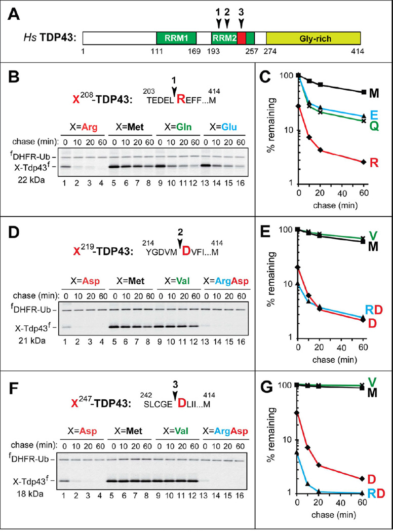
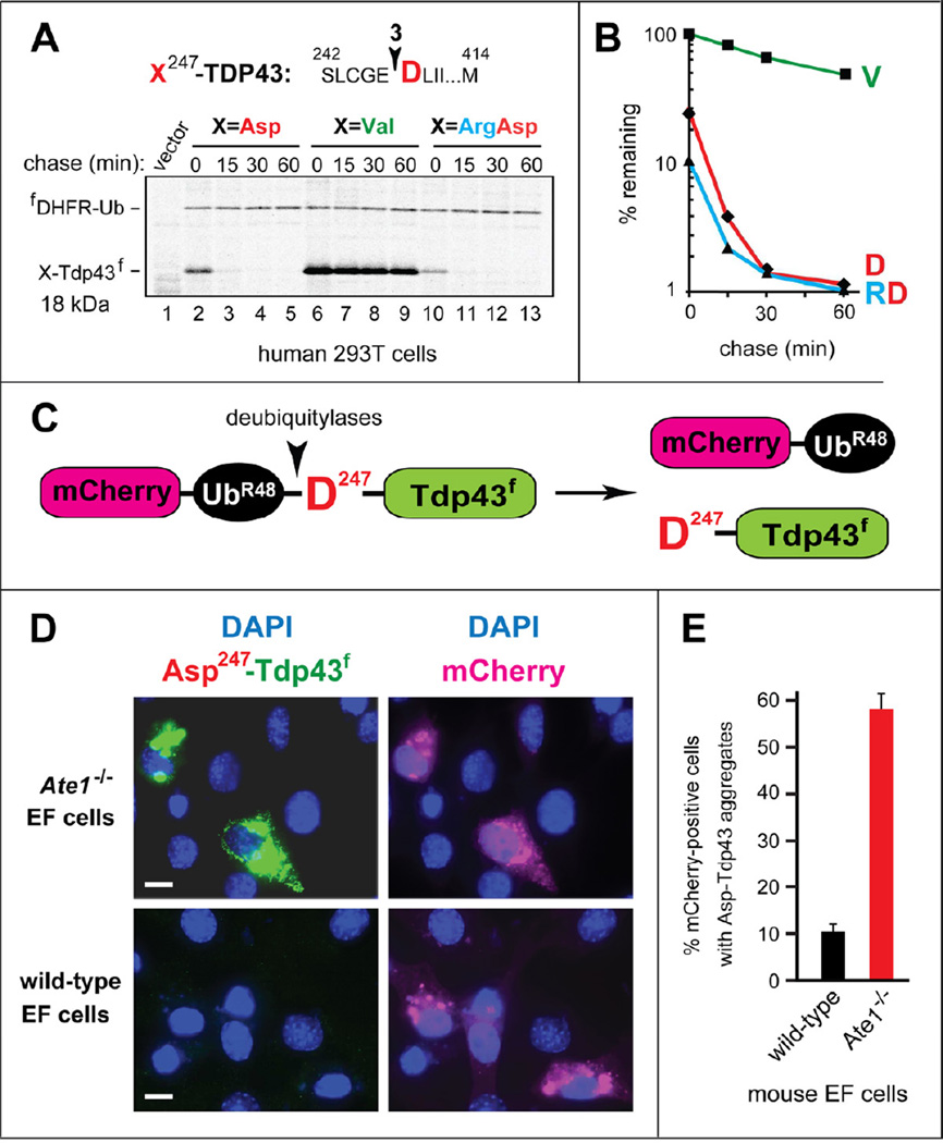
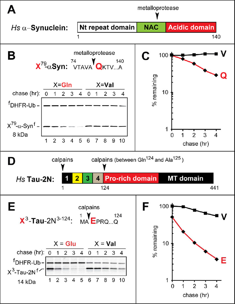
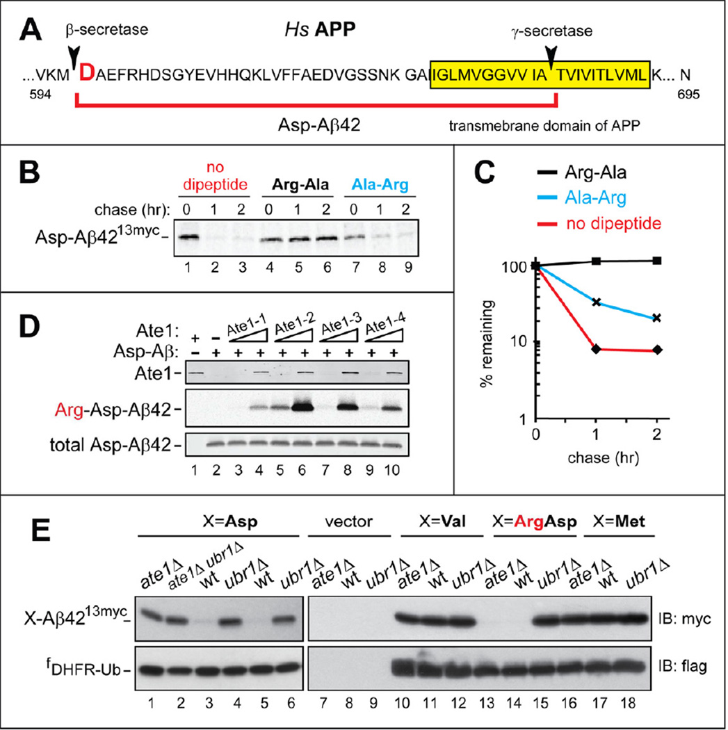
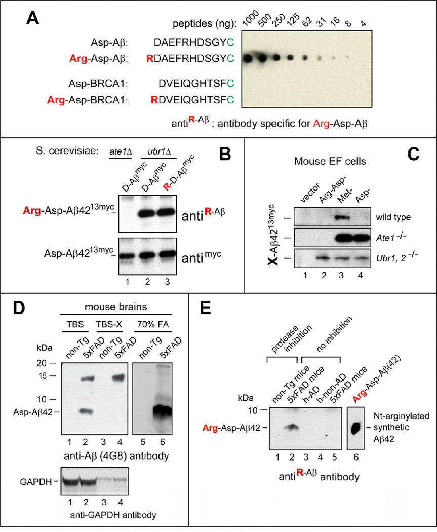
Protein aggregates are a common feature of neurodegenerative syndromes. Specific protein fragments were found to be aggregated in disorders including Alzheimer's disease, amyotrophic lateral sclerosis, and Parkinson's disease. Here, we show that the natural C-terminal fragments of Tau, TDP43, and α-synuclein are short-lived substrates of the Arg/N-end rule pathway, a processive proteolytic system that targets proteins bearing "destabilizing" N-terminal residues. Furthermore, a natural TDP43 fragment is shown to be metabolically stabilized in Ate1(-/-) fibroblasts that lack the arginylation branch of the Arg/N-end rule pathway, leading to accumulation and aggregation of this fragment. We also found that a fraction of Aβ42, the Alzheimer's disease-associated fragment of APP, is N-terminally arginylated in the brains of 5xFAD mice and is degraded by the Arg/N-end rule pathway. The discovery that neurodegeneration-associated natural fragments of TDP43, Tau, α-synuclein, and APP can be selectively destroyed by the Arg/N-end rule pathway suggests that this pathway counteracts neurodegeneration.


Figures

Figure 1
Figure 1. C-Terminal TDP43 Fragments From Aggregates in the Brains of Patients with Frontotemporal Lobal Degeneration (FTLD-TDP) As Short-Lived N-End Rule Substrates
(A) Domain organization of human TDP43. Arrowheads indicate the cleavage sites. RRM1, RRM2, RNA-binding domains. (B) The cleavage site 1 is indicated by an arrowhead, with the P1’ residue (Arg) in red. X208-TDP43f fragments, produced from fDHFR-UbR48-X208-TDP43f (X=Arg, Gln, Glu, Met), were expressed in reticulocyte extract and labeled with 35S-Met/Cys for 10 min at 30°C, followed by a chase, immunoprecipitation with anti-flag antibody, SDS-PAGE, and autoradiography. Indicated molecular masses of proteins in this and other panels include their ~1 kDa flag epitope. (C) Quantification of B using the 33 kDa reference protein fDHFR-UbR48. (D) Same as B but with X219-TDP43f fragments (X=Asp, Arg-Asp, Met, Val). (E) Quantification of D. (F) Same as B but with X247-TDP43f fragments (X=Asp, Arg-Asp, Met, Val). (G) Quantification of F. See also Figure S1.
Figure 2
Figure 2. In Vivo Degradation and Aggregation of X247-TDP43f fragments
(A) Pulse-chase assays with X247-TDP43f, produced from fDHFR-UbR48-X247-TDP43f (X=Asp, Val, Arg-Asp). The fusion proteins were expressed in transiently transfected human HEK-293T cells, which were labeled with 35S-Met/Cys for 15 min at 37°C, followed by a chase, preparation of extracts, immunoprecipitation with anti-flag, SDS-PAGE, and autoradiography. For designations, see the legend to Figure 1B. (B) Quantification of A. (C) Diagram of the mCherry-UbR48-Asp247-TDP43f fusion. The arrowhead indicates the site of cleavage by deubiquitylases (Figure S1C). (D) Representative images of either Ate1−/−(upper panels) or wt (lower panels) mouse EF cells transfected with a plasmid expressing mCherry-UbR48-Asp247-TDP43f. The mCherry moiety of mCherry-UbR48 was detected by red fluorescence, and the Asp247-TDP43f fragment was detected by indirect immunofluorescence, using anti-flag antibody and a fluorescein-conjugated secondary antibody. White bars indicate 10 µm. (E) Percentage of mCherry-positive cells containing Asp247-TDP43f aggregates, with surveys of > 1,000 mCherry-positive cells of the wt and Ate1−/− genotypes. The error bars indicate SEM (standard error of measurement). Statistical analyses employed the unpaired t-test (p <10−12). See also Figure S2A.
Figure 3
Figure 3. Neurodegeneration-Associated C-Terminal Fragments of Human α-Synuclein and Tau As Short-Lived N-End Rule Substrates
(A) Domain organization of human α-Synuclein. Arrowhead indicates the metalloprotease cleavage site. (B) The cleavage site is indicated by an arrowhead, with the P1’ Gln (Q) residue in red. X79-αSynf, produced from fDHFR-UbR48-X-αSynf (X= Gln, Val) in reticulocyte extract, were assayed as described in the legend to Figure 1B. (C) Quantification of B using the reference fDHFR-UbR48. (D) Domain organization of human Tau-2N. Arrowheads indicate the calpain cleavage sites. (E) Same as B but with X3-Tau-2Nf, produced from fDHFR-UbR48-X3-Tau-2Nf (X=Glu, Val). (F) Quantification of E. See also Figure S3.
Figure 4
Figure 4. N-Terminal Arginylation of Aβ42, and the Degradation of C-terminally Tagged Aβ42 by the Arg/N-End Rule Pathway
(A) Human APP near its transmembrane domain, indicated by yellow rectangle. Arrowheads indicate the cleavage sites by secretases that yield Aβ42, termed Asp-Aβ42 and indicated by a red square parenthesis. (B) C-terminally tagged Asp-Aβ4213myc (produced from fDHFR-UbR48-Asp-Aβ4213myc) was assayed in reticulocyte extract as described in the legend to Figure 1B, with either no added dipeptides (lanes 1–3), or 5 mM Arg-Ala (lanes 4–6), or Ala-Arg (lanes 7–9). (C) Quantification of B. (D) Nt-arginylation of Asp-Aβ42 by isoforms of mouse Ate1 R-transferase. 0.1 or 1 µg of Ate1-1 (lanes 3, 4), Ate1-2 (lanes 5, 6), Ate1-3 (lanes 7, 8), and Ate1-4 (lanes 9, 10) were incubated for 30 min at 37 °C with 20 µg of chemically synthesized Asp-Aβ42, [14C]-L-arginine, and other components of the Nt-arginylation assay, followed by SDS/PAGE and autoradiography. Lane 1, complete reaction (including equal amounts of all four Ate1 isoforms, 1 µg total) but no added Asp-Aβ42. Lane 2, same as lane 3 but without Ate1. Upper panel: immunoblotting with antibody to mouse Ate1. Middle panel: autoradiography to detect 14C-labeled Arg-Asp-Aβ42. Lower panel: immunoblotting with anti-4G8 antibody to detect Asp-Aβ42 or Arg-Asp-Aβ42. (E) Steady-state levels of X-Aβ4213myc, produced from fDHFR-UbR48-X-Aβ4213myc (X=Asp, Val, Arg-Asp, Met), in wt, ate1Δ, and ubr1Δ strains of S. cerevisiae. Upper panels: immunoblotting with anti-myc antibody. Lower panels: immunoblotting with anti-flag antibody. See also Figure S4.
Figure 5
Figure 5. Untagged, N-terminally Arginylated Human Aβ42 in Mouse Brains
(A) Antibody (antiR-Aβ) specific for RDAEFRHDSGYC was examined by dot assay with Nt-arginylated Aβ and BRCA1 peptides (see the main text). (B) Immunoblotting of extracts from the indicated ate1Δ or ubr1Δ yeast strains expressing X-Aβ4213myc, produced in vivo from fDHFR-UbR48-X-Aβ4213myc (X=Asp, Arg-Asp). Upper panel: immunoblotting with antiR-Aβ antibody, characterized in A. Lower panel: immunoblotting with anti-myc antibody. (C) X-Aβ4213myc was produced from fDHFR-UbR48-X-Aβ4213myc (X=Arg-Asp, Met, Asp) in the wt, Ate1−/−, or Ubr1−/− Ubr2−/− mouse EF cell lines, followed by SDS-PAGE of cell extracts and immunoblotting with anti-myc antibody. (D) Immunoblotting with anti-4G8 (recognizing total Aβ) of the indicated brain extracts from either 5xFAD or non-Tg (nontransgenic) mice. Also shown are immunoblots with antibody to GAPDH (loading controls). (E) Untagged, Nt-arginylated human Aβ42 in 70% formic acid (FA) extracts from mouse and human brains, detected by immunoblotting with antiR-Aβ. Lanes 1 and 2, FA extracts from brains of non-Tg and 5xFAD mice, respectively, that were treated with inhibitors of proteasome and neprilysin 12 hrs before harvesting brains. Lane 3 and 4, FA extracts from the cortices of a human AD patient and a non-AD control, respectively. Lane 5, same as lane 2 but from 5xFAD mice that had not been treated with protease inhibitors. Lane 6, in vitro Nt-arginylated Arg-Asp Aβ42 (see Figure 4D), a positive control. See also Figure S4.

References

    1. Balch WE, Morimoto RI, Dillin A, Kelly JW. Adapting proteostasis for disease intervention. Science. 2008;319:916–919. - PubMed
    1. Brower CS, Varshavsky A. Ablation of arginylation in the mouse N-end rule pathway: loss of fat, higher metabolic rate, damaged spermatogenesis, and neurological perturbations. PLoS ONE. 2009;4:e7757. - PMC - PubMed
    1. Chen B, Retzlaff M, Roos T, Frydman J. Cellular strategies of protein quality control. Cold Spring Harb Perspect Biol. 2011;3:a004374. - PMC - PubMed
    1. Choi DH, Kim YJ, Kim YG, Joh TH, Beal MF, Kim YS. Role of matrix metalloproteinase 3-mediated alpha-synuclein cleavage in dopaminergic cell death. J Biol Chem. 2011;286:14168–14177. - PMC - PubMed
    1. Cremades N, Cohen SI, Deas E, Abramov AY, Chen AY, Orte A, Sandal M, Clarke RW, Dunne P, Aprile FA, et al. Direct observation of the interconversion of normal and toxic forms of α-synuclein. Cell. 2012;149:1048–1059. - PMC - PubMed

Publication types

MeSH terms