. 2013 Apr 2;21(4):650-7.
doi: 10.1016/j.str.2013.02.001. Epub 2013 Mar 14.

The three-dimensional structure of the biotin carboxylase-biotin carboxyl carrier protein complex of E. coli acetyl-CoA carboxylase

Affiliations

The three-dimensional structure of the biotin carboxylase-biotin carboxyl carrier protein complex of E. coli acetyl-CoA carboxylase

Tyler C Broussard et al. Structure. .

Abstract

Acetyl-coenzyme A (acetyl-CoA) carboxylase is a biotin-dependent, multifunctional enzyme that catalyzes the regulated step in fatty acid synthesis. The Escherichia coli enzyme is composed of a homodimeric biotin carboxylase (BC), biotinylated biotin carboxyl carrier protein (BCCP), and an α2β2 heterotetrameric carboxyltransferase. This enzyme complex catalyzes two half-reactions to form malonyl-coenzyme A. BC and BCCP participate in the first half-reaction, whereas carboxyltransferase and BCCP are involved in the second. Three-dimensional structures have been reported for the individual subunits; however, the structural basis for how BCCP reacts with the carboxylase or transferase is unknown. Therefore, we report here the crystal structure of E. coli BCCP complexed with BC to a resolution of 2.49 Å. The protein-protein complex shows a unique quaternary structure and two distinct interfaces for each BCCP monomer. These BCCP binding sites are unique compared to phylogenetically related biotin-dependent carboxylases and therefore provide novel targets for developing antibiotics against bacterial acetyl-CoA carboxylase.


Figures

Figure 1
Figure 1. Reaction Catalyzed by Acetyl-CoA Carboxylase
Reaction 1 is catalyzed by biotin carboxylase and Reaction 2 is catalyzed by carboxyltransferase. Biotin is shown covalently attached to the biotin carboxyl carrier protein.
Figure 2
Figure 2. Quaternary Structure of the BCCP-BC Complex
(A–C) Surface rendering of the BCCP-BC complex showing the BC homodimer in purple (top) and yellow (bottom) with individual BCCPs shown in blue, green, orange, and red. (B) is (A) rotated 90° to the left and (C) is (A) rotated 90° around the × axis in a clockwise motion. The local symmetry axes of the BCCP-BC complex are also indicated in (A) and (C). (D) Cartoon representation of a monomer of BC with the two interfacing BCCP molecules in orange and green, respectively. The N-terminal domain, B-domain, and the C-terminal domain of the BC monomer are labeled and colored in light blue, dark blue, and yellow, respectively. Lys122 is represented as black sticks in each BCCP molecule with oxygen and nitrogen atoms in red and blue, respectively. The thumb of each BCCP molecule is designated by dashed circles.
Figure 3
Figure 3. Superposition of BCCP
Superposition of E. coli BCCP from the BCCP-BC complex (purple) with the BCCP domain from human acetyl-CoA carboxylase (yellow) and the 1.3S subunit of transcarboxylase (green). The β-hairpin turns can be seen pointing to the left and the thumb of the E. coli BCCP is “thumbs up” toward the top of the figure.
Figure 4
Figure 4. The BCCP-BC Complex Interfaces
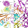
Oxygen, nitrogen, sulfur atoms, and water are depicted in red, blue, gold, and red spheres, respectively. Residues are indicated by their single-letter abbreviation followed by their residue number in E. coli. Dashed lines depict hydrogen bonds and fall within 2.41 to 3.25 Å. The color scheme remains constant throughout the figure. (A) BC homodimers are shown in purple (top) and yellow (bottom) of the complex. The N-terminal domain, B-domain, and C-terminal domain of one monomer of the yellow homodimer are labeled and colored light blue, dark blue, and yellow, respectively. One of the BCCPs binding these two homodimers together is shown in orange with the β-hairpin turn pointing to the purple homodimer. (A)–(D) correspond to the area that is enlarged in (B), (C), and (D), respectively. (B and C) A magnified view of the two BCCP-BC interfaces. (D) A magnified view of the BC-BC interface.
Figure 5
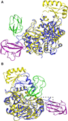
Figure 5. Biotin Carboxylase from E. coli Superimposed onto the Biotin Carboxylase Domain of R. etli Pyruvate Carboxylase
The BCCP-BC complex is shown in purple and gold, respectively, and the BCCP and BC domains of R. etli pyruvate carboxylase are shown in blue and green. (A) Overlay of BC from E. coli with the BC domain from R. etli pyruvate carboxylase (Protein Data Bank ID 2F9Y). (B) Overlay of BC from E. coli with the BC domain from R. etli pyruvate carboxylase (Protein Data Bank ID 3TW6). The dashed box indicates the loop in R. etli pyruvate carboxylase that would hinder E. coli BCCP binding.

References

    1. Adams PD, Afonine PV, Bunkóczi G, Chen VB, Davis IW, Echols N, Headd JJ, Hung LW, Kapral GJ, Grosse-Kunstleve RW, et al. PHENIX: a comprehensive Python-based system for macromolecular structure solution. Acta Crystallogr. D Biol. Crystallogr. 2010;66:213–221. - PMC - PubMed
    1. Alves J, Westling L, Peters EC, Harris JL, Trauger JW. Cloning, expression, and enzymatic activity of Acinetobacter baumannii and Klebsiella pneumoniae acetyl-coenzyme A carboxylases. Anal. Biochem. 2011;417:103–111. - PubMed
    1. Athappilly FK, Hendrickson WA. Structure of the biotinyl domain of acetyl-coenzyme A carboxylase determined by MAD phasing. Structure. 1995;3:1407–1419. - PubMed
    1. Bagautdinov B, Matsuura Y, Bagautdinova S, Kunishima N. Protein biotinylation visualized by a complex structure of biotin protein ligase with a substrate. J. Biol. Chem. 2008;283:14739–14750. - PubMed
    1. Bilder P, Lightle S, Bainbridge G, Ohren J, Finzel B, Sun F, Holley S, Al-Kassim L, Spessard C, Melnick M, et al. The structure of the carboxyltransferase component of acetyl-coA carboxylase reveals a zinc-binding motif unique to the bacterial enzyme. Biochemistry. 2006;45:1712–1722. - PubMed

Publication types

MeSH terms

Associated data