. 2013 Apr 4;92(4):565-74.
doi: 10.1016/j.ajhg.2013.02.010. Epub 2013 Mar 14.

Mutations in WNT1 cause different forms of bone fragility

Affiliations

Mutations in WNT1 cause different forms of bone fragility

Katharina Keupp et al. Am J Hum Genet. .

Abstract

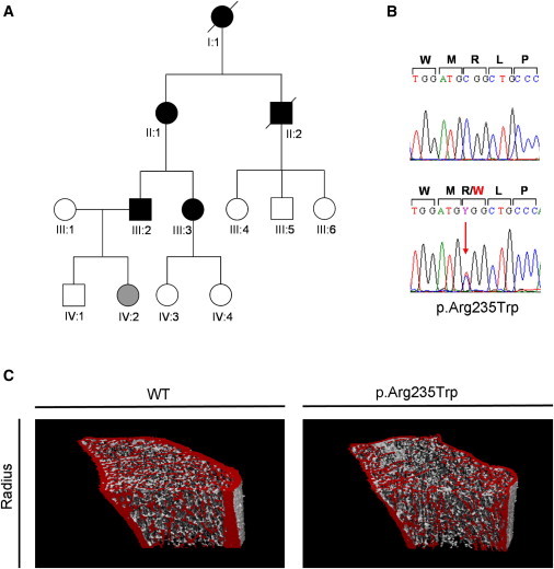
We report that hypofunctional alleles of WNT1 cause autosomal-recessive osteogenesis imperfecta, a congenital disorder characterized by reduced bone mass and recurrent fractures. In consanguineous families, we identified five homozygous mutations in WNT1: one frameshift mutation, two missense mutations, one splice-site mutation, and one nonsense mutation. In addition, in a family affected by dominantly inherited early-onset osteoporosis, a heterozygous WNT1 missense mutation was identified in affected individuals. Initial functional analysis revealed that altered WNT1 proteins fail to activate canonical LRP5-mediated WNT-regulated β-catenin signaling. Furthermore, osteoblasts cultured in vitro showed enhanced Wnt1 expression with advancing differentiation, indicating a role of WNT1 in osteoblast function and bone development. Our finding that homozygous and heterozygous variants in WNT1 predispose to low-bone-mass phenotypes might advance the development of more effective therapeutic strategies for congenital forms of bone fragility, as well as for common forms of age-related osteoporosis.


Figures

Figure 1
Figure 1
Families Affected by Autosomal-Recessive OI (A) Pedigrees of four Turkish families (families 1–4) and one Egyptian family (family 5) affected by autosomal-recessive OI. WES was done on DNA from affected member V:1 of family 1. (B) X-rays of family 1 individual IV:1, who harbors the homozygous p.His287Profs30 WNT1 variant, show a fracture. (C) Lateral spine X-rays of the family 2 member with the homozygous WNT1 p.Gly177Cys substitution. An X-ray was taken at the start of treatment with bisphosphonates (left). The vertebrae are severely compressed and deformed, and even after 4 years of treatment with bisphosphonates, no improvement in size or shape is visible in the lumbar spine (right). (D) An X-ray of individual II:3 (family 5), who has p.Phe298Cys in WNT1, shows severe deformities of the legs.
Figure 2
Figure 2
Homozygous Mutations in WNT1 in Autosomal-Recessive Families (A) Chromatograms of identified recessive WNT1 mutations (red arrows) in comparison to the respective sequence. (B) mRNA analysis of WNT1 transcripts in the affected individual harboring the homozygous c.624+4A>G splice-site mutation. The PCR product of analyzed cDNA shows a drastically reduced amount of WNT1 transcripts. (C and D) A schematic genomic overview of WNT1 (C) and WNT1 (D) indicates the localization of the identified WNT1 mutations. WNT1 domains were predicted on the crystal structure of XWnt8.
Figure 3
Figure 3
A Dominant WNT1 Mutation in a Family Affected by Early-Onset Osteoporosis (A and B) Pedigree of the family with autosomal-dominant early-onset osteoporosis (A) and chromatogram of the identified heterozygous WNT1 substitution p.Arg235Trp (B). Note that individual IV:2 has the p.Arg235Trp variant but is too young to be symptomatic. (C) Analysis of bone microarchitecture. Bone microarchitecture of the distal radius as assessed by high-resolution peripheral quantitative CT (XtremeCT, Scanco Medical, Switzerland) is observed in the family harboring the dominant heterozygous p.Arg235Trp substitution. Combined bone loss affecting both the cortical and the trabecular compartments was detected in family members with heterozygous p.Arg235Trp changes, but not in unaffected family members.
Figure 4
Figure 4
Functional Analysis of WNT1 Mutations Dual luciferase reporter assay measuring the canonical WNT signaling activity in HEK293T cells after coexpression of LRP5MESDC2, and WT or mutant WNT1 variants (encoding p.Gly177Cys, p.His287Profs30, or p.Arg235Trp) or LacZ-pcDNA3 (pcDNA3) as negative controls. The graph displays relative luciferase activity (average and SDs) of four independent experiments, which were performed in triplicate each time.
Figure 5
Figure 5
Consequences of WNT1 Mutations on Crystal Structure Modeling of the recessive WNT1 p.Gly177Cys and dominant p.Arg235Trp substitutions. The highly conserved Gly at position 177 (top right panel) allows close packing of helices C and D within the Wnt 7 α-helical core. The mutant Cys177 side chain is likely to clash with helix D, impairing the helix C and D interface and destabilizing the Wnt1 core. Arg235 is located within the AB β-hairpin, and the Trp side chain of the p.Arg235Trp mutant is likely to clash with the Trp233, Arg141, and Asp172 side chains, destabilizing the AB β-hairpin and Fz-ECD binding site. The altered protein could retain Fz and LRP5/6 binding but lose its ability to activate signaling, resulting in a dominant-negative effect.
Figure 6
Figure 6
Expression of Wnt1 in Cultured Osteoblasts and Long Bones Wnt1 expression analysis by quantitative PCR. (A) With advancing differentiation, in vitro cultured calvarial osteoblasts show moderately increasing Wnt1 expression. Osteoblast differentiation markers Sp7 and Sost prove efficient differentiation of osteoblast cultures used for measurement of Wnt1 expression. (B) Quantitative Wnt1 mRNA analysis in long bones shows enhanced expression with increasing age from embryonic day 14.5 (E14.5) to postnatal week 6 (6W). Error bars represent SDs; n=3 for each specimen analyzed.

References

    1. Forlino A., Cabral W.A., Barnes A.M., Marini J.C. New perspectives on osteogenesis imperfecta. Nat Rev Endocrinol. 2011;7:540–557. - PMC - PubMed
    1. Marini J.C., Forlino A., Cabral W.A., Barnes A.M., San Antonio J.D., Milgrom S., Hyland J.C., Körkkö J., Prockop D.J., De Paepe A. Consortium for osteogenesis imperfecta mutations in the helical domain of type I collagen: regions rich in lethal mutations align with collagen binding sites for integrins and proteoglycans. Hum. Mutat. 2007;28:209–221. - PMC - PubMed
    1. Morello R., Bertin T.K., Chen Y., Hicks J., Tonachini L., Monticone M., Castagnola P., Rauch F., Glorieux F.H., Vranka J. CRTAP is required for prolyl 3- hydroxylation and mutations cause recessive osteogenesis imperfecta. Cell. 2006;127:291–304. - PubMed
    1. Cabral W.A., Chang W., Barnes A.M., Weis M., Scott M.A., Leikin S., Makareeva E., Kuznetsova N.V., Rosenbaum K.N., Tifft C.J. Prolyl 3-hydroxylase 1 deficiency causes a recessive metabolic bone disorder resembling lethal/severe osteogenesis imperfecta. Nat. Genet. 2007;39:359–365. - PMC - PubMed
    1. Christiansen H.E., Schwarze U., Pyott S.M., AlSwaid A., Al Balwi M., Alrasheed S., Pepin M.G., Weis M.A., Eyre D.R., Byers P.H. Homozygosity for a missense mutation in SERPINH1, which encodes the collagen chaperone protein HSP47, results in severe recessive osteogenesis imperfecta. Am. J. Hum. Genet. 2010;86:389–398. - PMC - PubMed

Publication types

MeSH terms