. 2013 Jun;31(4):258-66.
doi: 10.1016/j.ijdevneu.2013.02.009. Epub 2013 Mar 7.

Morphine-enhanced apoptosis in selective brain regions of neonatal rats

Affiliations

Morphine-enhanced apoptosis in selective brain regions of neonatal rats

Dusica Bajic et al. Int J Dev Neurosci. 2013 Jun.

Abstract

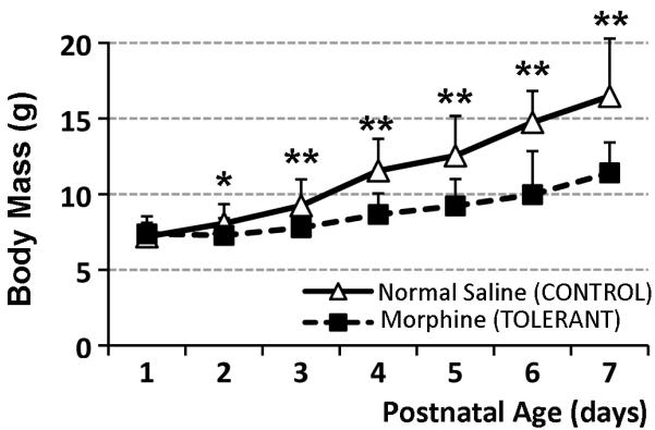
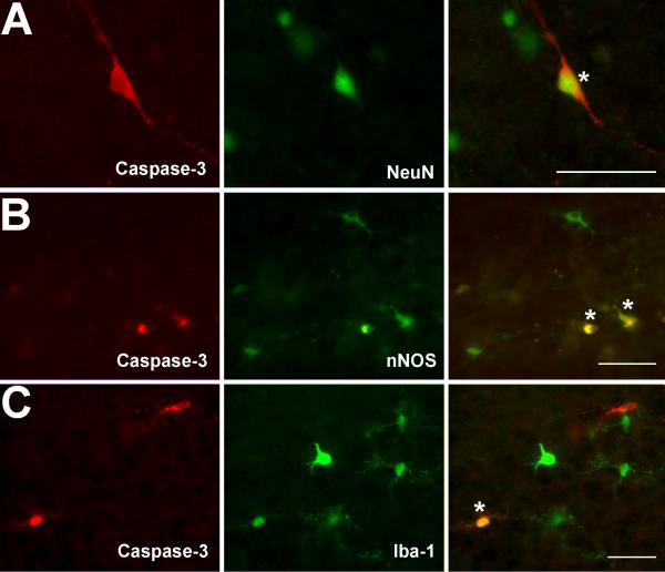
Prolonged neonatal opioid exposure has been associated with: antinociceptive tolerance, long-term neurodevelopmental delay, cognitive, and motor impairment. Morphine has also been shown to induce apoptotic cell death in vitro studies, but its in vivo effect in developing rat brain is unknown. Thus, we hypothesized that prolongued morphine administration in neonatal rats in a model of antinociceptive tolerance and dependence is associated with increased neuroapoptosis. We analyzed neonatal rats from the following groups (1) naïve group (n=6); (2) control group (normal saline (NS), n=5), and (3) morphine group (n=8). Morphine sulfate or equal volume of NS was injected subcutaneously twice daily for 6½ days starting on postnatal day (PD) 1. Development of antinociceptive tolerance was previously confirmed by Hot Plate test on the 7th day. Evidence of neuronal and glial apoptosis was determined by cleaved caspase-3 immunofluorescence combined with specific markers. At PD7, morphine administration after 6½ days significantly increased the density of apoptotic cells in the cortex and amygdala, but not in the hippocampus, hypothalamus, or periaqueductal gray. Apoptotic cells exhibited morphology analogous to neurons. Irrespective of the treatment, only a very few individual microglia but not astrocytes were caspase-3 positive. In summary, repeated morphine administration in neonatal rats (PD1-7) is associated with increased supraspinal apoptosis in distinct anatomical regions known to be important for sensory (cortex) and emotional memory processing (amygdala). Brain regions important for learning (hippocampus), and autonomic and nociceptive processing (hypothalamus and periaqueductal gray) were not affected. Lack of widespread glial apoptosis or robust glial activation following repeated morphine administration suggests that glia might not be affected by chronic morphine at this early age. Future studies should investigate long-term behavioral sequelae of demonstrated enhanced apoptosis associated with prolonged morphine administration in a neonatal rat model.


Figures

Figure 1
Figure 1. Chronic Morphine Administration in Newborn Rats
Graph illustrates the effect of repeated morphine exposure on the body mass of newborn rats. Day of birth was considered day 0 of life. Morphine sulfate (10 mg/kg sc twice-daily) or equivalent volume of normal saline was injected for 6 ½ days starting on the postnatal day (PD) 1. Systemic morphine exposure (n=17) significantly decreased body mass (± SD) in newborn rats in comparison to normal saline control group (n=30). There were no differences between naïve (n=6) and normal saline groups (not shown). Two-tailed t-Test: *, p<0.05; **, p<0.01.
Figure 2
Figure 2. Anatomical Regions of Analysis in PD7 Rats
Representative coronal forebrain (A) and brainstem (B) sections of newborn rat at the postnatal day (PD) 7 schematically illustrate anatomical regions of interest. Sections were drawn from a representative animal that was treated with morphine for 6 ½ days. Solid lines outline the brain sections, while dashed lines illustrate approximate location of anatomical landmarks. Dots illustrate distribution of caspase-3 immunoreactive cells. Density of caspase-3 immunoreactive cells was analyzed in anatomical regions shown in grey. These areas include: (1) cortex, (2) hippocampus (Hipp), (3) amygdala (Amyg), (4) hypothalamus (Hypoth), and (5) periaqueductal gray of the midbrain. Region of cortex included visual cortex (VCtx), somatosensory cortex (SCtx), auditory (AuCtx) and piriform (Pir) cortex at the level of the thalamus (Th). Hypothalamus included both posterior hypothalamic area (PH) and ventromedial hypothalamic nucleus (VMH). Periaqueductal gray included both of its divisions: ventrolateral (vlPAG) and dorsolateral (dlPAG). Abbreviations: IC, inferior colliculus; PnO, pontine reticular nucleus, oral part; py, pyramidal tract; scp, superior cerebellar peduncle. Abbreviations were adapted from The Rat Brain Atlas (Paxinos and Watson, 1998).
Figure 3
Figure 3. Density of Caspase-3 Immunoreactive Cell in Newborn Rat at Postnatal Day 7 (PD7)
Graph illustrates average density of apoptotic cells (#caspase-3 cell/section/brain) ± SD among three treatment groups (naïve, normal saline (control), and morphine (tolerant) group) in five different anatomical regions of interest. There are no differences in density between naïve and control groups in any of the regions analyzed. Morphine treatment was associated with increased density of apoptotic cells only in the cortex (F(2,15)=5.82; p=0.013) and amygdala (F(2,13)=5.84; p=0.006) of newborn rats. Pairwise comparisons of all groups showed statistically significant increase in density of caspase-3 immunoreactive cells in tolerant group compared to naïve (p<0.05) and control groups (p<0.05 for cortex; p<0.01 for amygdala). We found no statistical difference in density of apoptotic cells in hippocampus (F(2,13)=0.47, p=0.63), hypothalamus (F(2,10)=1.68; p=0.23), and periaqueductal gray (PAG; F(2,12)=0.34, p=0.71) among different treatments. For schematic representation of anatomical regions of analysis, refer to Fig. 2. One-way ANOVA with Tukey post-hoc test. Asterisks (*) denote statistically significant increase in values.
Figure 4
Figure 4. Identity of Apoptotic Cells in PD7 Rats
Photomicrographs show double-labeling of caspase-3 immunoreactive cells with neuronal nuclear label (NeuN; A), neuronal nitric oxide synthase (nNOS; B), and microglial label (Iba-1; C). Individual double-labeled cells were marked with asterisks. Scale Bars = 50 μm.
Figure 5
Figure 5. Clusters of Caspase-3 Immunoreactive Cells
Photomicrographs show double-labeling of sporadic clusters of caspase-3 immunoreactive cells with neuronal nitric oxide synthase (nNOS; A), and label for astrocytes (GFAP; B). Clusters of caspase-3 immunoreactive cells that were losing morphological features were noted only in morphine group in the regions of cortex and hippocampus. Scale Bars = 50 μm.

References

    1. Adolphs R, Tranel D, Damasio H, Damasio AR. Fear and the human amygdala. J. Neurosci. 1995;15:5879–5891. - PMC - PubMed
    1. Anand KJ. Consensus statement for the prevention and management of pain in the newborn. Arch. Pediatr. Adolesc. Med. 2001;155:173–180. - PubMed
    1. Anand KJ, Garg S, Rovnaghi CR, Narsinghani U, Bhutta AT, Hall RW. Ketamine reduces the cell death following inflammatory pain in newborn rat brain. Pediatr. Res. 2007;62:283–290. - PubMed
    1. Anand KJ, Hall RW, Desai N, Shephard B, Bergqvist LL, Young TE, Boyle EM, Carbajal R, Bhutani VK, Moore MB, Kronsberg SS, Barton BA. Effects of morphine analgesia in ventilated preterm neonates: primary outcomes from the NEOPAIN randomised trial. Lancet. 2004;363:1673–1682. - PubMed
    1. Anand KJ, Willson DF, Berger J, Harrison R, Meert KL, Zimmerman J, Carcillo J, Newth CJ, Prodhan P, Dean JM, Nicholson C. Tolerance and Withdrawal From Prolonged Opioid Use in Critically Ill Children. Pediatrics. 2010;125:1208–1225. - PMC - PubMed

Publication types