. 2013 Apr 8;23(7):599-606.
doi: 10.1016/j.cub.2013.02.022. Epub 2013 Mar 14.

Cohesin-dependent association of scc2/4 with the centromere initiates pericentromeric cohesion establishment

Affiliations

Cohesin-dependent association of scc2/4 with the centromere initiates pericentromeric cohesion establishment

Josefin Fernius et al. Curr Biol. .

Abstract

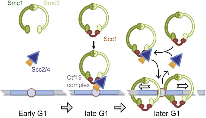
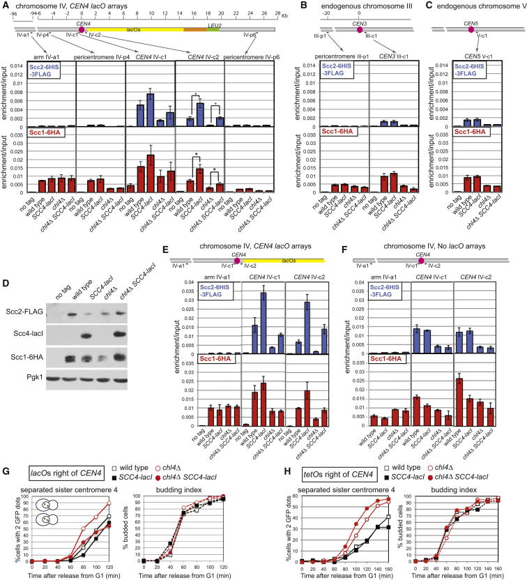
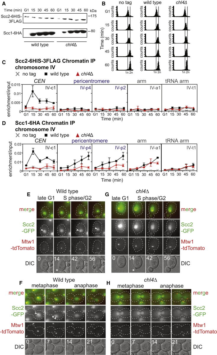
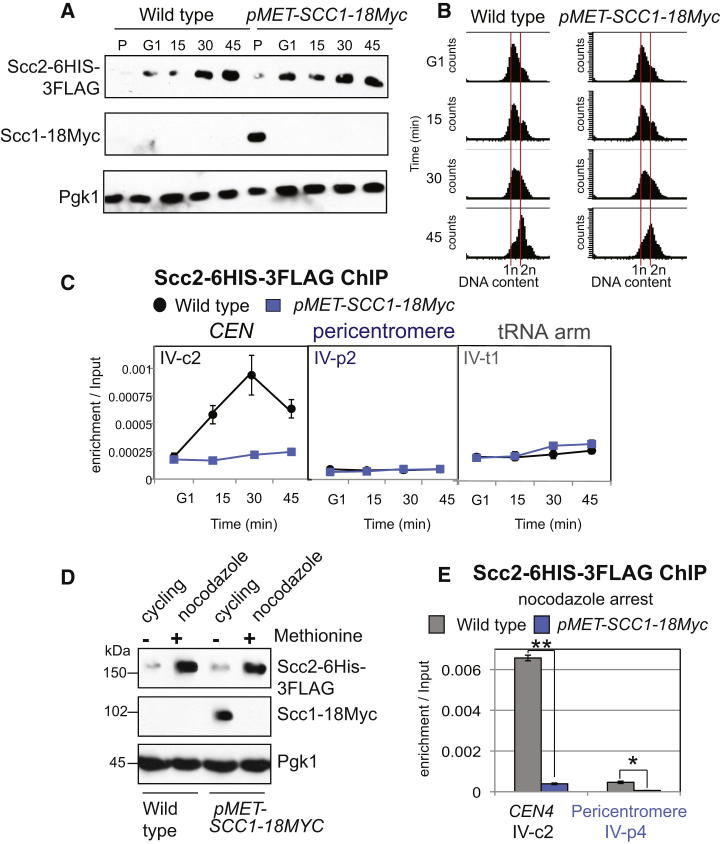
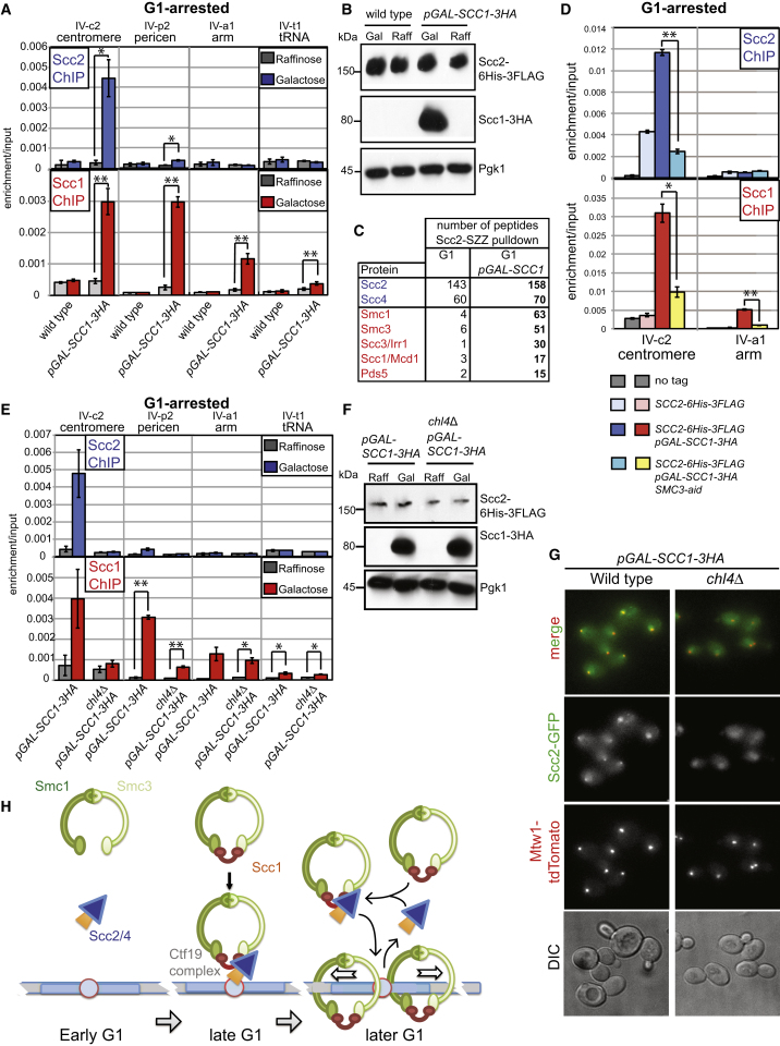
Cohesin is a conserved ring-shaped multiprotein complex that participates in chromosome segregation, DNA repair, and transcriptional regulation [1, 2]. Cohesin loading onto chromosomes universally requires the Scc2/4 "loader" complex (also called NippedBL/Mau2), mutations in which cause the developmental disorder Cornelia de Lange syndrome in humans [1-9]. Cohesin is most concentrated in the pericentromere, the region surrounding the centromere [10-15]. Enriched pericentromeric cohesin requires the Ctf19 kinetochore subcomplex in budding yeast [16-18]. Here, we uncover the spatial and temporal determinants for Scc2/4 centromere association. We demonstrate that the critical role of the Ctf19 complex is to enable Scc2/4 association with centromeres, through which cohesin loads and spreads onto the adjacent pericentromere. We show that, unexpectedly, Scc2 association with centromeres depends on cohesin itself. The absence of the Scc1/Mcd1/Rad21 cohesin subunit precludes Scc2 association with centromeres from anaphase until late G1. Expression of SCC1 is both necessary and sufficient for the binding of cohesin to its loader, the association of Scc2 with centromeres, and cohesin loading. We propose that cohesin triggers its own loading by enabling Scc2/4 to connect with chromosomal landmarks, which at centromeres are specified by the Ctf19 complex. Overall, our findings provide a paradigm for the spatial and temporal control of cohesin loading.


Figures

None
Graphical abstract
Figure 1
Figure 1
The Ctf19 Complex Promotes Cohesion Establishment by Enabling Association of the Scc2/4 Loader with Centromeres (A–D) Artificial recruitment of Scc2/4 to a centromere leads to a modest increase in Scc1 levels at the tethering site. An approximately 10 kb array of lacO repeats was integrated approximately 500 bp to the right of CEN4 in strains that were otherwise wild-type (AM10285), pGAL-SCC4-lacI (AM10287), chl4Δ (AM10289), or chl4Δ pGAL-SCC4-lacI (AM10291) and carried SCC2-6HIS-3FLAG and SCC1-6HA as well as a no-tag control (AM8387). Strains were precultured in raffinose-containing medium and then arrested in nocodazole and benomyl for 2.5 hr in medium containing raffinose and galactose before harvesting for Scc1 and Scc2 ChIP. qPCR analysis (A–C) using the indicated primer sets on chromosome IV (A), III (B), or V (C). The mean of four independent experiments is shown with error bars representing standard error (p < 0.5, two-tailed paired t test). Anti-FLAG, anti-lacI, anti-HA, and anti-Pgk1 (D) immunoblots showing relative levels of Scc2-6HIS-3FLAG, Scc4-LacI, Scc1-6HA, and Pgk1 (loading control) in the samples analyzed from a representative experiment. (E and F) Recruitment of Scc1 and Scc2 by Scc4-LacI is dependent on lacOs. Strains with a 10 kb array of lacO repeats ∼500 bp from CEN4 (E) or without lacOs (F) and with the indicated genotypes were grown and analyzed as in (A), and Scc1 and Scc2 ChIP is shown for the indicated primer sets. Strains with lacOs (E) are as in (A). Strains without lacOs (F) all carried SCC2-6HIS-3FLAG and SCC1-6HA and are otherwise wild-type (AM8413), pGAL-SCC4-lacI (AM11066), chl4Δ (AM8415), chl4Δ pGAL-SCC4-lacI (AM11065). Note that the presence of lacO arrays adjacent to CEN4 reduces cohesin association with IV-c2. Mean of three independent experiments is shown and error bars indicate standard error. (G) Tethering Scc2/4 to centromeres rescues the centromeric cohesion defect of chl4Δ cells. Strains with approximately 10 kb lacO arrays integrated adjacent to CEN4, producing LacI-GFP and carrying pMET-CDC20, were arrested in G1 in raffinose-containing medium lacking methionine by treatment with alpha factor before being released into medium containing raffinose, galactose, and methionine to induce a metaphase arrest and induce Scc4-LacI. Samples were taken at the indicated time points after release from alpha factor, and the number of GFP foci per cell and budding was scored. A representative experiment is shown. Strains used were AM10570 (wild-type), AM10571 (pGAL-SCC4-lacI), AM10572 (chl4Δ), and AM10573 (chl4Δ pGAL-SCC4-lacI). (H) Scc4-lacI requires lacO arrays at CEN4 to rescue the cohesion defect of chl4Δ cells. To visualize CEN4, while preventing Scc4-lacI binding, tetO arrays were integrated approximately 2.4 kb right of CEN4 in pMET-CDC20 strains producing TetR-GFP. Wild-type (AM4643), SCC4-lacI (AM11162), chl4Δ (AM4644), and chl4Δ SCC4-lacI (AM11163) were treated as described in (G), and a representative experiment is shown. See also Figure S1.
Figure 2
Figure 2
Scc2 Associates with Centromeres from Late G1 until Anaphase (A–D) Sites of association of Scc1 and Scc2 as cells progress into the cell cycle and their dependence on Chl4. Strains AM1176 (no tag), AM8414 (SCC2-6HIS-3FLAG), and AM8415 (SCC2-6HIS-3FLAG, chl4Δ) were arrested in G1 in alpha factor at room temperature, then released into medium containing benomyl and nocodazole at 18°C. Samples were extracted prerelease (G1) and at 15, 30, 45, and 60 min following release for analysis of Scc1-6HA and Scc2-6His-3FLAG levels by western blotting (A), analysis of DNA content by FACS (B), and association of Scc1-6HA and Scc2-6His-3FLAG with the indicated sites by ChIP-qPCR (C and D). The mean of three independent experiments is shown, with error bars representing standard error. (E–H) Live-cell imaging showing Scc2 localization during the cell cycle and its dependence on Chl4. Still images from movies of wild-type (E and F; AM10713) and chl4Δ (G and H; AM10714) cells carrying Scc2-GFP and Mtw1-tdTomato. The numbers on the DIC images represent minutes relative to the first image. Arrowheads and asterisks indicate the position of the Scc2-GFP associated with kinetochores and the newly forming bud, respectively. See also Figure S2 and Movies S1, S2, S3, and S4.
Figure 3
Figure 3
Scc1 Is Required for Scc2 Association with Centromeres (A–C) Analysis of Scc2 association with centromeres upon cell-cycle entry in the absence of Scc1. Strains AM6006 (SCC2-6HIS-3FLAG) and AM9519 (SCC2-6HIS-3FLAG pMET-SCC1-18MYC) were arrested in G1 with alpha factor in rich medium containing methionine to deplete Scc1 for 3 hr and then released into methionine-containing medium. Samples were extracted at 0 (G1), 15, 30, and 45 min after release for analysis of protein levels by anti-FLAG, anti-Myc, and anti-Pgk1 immunoblot (A), analysis of DNA content by FACS (B), and protein localization at the indicated sites by ChIP-qPCR (C). (D and E) Analysis of Scc2 association with centromeres in nocodazole-arrested cells after Scc1 depletion. Strains as in (A)–(C) were arrested in alpha factor in the presence of methionine for 3 hr before being released into methionine- and nocodazole-containing medium for 2 hr. Immunoblot (D) showing protein levels at the time of harvesting (nocodazole) and preincubation with alpha factor (cycling). Analysis of ChIP samples by qPCR (E) at the indicated sites. The mean of three independent experiments is shown. Error bars represent standard error (∗∗p < 0.01; p < 0.05, two-tailed paired t test). See also Figure S3.
Figure 4
Figure 4
SCC1 Expression Is Sufficient for Scc2 to Associate with Centromeres and for Scc1 Loading in G1 (A and B) Strains AM9335 (SCC2-6HIS-3FLAG) and AM9334 (SCC2-6HIS-3FLAG pGAL-SCC1-3HA) were arrested in G1 in raffinose-containing medium with alpha factor for 3 hr. Samples were extracted for anti-FLAG, anti-HA, and anti-Pgk1 immunoblotting (B) and ChIP-qPCR (A; raffinose), then 2% galactose was added to the remainder, while maintaining the G1 arrest. After 45 min the remainder of the culture was harvested for immunoblotting (B) and ChIP-qPCR (A; galactose). (C) Mass spectrometry analysis of proteins copurifying with Scc2 after immunoprecipitation from G1-arrested cells with and without SCC1 expression. The number of identified peptides for the indicated proteins is shown. (D) Smc3 is required for Scc2 and Scc1 to associate with centromeres. Strains AM1176 (no tag), AM6006 (SCC2-6HIS-3FLAG), AM9334 (SCC2-6HIS-3FLAG pGAL-SCC1), and AM11092 (SCC2-6HIS-3FLAG pGAL-SCC1 SMC3-aid) were arrested in G1 in raffinose-containing medium with alpha factor for 3 hr before addition of 0.5 mM NAA. After 30 min, while maintaining G1 arrest, 2% galactose was added, and cultures were harvested for Scc2 and Scc1 ChIP after 45 min. (E and F) Ectopic loading of cohesin in G1 is dependent on Chl4. Analysis of Scc1 and Scc2 loading at centromeres in G1-arrested wild-type and chl4Δ mutant cells, following ectopic expression of Scc1. Strains AM9334 (SCC2-6HIS-3FLAG pGAL-SCC1-3HA) and AM9613 (SCC2-6HIS-3FLAG, pGAL-SCC1-3HA chl4Δ) were treated as in (A) and (B), and qPCR analysis of ChIP samples (E) and immunoblot (F) are shown. (G) Scc1 production enables Scc2 association with kinetochores throughout the cell cycle. Images are shown of live wild-type (AM10715) and chl4Δ cells (AM10716) carrying pGAL-SCC1-3HA, SCC2-GFP, and MTW1-tdTomato after culturing in galactose-containing medium for 2 hr. (H) Model for the spatial and temporal control of cohesin loading by Scc1 and the Ctf19 complex. In (A), (D), and (E) the mean of three independent experiments is shown with error bars representing standard error (∗∗p < 0.01; p < 0.05, two-tailed paired t test). See also Figure S4 and Table S3.

References

    1. Haering C.H., Löwe J., Hochwagen A., Nasmyth K. Molecular architecture of SMC proteins and the yeast cohesin complex. Mol. Cell. 2002;9:773–788. - PubMed
    1. Dorsett D., Ström L. The ancient and evolving roles of cohesin in gene expression and DNA repair. Curr. Biol. 2012;22:R240–R250. - PMC - PubMed
    1. Bernard P., Drogat J., Maure J.-F., Dheur S., Vaur S., Genier S., Javerzat J.-P. A screen for cohesion mutants uncovers Ssl3, the fission yeast counterpart of the cohesin loading factor Scc4. Curr. Biol. 2006;16:875–881. - PubMed
    1. Ciosk R., Shirayama M., Shevchenko A., Tanaka T., Toth A., Shevchenko A., Nasmyth K. Cohesin’s binding to chromosomes depends on a separate complex consisting of Scc2 and Scc4 proteins. Mol. Cell. 2000;5:243–254. - PubMed
    1. Lightfoot J., Testori S., Barroso C., Martinez-Perez E. Loading of meiotic cohesin by SCC-2 is required for early processing of DSBs and for the DNA damage checkpoint. Curr. Biol. 2011;21:1421–1430. - PubMed

Publication types

MeSH terms