Comparative Study
. 2013 May 7;61(18):1884-93.
doi: 10.1016/j.jacc.2013.02.012. Epub 2013 Mar 6.

Heme levels are increased in human failing hearts

Affiliations
Comparative Study

Heme levels are increased in human failing hearts

Arineh Khechaduri et al. J Am Coll Cardiol. .

Abstract

Objectives: The goal of this study was to characterize the regulation of heme and non-heme iron in human failing hearts.

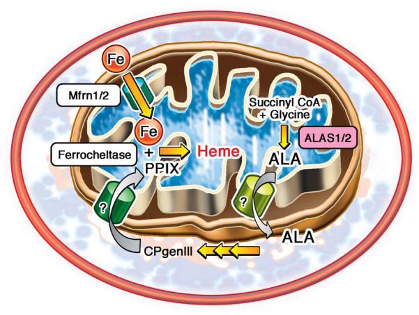
Background: Iron is an essential molecule for cellular physiology, but in excess it facilitates oxidative stress. Mitochondria are the key regulators of iron homeostasis through heme and iron-sulfur cluster synthesis. Because mitochondrial function is depressed in failing hearts and iron accumulation can lead to oxidative stress, we hypothesized that iron regulation may also be impaired in heart failure (HF).

Methods: We measured mitochondrial and cytosolic heme and non-heme iron levels in failing human hearts retrieved during cardiac transplantation surgery. In addition, we examined the expression of genes regulating cellular iron homeostasis, the heme biosynthetic pathway, and micro-RNAs that may potentially target iron regulatory networks.

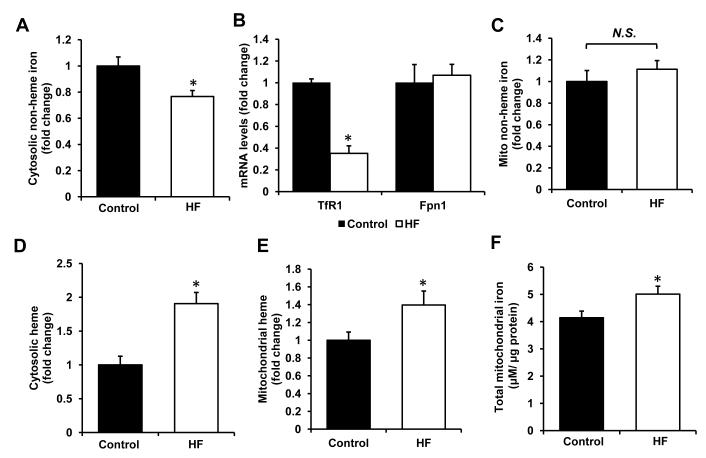
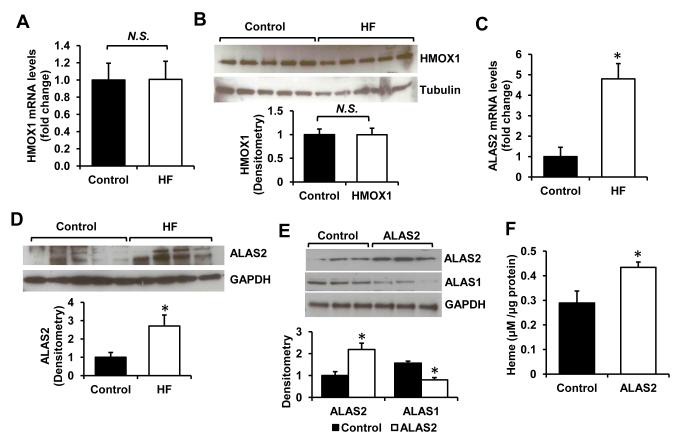
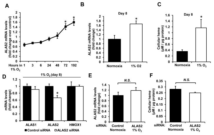
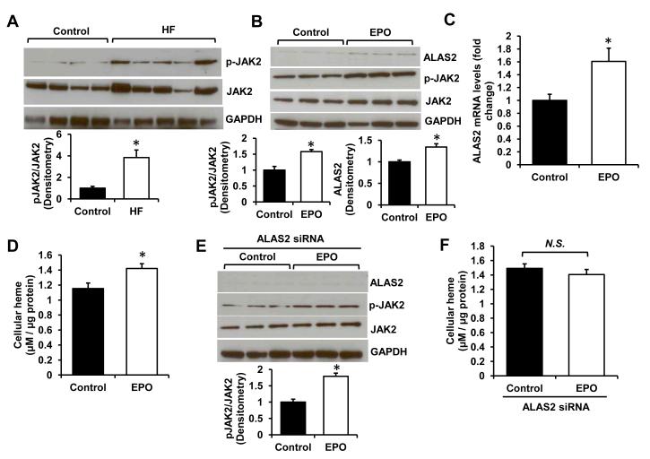
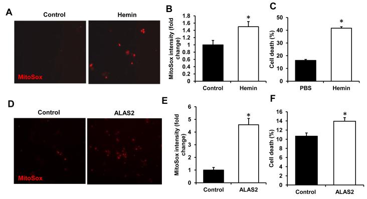
Results: Although cytosolic non-heme iron levels were reduced in HF, mitochondrial iron content was maintained. Moreover, we observed a significant increase in heme levels in failing hearts, with corresponding feedback inhibition of the heme synthetic enzymes and no change in heme degradation. The rate-limiting enzyme in heme synthesis, delta-aminolevulinic acid synthase 2 (ALAS2), was significantly upregulated in HF. Overexpression of ALAS2 in H9c2 cardiac myoblasts resulted in increased heme levels, and hypoxia and erythropoietin treatment increased heme production through upregulation of ALAS2. Finally, increased heme levels in cardiac myoblasts were associated with excess production of reactive oxygen species and cell death, suggesting a maladaptive role for increased heme in HF.

Conclusions: Despite global mitochondrial dysfunction, heme levels are maintained above baseline in human failing hearts.


Figures

Figure 1
Figure 1
Schematic representation of cellular heme synthesis pathway
Figure 2
Figure 2. Heme and non-heme iron regulation in HF
(A) Cytosolic non-heme iron levels in control and failing hearts normalized to the cytosolic protein concentration (n=10). (B) mRNA expression of TfR1, an iron importer, and Fpn1, an iron exporter, in HF and control groups (n=10). (C) Non-heme iron levels in the mitochondria of control and failing hearts normalized to mitochondrial protein content (n=10). Cytosolic (D) and mitochondrial (E) heme levels in control and failing hearts normalized to the protein concentration of cytosolic and mitochondrial fractions, respectively (n=10). (F) Total mitochondrial iron levels, obtained by addition of non-heme and heme iron content (n=10). Data are presented as mean ± SEM. * p<0.05 vs. control.
Figure 3
Figure 3. ALAS2 is upregulated in failing hearts
mRNA (A) and protein (B) levels of HMOX1 in control and failing hearts (n=5-6). mRNA (C) and protein levels (D) of ALAS2 in control and failing hearts (n=4-6). (E) Westen blot analysis of ALAS1/2 proteins in H9c2 with lentiviral overexpression of ALAS2 enzyme (n=3). Densitometry analyses are presented below the Western blots. (F) Heme levels in H9c2 cells transduced with ALAS2 or control lentiviral vector (n=6). Data are presented as mean ± SEM. * p<0.05 vs. control.
Figure 4
Figure 4. Regulation of ALAS2 by hypoxia in cardiac myoblasts
(A) Time-course of ALAS2 mRNA expression in H9c2 cardiac myoblasts subjected to hypoxia (n=6). (B) ALAS2 mRNA levels after 8 days in hypoxia or normoxia (n=6). (C) Cellular heme content after 8 days in hypoxia or normoxia (n=6). (D) mRNA levels of heme synthesis/degradation enzymes in hypoxic cardiac myoblasts with or without siRNA-mediated ALAS2 knockdown (n=6). (E) Comparison of ALAS2 mRNA levels in control normoxic and ALAS2 siRNA-treated hypoxic cells after 8 days (n=6). (F) Heme levels in control normoxic and ALAS2 siRNA hypoxic cells after 8 days (n=6). Data are presented as mean ± SEM. * p<0.05 vs. control.
Figure 5
Figure 5. ALAS2 is regulated by erythropoietin
(A) Western blot analysis of p-JAK2 and JAK2 protein in failing and control hearts (n=4-5). (B) ALAS2 and pJAK2/JAK2 protein levels in H9c2 cells treated with 0.6mg/mL EPO (n=3). (C) qRT-PCR analysis of ALAS2 mRNA expression in H9c2 cells with EPO or vehicle control treatment (n=6). (D) Cellular heme content with EPO treatment in cardiac myoblasts (n=6). (E) Western blot analysis of ALAS2 and pJAK2/JAK2 in ALAS2 siRNA-treated cells incubated with EPO or vehicle control (n=3). (F) Cellular heme content in EPO- or vehicle-treated cells with ALAS2 siRNA knockdown (n=6). Data are presented as mean ± SEM. * p<0.05 vs. control.
Figure 6
Figure 6. Heme and ALAS2 overexpression increase oxidative stress
(A) Representative images of H9c2 incubated with the vehicle control or 10μM hemin for 6 hours and stained with mitochondria-specific ROS-sensitive dye MitoSox. (B) Image J analysis of MitoSox-stained H9c2 following the treatment with vehicle or hemin (n=4-5, 4 fields per sample). (C) Cell death assessed by propidium iodine/Annexin V double-labeling with hemin treatment as in A (n=6). (D) Representative MitoSox images of H9c2 with or without ALAS2 overexpression. (E) Quantification of MitoSox staining with ALAS2 overexpression (n=4-5, 4 fields per sample). (F) Cell death with ALAS2 overexpression assessed as in C (n=6). Data are presented as mean ± SEM. * p<0.05 vs. control.

References

    1. Lloyd-Jones D, Adams RJ, Brown TM, Carnethon M, et al. Heart disease and stroke statistics--2010 update: a report from the American Heart Association. Circulation. 2010;121:e46–e215. - PubMed
    1. Mudd JO, Kass DA. Tackling heart failure in the twenty-first century. Nature. 2008;451:919–928. - PubMed
    1. Karamanlidis G, Bautista-Hernandez V, Fynn-Thompson F, Del Nido P, et al. Impaired mitochondrial biogenesis precedes heart failure in right ventricular hypertrophy in congenital heart disease. Circ Heart Fail. 2011;4:707–713. - PMC - PubMed
    1. Bayeva M, Gheorghiade M, Ardehali H. Mitochondria as Therapeutic Target in Heart Failure. J Am Coll Cardiol. 2012 doi: 10.1016/j.jacc.2012.08.1021. - PMC - PubMed
    1. Sihag S, Cresci S, Li AY, Sucharov CC, et al. PGC-1alpha and ERRalpha target gene downregulation is a signature of the failing human heart. J Mol Cell Cardiol. 2009;46:201–212. - PMC - PubMed

Publication types

MeSH terms