

Drosophila melanogaster as an emerging translational model of human nephrolithiasis
- PMID: 23500641
- PMCID: PMC3842186
- DOI: 10.1016/j.juro.2013.03.010
Drosophila melanogaster as an emerging translational model of human nephrolithiasis
Abstract
Purpose: The limitations imposed by human clinical studies and mammalian models of nephrolithiasis have hampered the development of effective medical treatments and preventive measures for decades. The simple but elegant Drosophila melanogaster is emerging as a powerful translational model of human disease, including nephrolithiasis. It may provide important information essential to our understanding of stone formation. We present the current state of research using D. melanogaster as a model of human nephrolithiasis.
Materials and methods: We comprehensively reviewed the English language literature using PubMed®. When necessary, authoritative texts on relevant subtopics were consulted.
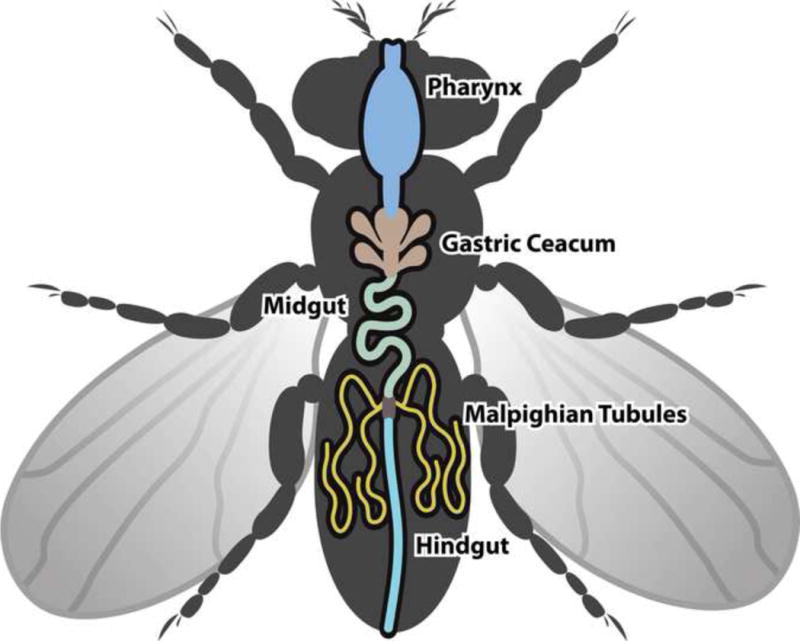
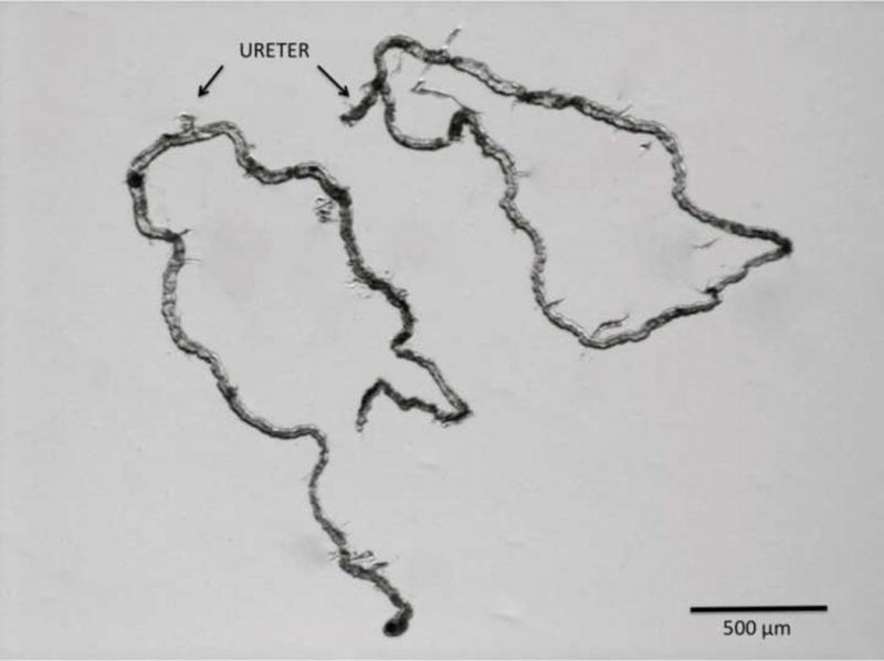
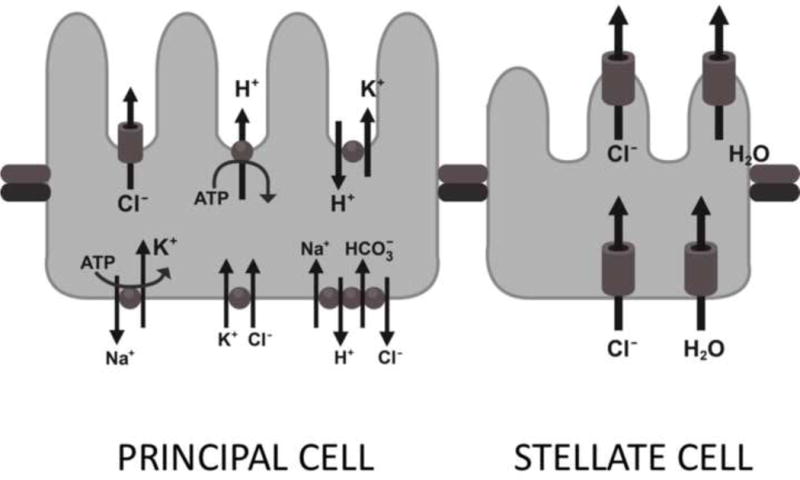
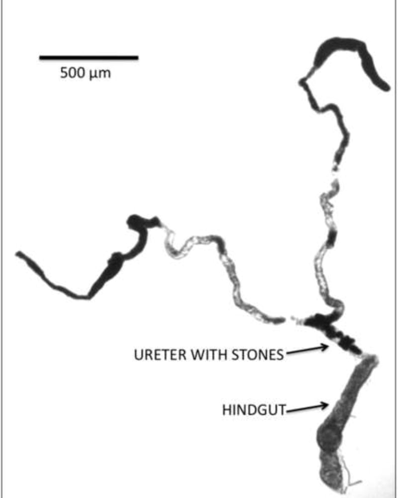
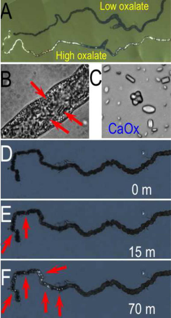
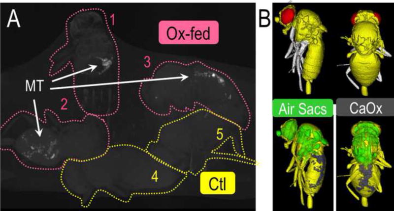
Results: The genetic composition, anatomical structure and physiological function of Drosophila malpighian tubules are remarkably similar to those of the human nephron. The direct effects of dietary manipulation, environmental alteration and genetic variation on stone formation can be observed and quantified in a matter of days. Several Drosophila models of human nephrolithiasis have been developed, including genetically linked and environmentally induced stones. A model of calcium oxalate stone formation is among the most recent fly models of human nephrolithiasis.
Conclusions: The ability to readily manipulate and quantify stone formation in D. melanogaster models of human nephrolithiasis presents the urological community with a unique opportunity to increase our understanding of this enigmatic disease.
Keywords: ATPase; CT; Drosophila melanogaster; UAS; XDH; adenosine triphosphatase; animal; computerized tomography; disease models; kidney; malpighian tubules; nephrolithiasis; upstream activation sequence; xanthine dehydrogenase.
Copyright © 2013 American Urological Association Education and Research, Inc. Published by Elsevier Inc. All rights reserved.
Figures
References
-
- Wessing A, Eichelberg D. Malpighian tubules, rectal papillae and excretion. In: Ashburner M, Wright TRF, editors. The genetics and biology of Drosophila. 2c. New York: Academic Press Inc; 1978. pp. 1–42.
-
- Dow JAT, Maddrell SHP, Görtz A, et al. The Malpighian tubules of Drosophila melanogaster: a novel phenotype for studies of fluid secretion and its control. J Exp Biol. 1994;197:421. - PubMed
-
- Romero MF, Henry D, Nelson S, et al. Cloning and characterization of a Na+-driven anion exchanger (NDAE1). A new bicarbonate transporter. J Biol Chem. 2000;275:24552. - PubMed
Publication types
MeSH terms
Grants and funding
- R21 DK091727/DK/NIDDK NIH HHS/United States
- EY017732/EY/NEI NIH HHS/United States
- K12 DK083021/DK/NIDDK NIH HHS/United States
- P50-DK083007/DK/NIDDK NIH HHS/United States
- R13 AG044873/AG/NIA NIH HHS/United States
- AG038012-01/AG/NIA NIH HHS/United States
- RL1 AG032113/AG/NIA NIH HHS/United States
- K12-DK-07-006/DK/NIDDK NIH HHS/United States
- DK092408/DK/NIDDK NIH HHS/United States
- P50 DK083007/DK/NIDDK NIH HHS/United States
- DK091727-01A1/DK/NIDDK NIH HHS/United States
- U54 DK100227/DK/NIDDK NIH HHS/United States
- R01 DK092408/DK/NIDDK NIH HHS/United States
- R01 AG031337/AG/NIA NIH HHS/United States
- R01 AG038012/AG/NIA NIH HHS/United States
- R21 AG028241/AG/NIA NIH HHS/United States
- R01 AG038688/AG/NIA NIH HHS/United States
- R01 EY017732/EY/NEI NIH HHS/United States
LinkOut - more resources
Full Text Sources
Other Literature Sources
Medical
Molecular Biology Databases
Miscellaneous
