Review
. 2013 Oct;1831(10):1533-41.
doi: 10.1016/j.bbalip.2013.02.010. Epub 2013 Mar 14.

Adipose tissue and adipocytes support tumorigenesis and metastasis

Affiliations
Review

Adipose tissue and adipocytes support tumorigenesis and metastasis

Kristin M Nieman et al. Biochim Biophys Acta. 2013 Oct.

Abstract

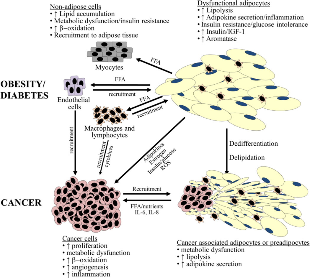
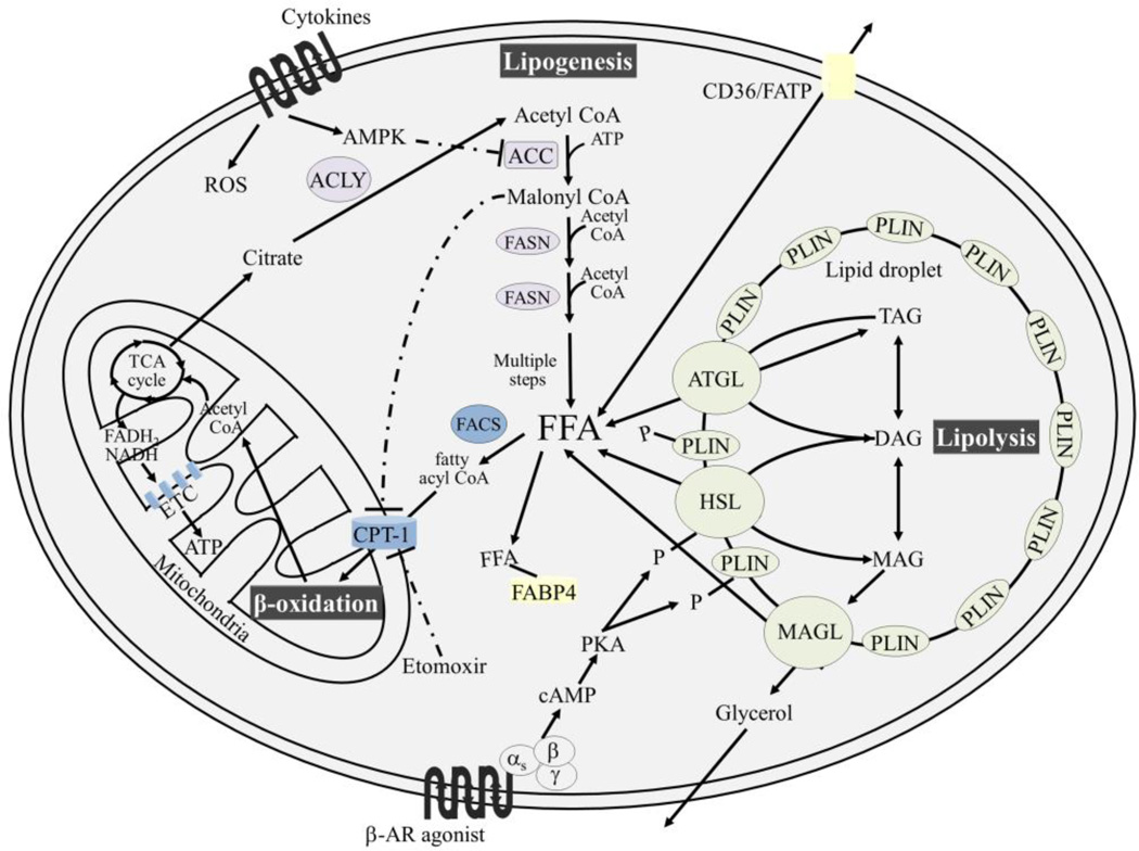
Adipose tissue influences tumor development in two major ways. First, obese individuals have a higher risk of developing certain cancers (endometrial, esophageal, and renal cell cancer). However, the risk of developing other cancers (melanoma, rectal, and ovarian) is not altered by body mass. In obesity, hypertrophied adipose tissue depots are characterized by a state of low grade inflammation. In this activated state, adipocytes and inflammatory cells secrete adipokines and cytokines which are known to promote tumor development. In addition, the adipocyte mediated conversion of androgens to estrogen specifically contributes to the development of endometrial cancer, which shows the greatest relative risk (6.3-fold) increase between lean and obese individuals. Second, many tumor types (gastric, breast, colon, renal, and ovarian) grow in the anatomical vicinity of adipose tissue. During their interaction with cancer cells, adipocytes dedifferentiate into pre-adipocytes or are reprogrammed into cancer-associated adipocytes (CAA). CAA secrete adipokines which stimulate the adhesion, migration, and invasion of tumor cells. Cancer cells and CAA also engage in a dynamic exchange of metabolites. Specifically, CAA release fatty acids through lipolysis which are then transferred to cancer cells and used for energy production through β-oxidation. The abundant availability of lipids from adipocytes in the tumor microenvironment, supports tumor progression and uncontrolled growth. Given that adipocytes are a major source of adipokines and energy for the cancer cell, understanding the mechanisms of metabolic symbiosis between cancer cells and adipocytes, should reveal new therapeutic possibilities. This article is part of a Special Issue entitled Lipid Metabolism in Cancer.

Keywords: Adipocytes; Cancer; Metabolic symbiosis; Metastasis; Obesity; Visceral adipose tissue.


Conflict of interest statement

Conflict of interest

None.

Figures

Figure 1
Figure 1. Adipocytes in metabolic disease and cancer
Striking similarities exist between the involvement of adipocytes in both tumorigenesis and obesity/type 2 diabetes. These disease states result in the recruitment of immune cells including macrophages and lymphocytes. Cytokine secretion from adipocytes (e.g., leptin, IL-6, IL-8, and TNF-α) are also involved in both disease states. Activated adipocytes in obesity/diabetes and/or cancer are delipidated and potentially dedifferentiate to fibroblast-like cells. Expelled nutrients (e.g. FFA) can be taken up by both cancer cells as well as other non-adipose cells (e.g., myocytes, macrophages, and vascular endothelial cells) resulting in metabolic dysfunction. Abbreviations: CAA, cancer-associated adipocytes; FFA, free-fatty acid(s); IGF-1, insulin-like growth factor protein-1; IL; interleukin; MCP-1, monocyte chemoattractant protein-1; ROS, reactive oxygen species; TNF-α, tumor necrosis factor-α.
Figure 2
Figure 2. An overview of adipocyte lipid metabolism
Lipogenesis occurs in the cytoplasm of the cell and requires acetyl CoA which can be generated from citrate, through the activity of ACLY. The irreversible formation of malonyl CoA from acetyl CoA initiates lipogenesis and requires the activity of ACC. AMPK phosphorylates and inactivates ACC. Cytokines (e.g., leptin, IL-6, IL-8, MCP1, and TNF-α) can trigger both ROS production and AMPK activation. Lipogenesis proceeds in a repeating reaction sequence which involves the addition of two carbon units to a growing fatty acid chain, through the activity FASN. Accumulation of malonyl CoA during energy surplus results in allosteric inhibition of CPT-1, the rate-limiting enzyme involved in shuttling acylated free fatty acids (via FACS) into the mitochondria for β-oxidation. Etomoxir prevents β-oxidation by inhibiting CPT-1. In contrast, under the condition of energy deficit, citrate becomes limited and malonyl CoA concentrations decrease within the cell, lifting the allosteric regulation of CPT-1 and allowing ATP generation through β-oxidation. To prevent lipotoxicity, fatty acids bound to glycerol are stored in lipid droplets, primarily as TAG. TAG can be mobilized by β-AR agonists (e.g., epinephrine and norepinephrine) which initiate a G-protein coupled cascade leading to the phosphoryation of HSL and PLIN. Phosphorylation results in a conformational change in PLIN, allowing HSL access to the lipid droplet. Triacylglycerols are completely hydrolyzed by the activity of (i) ATGL which removes one fatty acid forming DAG, (ii) HSL which remove another fatty acid from DAG resulting in MAG, and (iii) MAGL which removes the final fatty acid from the glycerol backbone. FFA can be exported and imported through fatty acid receptors such as CD36 and FATPs. FABP4 chaperones free fatty acid within the cell. Abbreviations: ACC, acetyl CoA carboxylase; ACSL, acyl CoA synthetase lyase; ACLY, ATP citrate lyase; AMPK, AMP kinase; AR, adrenergic; ATGL, adipose triglyceride lipase; CPT-1, carnitine-palmitoyltransferase 1; DAG, diacylglycerol(s); ETC, electron transport chain; FABP4, fatty acid binding protein 4; FACS, fatty acyl CoA synthetase; FASN, fatty acid synthase; FATP, fatty acid transport proteins; FFA, free-fatty acids; HSL, hormone-sensitive lipase; MAG, monacylglycerol(s); MAGL, monacylglycerol lipase; MCP-1, monocyte chemoattractant protein-1; P, phosphorylation; PKA, protein kinase A; PLIN, perilipin; TAG, triacylglycerol(s); TCA; tricarboxylic acid; TNF-α, tumor necrosis factor-α.
Figure 3
Figure 3. Breast and renal cell cancer invading adipose tissue
Similar to ovarian cancer, breast and renal cancers both invade and devour neighboring adipose tissue. (A) Ductal breast cancer in a postmenopausal patient infiltrating the adjacent breast adipose tissue. The adipocytes farther away from the tumor are of normal size, while those in close proximity are much smaller. This observation suggests dedifferentiation and delipidation of mature adipocytes to pre-adipocytes (400x). (B) Renal cell cancer invading the perirenal fat pad (100x).

References

    1. Dirat B, Bochet L, Escourrou G, Valet P, Muller C. Unraveling the obesity and breast cancer links: A role for cancer-associated adipocytes. Endocr Dev. 2010;19:45–52. - PubMed
    1. Hanahan D, Coussens LM. Accessories to the crime: Functions of cell recruited to the tumor microenvironment. Cancer Cell. 2012;21:309–322. - PubMed
    1. Nakajima EC, Van Houten B. Metabolic symbiosis in cancer: Refocusing the Warburg lens. Mol Carcinog. 2012:1–9. - PMC - PubMed
    1. Martinez-Outschoorn UE, Whitaker-Menezes D, Pavlides S, Chiavarina B, Bonuccelli G, Trimmer C, Tsirigos A, Migneco G, Witkiewicz AK, Balliet R, Mercier I, Wang C, Flomenberg N, Howell A, Lin Z, Caro J, Pestell RG, Sotgia F, Lisanti MP. The autophagic tumor stroma model of cancer or "battery-operated tumor growth" a simple solution to the autophagy paradox. Cell Cycle. 2010;9:4297–4306. - PMC - PubMed
    1. Santos CR, Schulze A. Lipid metabolism in cancer. FEBS Journal. 2012;279:2610–2623. - PubMed

Publication types

LinkOut - more resources