. 2013 May 10;112(10):1345-54.
doi: 10.1161/CIRCRESAHA.111.300581. Epub 2013 Mar 15.

Apolipoprotein AI and high-density lipoprotein have anti-inflammatory effects on adipocytes via cholesterol transporters: ATP-binding cassette A-1, ATP-binding cassette G-1, and scavenger receptor B-1

Affiliations

Apolipoprotein AI and high-density lipoprotein have anti-inflammatory effects on adipocytes via cholesterol transporters: ATP-binding cassette A-1, ATP-binding cassette G-1, and scavenger receptor B-1

Tomio Umemoto et al. Circ Res. .

Abstract

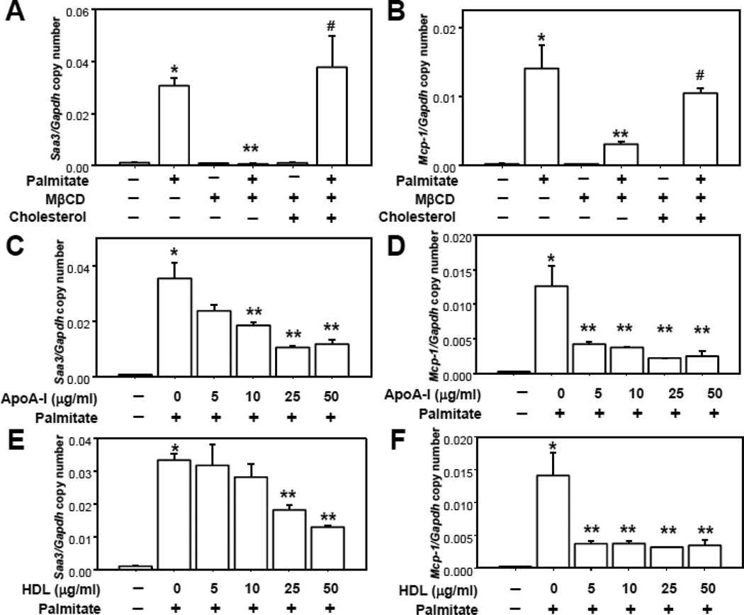
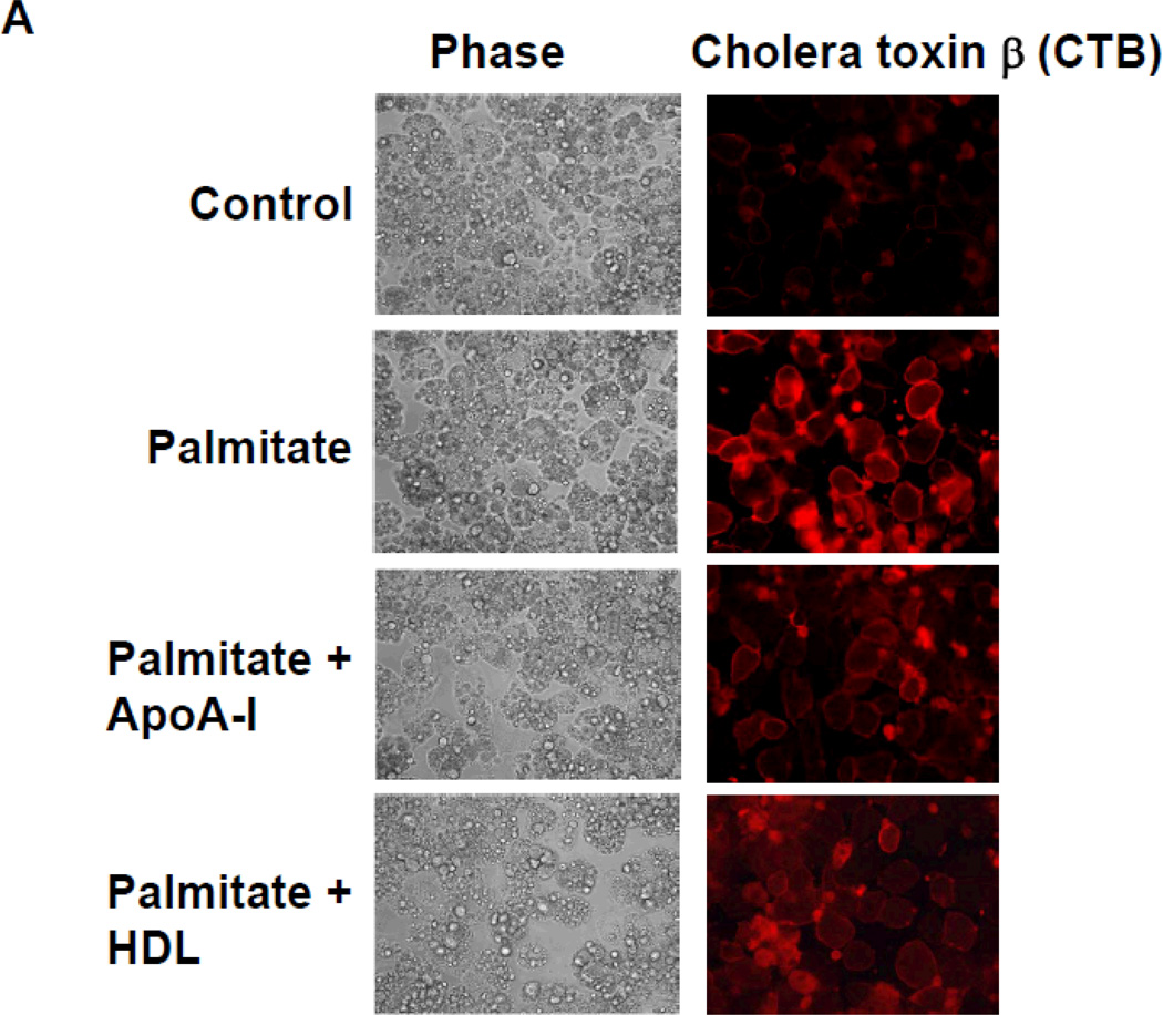
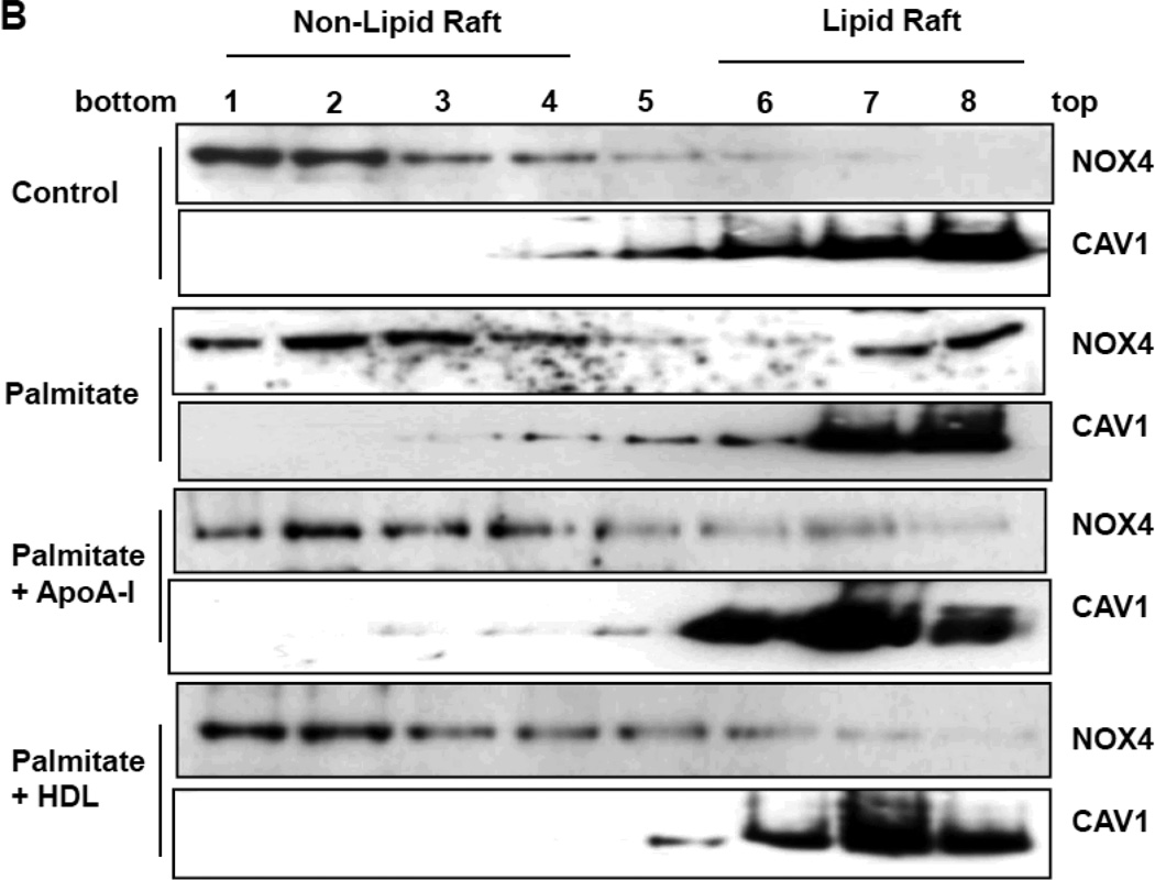
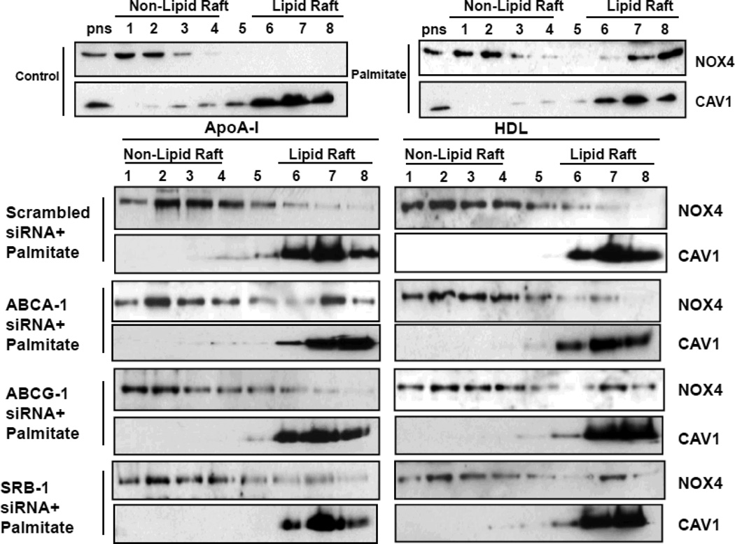
Rationale: Macrophage accumulation in adipose tissue associates with insulin resistance and increased cardiovascular disease risk. We previously have shown that generation of reactive oxygen species and monocyte chemotactic factors after exposure of adipocytes to saturated fatty acids, such as palmitate, occurs via translocation of NADPH oxidase 4 into lipid rafts (LRs). The anti-inflammatory effects of apolipoprotein AI (apoAI) and high-density lipoprotein (HDL) on macrophages and endothelial cells seem to occur via cholesterol depletion of LRs. However, little is known concerning anti-inflammatory effects of HDL and apoAI on adipocytes.

Objective: To determine whether apoAI and HDL inhibit inflammation in adipocytes and adipose tissue, and whether this is dependent on LRs.

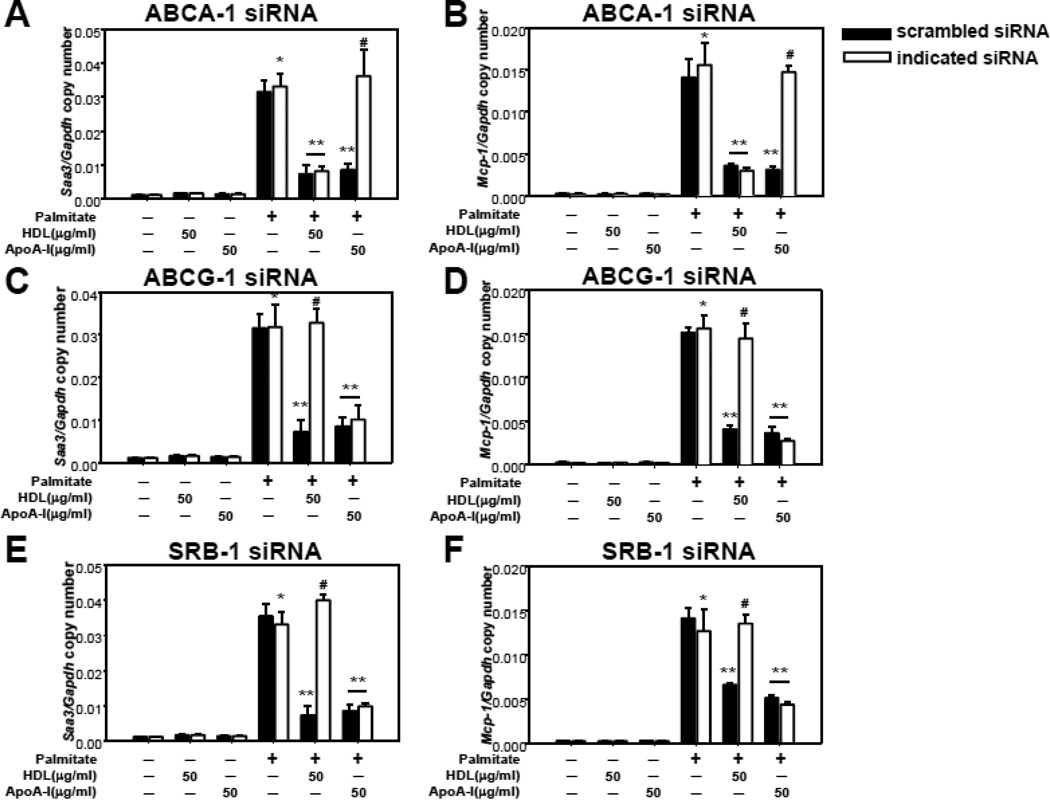
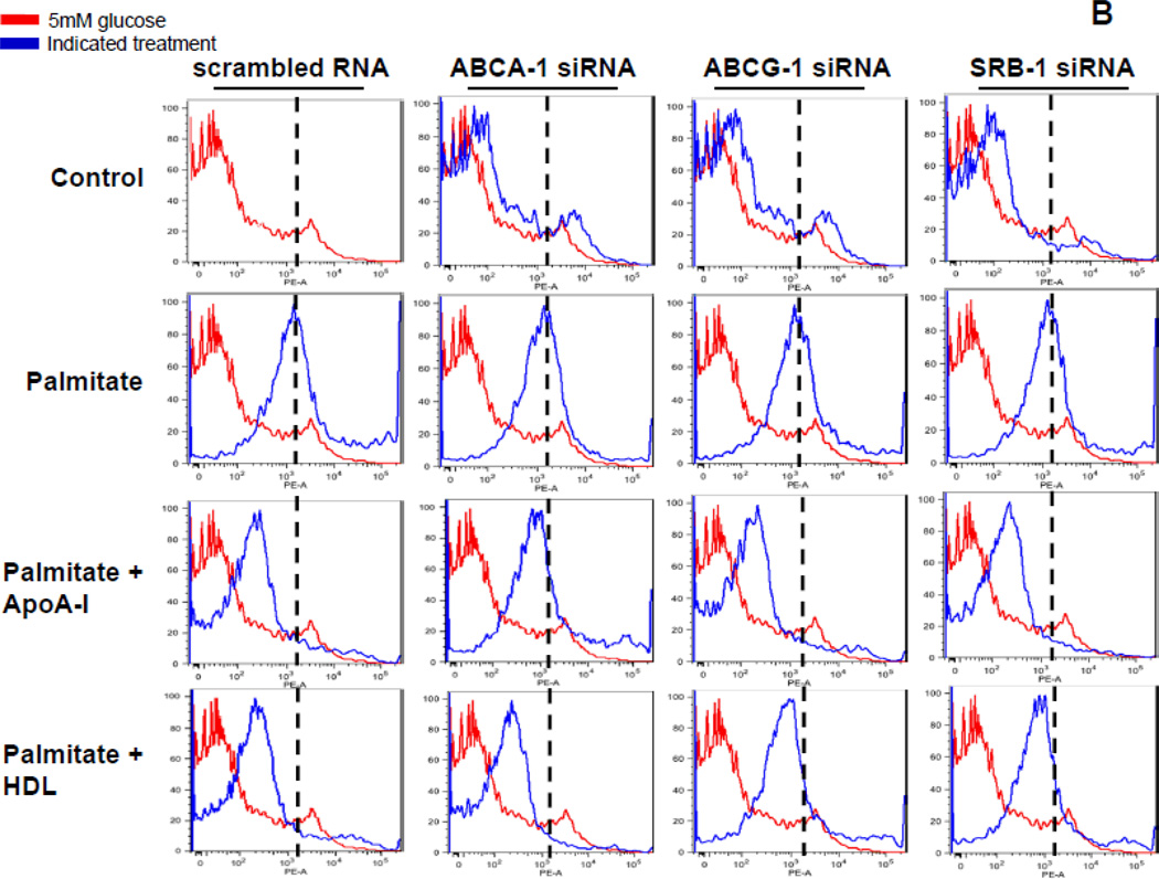
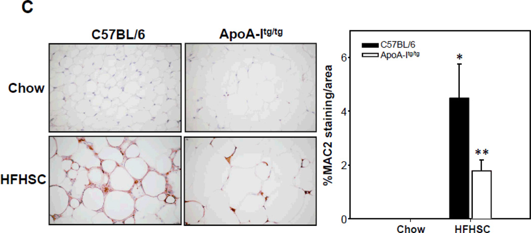
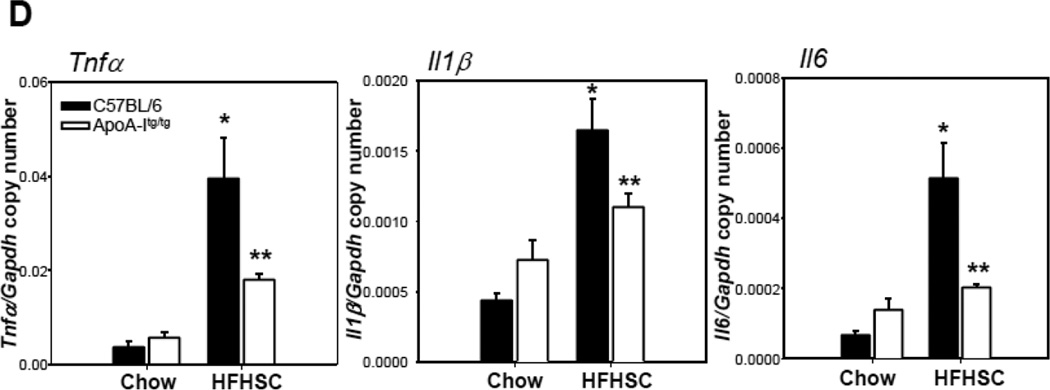
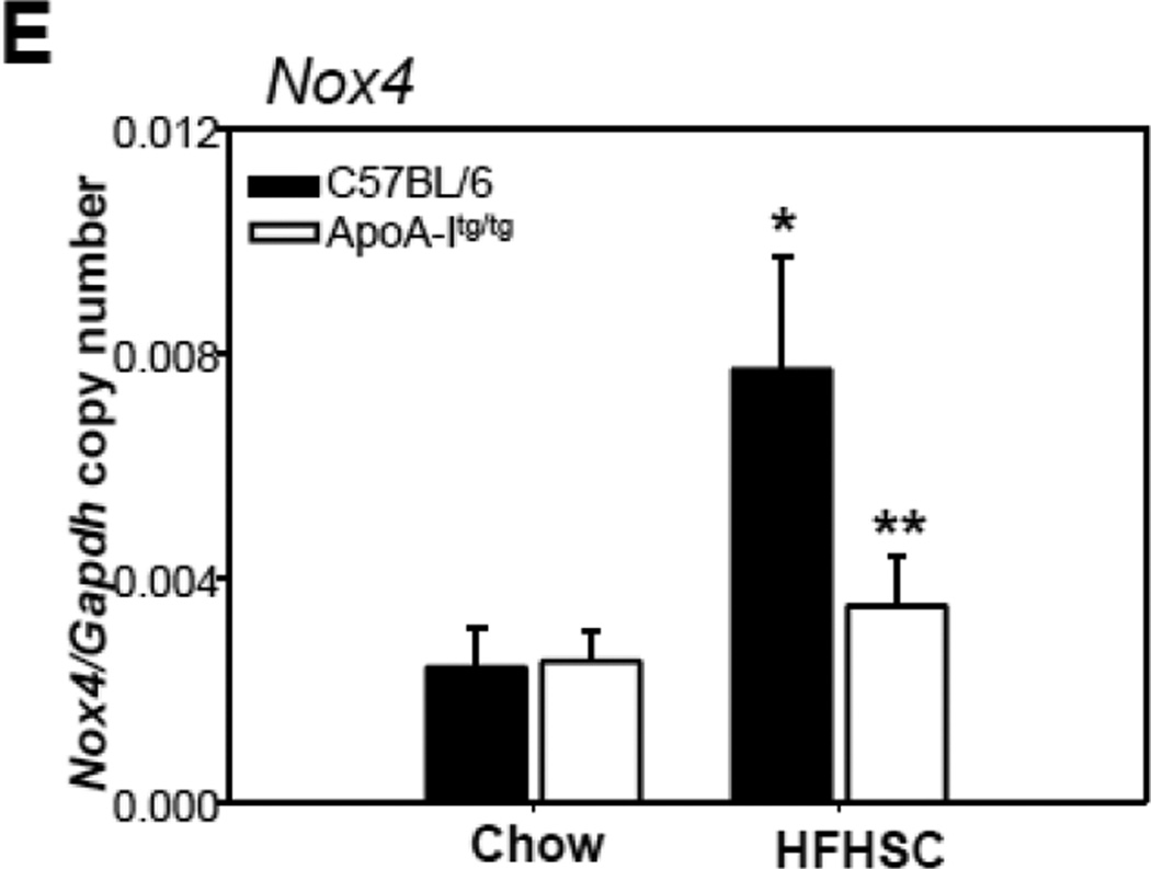
Methods and results: In 3T3L-1 adipocytes, apoAI, HDL, and methyl-β-cyclodextrin inhibited chemotactic factor expression. ApoAI and HDL also disrupted LRs, reduced plasma membrane cholesterol content, inhibited NADPH oxidase 4 translocation into LRs, and reduced palmitate-induced reactive oxygen species generation and monocyte chemotactic factor expression. Silencing ATP-binding cassette A-1 abrogated the effect of apoAI, but not HDL, whereas silencing ATP-binding cassette G-1 or scavenger receptor B-1 abrogated the effect of HDL but not apoAI. In vivo, apoAI transgenic mice fed a high-fat, high-sucrose, cholesterol-containing diet showed reduced chemotactic factor and proinflammatory cytokine expression and reduced macrophage accumulation in adipose tissue.

Conclusions: ApoAI and HDL have anti-inflammatory effects in adipocytes and adipose tissue similar to their effects in other cell types. These effects are consistent with disruption and removal of cholesterol from LRs, which are regulated by cholesterol transporters, such as ATP-binding cassette A-1, ATP-binding cassette G-1, and scavenger receptor B-1.

Keywords: ABC transporters; HDL; adipocytes; apolipoprotein AI; cholesterol.


Figures

Figure 1
Figure 1. MβCD, apoA-I and HDL inhibits chemotactic factor expression
3T3-L1 adipocytes were pre-exposed to MβCD (10µmol/ml, A and B), cholesterol loaded MβCD (A and B), apoA-I and/or HDL (at the indicated concentrations in µg protein/ml), C–F) for 6h. After that, adipocytes were incubated with or without 250 µmol/L palmitate for 24h. Saa3 and Mcp-1 gene expression was analyzed by multiplex real-time RT-PCR, normalized to GAPDH (A–F). *P < 0.001 vs. control media, **P < 0.001 vs. palmitate, #P < 0.001 vs. palmitate plus MβCD.
Figure 2
Figure 2. ApoA-I and HDL disrupt palmitate-induced LR formation and NOX4 translocation into LRs
3T3-L1 adipocytes were pre-exposed to apoA-I (50µg protein/ml) or HDL (50µg protein/ml) for 6h. After that, adipocytes were incubated with or without 250µmol/L palmitate for 24h. A. LRs were stained by Alexa Fluor 594 conjugated cholera toxin subunit β (CTB) and photographed by fluorescent microscopy (Nikon Eclipse 80i, original magnification ×400). B. LRs were isolated and fractionated by ultracentrifugation using a detergent-free fractionation method. Proteins from OptiPre-gradient fractions were immunoblotted with anti-NOX4 antibody and anti-caveolin-1 (CAV1) antibody (B). Fractions 6 to 8 contain LRs and fractions 1 to 4 are non-LR containing fractions.
Figure 3
Figure 3. The effects of apoA-I and HDL on palmitate-induced chemotactic factor gene expression are mediated by ABCA-1, ABCG-1 and SRB-1
3T3-L1 adipocytes were transfected with a siRNA specific for ABCA-1 (A and B), ABCG-1 (C and D), SRB-1 (E and F) or a scrambled siRNA (negative control) as indicated. 24h later the siRNA was removed and the cells were cultured for a further 3 days. After that, the cells were pre-exposed to apoA-I (50µg/ml) or HDL (50µg protein/ml) for 6h, and then incubated with or without added palmitate (250µmol/L) for 24h. Total RNA was isolated and analyzed by multiplex real-time RT-PCR using Saa3-specific (A, C and E) or Mcp-1-specific (B, D and F) primers and normalized to Gapdh. *P < 0.001 vs. control media, **P < 0.001 vs. palmitate, #P < 0.001 vs. palmitate plus HDL or apoA-I.
Figure 4
Figure 4. CTB-stained LRs are modulated by HDL and apoA-I via ABCA-1, ABCG-1 and SRB-1
3T3-L1 adipocytes were transfected with a siRNA specific for ABCA-1, ABCG-1, SRB-1 or a scrambled siRNA (negative control) as indicated. 24h later the siRNA was removed and the cells were cultured for a further 3 days. After that, the cells were pre-exposed to apoA-I (50µg/ml) or HDL (50µg protein/ml) for 6h, and then incubated with or without added palmitate (250 µmol/L) for 24h. A. LRs were stained by Alexa Fluor 594 conjugated cholera toxin subunit β (CTB) and photographed by fluorescent microscopy (Nikon Eclipse 80i, original magnification ×400). B. Cells in which LRs were stained by CTB were subjected to FACS analysis to quantify LR formation. Cells exposed to control media are shown in red and the peak value for cells exposed to 250µmol/L palmitate are indicated by the dashed lines. Cells exposed to the indicated treatments are shown in blue.
Figure 5
Figure 5. HDL and apoA-I block the palmitate-induced NOX4 translocation via ABCA-1, ABCG-1 and SRB-1
3T3-L1 adipocytes were transfected with a siRNA specific for ABCA-1, ABCG-1, SRB-1 or a scrambled siRNA (negative control) as indicated. 24h later siRNA was removed and the cells were cultured for a further 3 days. After that, the cells were pre-exposed to apoA-I (50µg/ml) or HDL (50µg protein/ml) for 6h, and then incubated with or without added palmitate (250µmol/L) for 24h. LRs were isolated and fractionated by ultracentrifugation using a detergent-free fractionation method. Proteins from OptiPre-gradient fractions were immunoblotted with anti-NOX4 and anti-caveolin-1 (CAV1) antibodis. PNS: the post-nuclear supernatant fraction.
Figure 6
Figure 6. ApoA-I and HDL affect palmitate-induced NOX4-derived ROS generation via ABCA-1, ABCG-1 and SRB-1
3T3-L1 adipocytes were transfected with a siRNA specific for ABCA-1, ABCG-1, SRB-1 or a scrambled siRNA (negative control) as indicated. 24h later the siRNA was removed and the cells were cultured for a further 3 days. After that, the cells were exposed to apoA-I (50µg/ml) or HDL (50µg protein/ml) for 6h, and then incubated with or without added palmitate (250µmol/L) for 24h. Cells were subjected to FACS analysis using CM-H2DCFDA. Results are plotted as counts (number of cells) on the vertical axis, versus DCF fluorescence intensity on the horizontal axis. Cells exposed to control media are shown in red and the peak value for cells exposed to 250µmol/L palmitate are indicated by the dashed lines. Cells exposed to the indicated treatments are shown in blue.
Figure 7
Figure 7. Overexpression of human apoA-I inhibits chemotactic factor expression and macrophage accumulation in adipose tissue of mice
ApoA-Itg/tg and C57BL/6 control mice were fed chow or a high-fat, high-sucrose, cholesterol-containing (HFHSC) diet for 24 weeks (A–E; n=9). Epididymal fat was isolated and analyzed by real-time RT-PCR using Saa3, Mcp-1 (A), Mac2, F4/80 (B), Tnfα, Il1β, Il6 (D) and Nox4 (E)-specific primers and probes and normalized to Gapdh. Epididymal fat was isolated and analyzed by immunohistochemistry using a Mac-2 antibody (C), which detects murine macrophages. Tissues were photographed using microscopy (original magnification ×60. *P < 0.005 vs. chow, **P < 0.005 vs. C57BL/6 or LDLR-/- in HFHSC.

Comment in

References

    1. Murdolo G, Smith U. The dysregulated adipose tissue: A connecting link between insulin resistance, type 2 diabetes mellitus and atherosclerosis. Nutr Metab Cardiovasc Dis. 2006;16(Suppl 1):S35–S38. - PubMed
    1. Mathieu P, Lemieux I, Despres JP. Obesity, inflammation, and cardiovascular risk. Clinical pharmacology and therapeutics. 2010;87:407–416. - PubMed
    1. Xu H, Barnes GT, Yang Q, Tan G, Yang D, Chou CJ, Sole J, Nichols A, Ross JS, Tartaglia LA, Chen H. Chronic inflammation in fat plays a crucial role in the development of obesity-related insulin resistance. J Clin Invest. 2003;112:1821–1830. - PMC - PubMed
    1. Weisberg SP, McCann D, Desai M, Rosenbaum M, Leibel RL, Ferrante AW., Jr Obesity is associated with macrophage accumulation in adipose tissue. J Clin Invest. 2003;112:1796–1808. - PMC - PubMed
    1. Strissel KJ, DeFuria J, Shaul ME, Bennett G, Greenberg AS, Obin MS. T-cell recruitment and th1 polarization in adipose tissue during diet-induced obesity in c57bl/6 mice. Obesity (Silver Spring) 2010;18:1918–1925. - PMC - PubMed

Publication types

MeSH terms