. 2013 Apr;19(5):891-903.
doi: 10.1097/MIB.0b013e3182813297.

Matrix stiffness corresponding to strictured bowel induces a fibrogenic response in human colonic fibroblasts

Affiliations

Matrix stiffness corresponding to strictured bowel induces a fibrogenic response in human colonic fibroblasts

Laura A Johnson et al. Inflamm Bowel Dis. 2013 Apr.

Abstract

Background: Crohn's disease is characterized by repeated cycles of inflammation and mucosal healing which ultimately progress to intestinal fibrosis. This inexorable progression toward fibrosis suggests that fibrosis becomes inflammation-independent and auto-propagative. We hypothesized that matrix stiffness regulates this auto-propagation of intestinal fibrosis.

Methods: The stiffness of fresh ex vivo samples from normal human small intestine, Crohn's disease strictures, and the unaffected margin were measured with a microelastometer. Normal human colonic fibroblasts were cultured on physiologically normal or pathologically stiff matrices corresponding to the physiological stiffness of normal or fibrotic bowel. Cellular response was assayed for changes in cell morphology, α-smooth muscle actin staining, and gene expression.

Results: Microelastometer measurements revealed a significant increase in colonic tissue stiffness between normal human colon and Crohn's strictures and between the stricture and adjacent tissue margin. In Ccd-18co cells grown on stiff matrices corresponding to Crohn's strictures, cellular proliferation increased. Pathologic stiffness induced a marked change in cell morphology and increased α-smooth muscle actin protein expression. Growth on a stiff matrix induced fibrogenic gene expression, decreased matrix metalloproteinase, and proinflammatory gene expression and was associated with nuclear localization of the transcriptional cofactor MRTF-A.

Conclusions: Matrix stiffness, representative of the pathologic stiffness of Crohn's strictures, activates human colonic fibroblasts to a fibrogenic phenotype. Matrix stiffness affects multiple pathways, suggesting that the mechanical properties of the cellular environment are critical to fibroblast function and may contribute to auto-propagation of intestinal fibrosis in the absence of inflammation, thereby contributing to the intractable intestinal fibrosis characteristic of Crohn's disease.


Figures

Figure 1
Figure 1. Mechanical characteristics of polyacrylamide stiffness gels and human ex vivo tissue samples from Crohn’s strictures, the unaffected margin, or normal human intestine
(A) Stress-strain curves of collagen-coated polyacrylamide gels corresponding to low (3 kPa) and high (28 kPa) elastic moduli. (B) Mathematical formula for the Young’s Modulus calculation where E = Young’s Modulus, F = the force applied to the material, A0 = the cross-section area through which the force was applied to the material, ΔL = amount the length of the material changes when the force is applied, L0 = original length of the material (before the force was applied). (C) Representative stress-strain curves of ex vivo normal bowel (NL, dashed line) compared to a Crohn’s ex vivo tissue section of stricture (CD-S, solid line) or the margin adjacent to the stricture (CD-un, dotted line). The stricture and margin are from the same resected specimen. Normalized strain was calculated from the linear range between the first and second inflection points on the stress-strain graph (50–60% of the strain value) as indicated by the red line. (D) Normalized strain values of ex vivo small intestine demonstrating tissue elastic moduli of normal (NL, n = 5) compared to Crohn’s stricture (CD-S, n =11) and the unaffected margin (CD-un, n =11). Representative photos of the ex vivo tissue samples are shown below the graph. The horizontal line in the box plot represents the median, the upper and lower box boundaries represent the interquartile range, and the ends of the whiskers represent the 5% and 95% confidence limits. Extreme values (> 2 standard deviations) are represented as individual dots. Horizontal bars denote statistical comparisons between normal tissue and CD-S or comparisons between CD-S and CD-un. Representative photos of normal colon compared to strictured colon are shown below the graph. (**p<0.01, *** p<0.001 compared to normal bowel)
Figure 2
Figure 2. Expression of extracellular matrix and matrix remodeling genes are increased in Crohn’s strictures compared to the unaffected margin or normal human intestine
(A–E) QRT-PCR gene expression of normal bowel (NL), Crohn’s stricture (CD-S) or the margin adjacent to the stricture (CD-un) from FFPE samples of the same patients. (A) Col1A1 (B) MMP-3, (C) MYLK (D) IL-1β (E) PTGS2. Statistical comparisons between the groups are denoted with an asterisk. Horizontal bars denote statistical comparisons between normal tissue and CD-S or comparisons between CD-S and CD-un. (* p <0.05, *** p < 0.001 compared to normal bowel)
Figure 3
Figure 3. Increasing matrix stiffness alters fibroblast morphology and actin stress fiber accumulation
Ccd-18co colonic fibroblasts were cultured on collagen-coated acrylamide gels of elastic moduli corresponding to normal (2.6–4.3 kPa) or strictured (16–28 kPa) bowel. (Left panel) Phase contrast micrographs (100× magnification) of Ccd-18co cells cultured on increasing matrix stiffness after 24 hr in culture, demonstrating morphological transition from rounded to activated, stellate morphology. Cells were plated at equal densities. (Middle panel) Confocal micrographs (600× magnification) of Ccd-18co cells plated on increasing matrix stiffness. Increased matrix stiffness induces accumulation of actin stress fibers (green) and focal adhesions (vinculin staining, red). Nuclei were visualized with DAPI (blue). Yellow scale bar represents 100 µm. Insets which are shown in detail (right panel) are indicated by yellow boxes. (Right panel) Insets from the confocal images, detailing focal adhesion staining. All inserts are the same magnification.
Figure 4
Figure 4. Increased matrix stiffness increases cell number and induces fibroblast proliferation
(A) Ccd-18co cell density increases in response to increased matrix stiffness. Cell density was determined from three 100x-magnifcation fields per matrix stiffness and graphed. Individual points represent biologically independent experiments while the horizontal represents the mean for each matrix stiffness. The horizontal bar denotes statistical comparisons between the transition point between soft (4.3 kPa) and stiff (16 kPa). (B) MTT cell proliferation assay of Ccd-18co cells cultured on low (2.3 and 4.6 kPa) or high (16 kPa and 28 kPa) matrix stiffness. Results are from 3 independent experiments with multiple replicates per experiment. (*p<0.05, **p<0.01, *** p<0.001 compared to the 2.6 kPa elastic modulus)
Figure 5
Figure 5. Increasing matrix stiffness induces myosin light chain kinase (MYLK) and represses matrix turnover gene expression
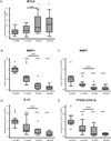
Increasing matrix elastic moduli from 2.6 to 28 kPa induces MYLK gene expression (A), represses matrixmetalloproteinase expression (MMP3 (B)), MMP1(C)), and represses inflammatory gene expression (IL-1β (D) and PTGS2 (E)) in Ccd-18co cells. Horizontal bars denote statistical comparisons between the transition point between soft (4.3 kPa) and stiff (16 kPa). (*** p<0.001 compared to the 4.3 kPa elastic modulus)
Figure 6
Figure 6. MMP-3 proteolytic activity is repressed by high matrix stiffness
(A) Representative zymographs of MMP-3 caseinolytic activity in culture supernatants from Ccd-18co cells cultured for 24 or 72 hours on soft (4.3 kPa) or stiff (28 kPa) matrices. (B) Densitrometric analysis from 3 independent experiments demonstrating MMP-3 proteolytic activity decreases over time in cells cultured on the stiff matrix. (*** p<0.001)
Figure 7
Figure 7. Fibroblast activation by increased matrix stiffness is associated with increased αSMA protein expression
(A) A representative Western illustrating αSMA protein expression of Ccd-18co colonic fibroblasts cultured on soft or stiff matrices for 24 hr. GAPDH is shown as protein loading control. (B) Densitrometric analysis of αSMA protein expression of Ccd-18co fibroblasts in response to increased matrix stiffness. Results are from 3 independent experiments with a total of 5 biological replicates. (* p<0.05)
Figure 8
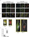
Figure 8. Increased matrix stiffness induces MKL1 (MRTF-A) transcription and promotes MRTF-A nuclear translocation
(A) Immunostaining for MRTF-A localization (red) and actin-stress fibers (green) in Ccd-18co cells cultured for 1, 4, and 24 hours on soft (4.3 kPa, left panel) or stiff (28 kPa, right panel) matrices. Nuclei are identified by DAPI staining (blue). The white scale bar represents 100 µm. Representative images are shown. (600× magnification). (B) Insets of individual cells cultured for 24 hours (identified by yellow boxes in (A)) demonstrating MRTF-A localization relative to the nucleus and cell membrane (outlined in white). The merged MRTF-A/actin/DAPI image is shown in comparison to the MRTF-A immunostaining. Insets are the same magnification. (C) MKL1 (MRTF-A) gene expression in Ccd-18co cells cultured on soft (4.3 kPa) or stiff (28 kPa) matrices. (*** p<0.001)

References

    1. Rieder F, Brenmoehl J, Leeb S, et al. Wound healing and fibrosis in intestinal disease. Gut. 2007;56:130–139. - PMC - PubMed
    1. Andres PG, Friedman LS. Epidemiology and the natural course of inflammatory bowel disease. Gastroenterol Clin North Am. 1999;28:255–281. vii. - PubMed
    1. Sands BE, Arsenault JE, Rosen MJ, et al. Risk of early surgery for Crohn's disease: implications for early treatment strategies. Am J Gastroenterol. 2003;98:2712–2718. - PubMed
    1. Louis E, Collard A, Oger AF, et al. Behaviour of Crohn's disease according to the Vienna classification: changing pattern over the course of the disease. Gut. 2001;49:777–782. - PMC - PubMed
    1. Johnson LA, Luke A, Sauder K, et al. Intestinal fibrosis is reduced by early elimination of inflammation in a mouse model of IBD: Impact of a "Top-Down" approach to intestinal fibrosis in mice. Inflamm Bowel Dis. 2011 - PMC - PubMed

Publication types

MeSH terms