. 2013 Mar 14:5:9.
doi: 10.3389/fnagi.2013.00009. eCollection 2013.

Copper metabolism of astrocytes

Affiliations

Copper metabolism of astrocytes

Ralf Dringen et al. Front Aging Neurosci. .

Abstract

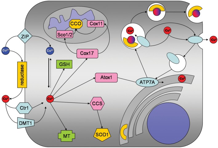
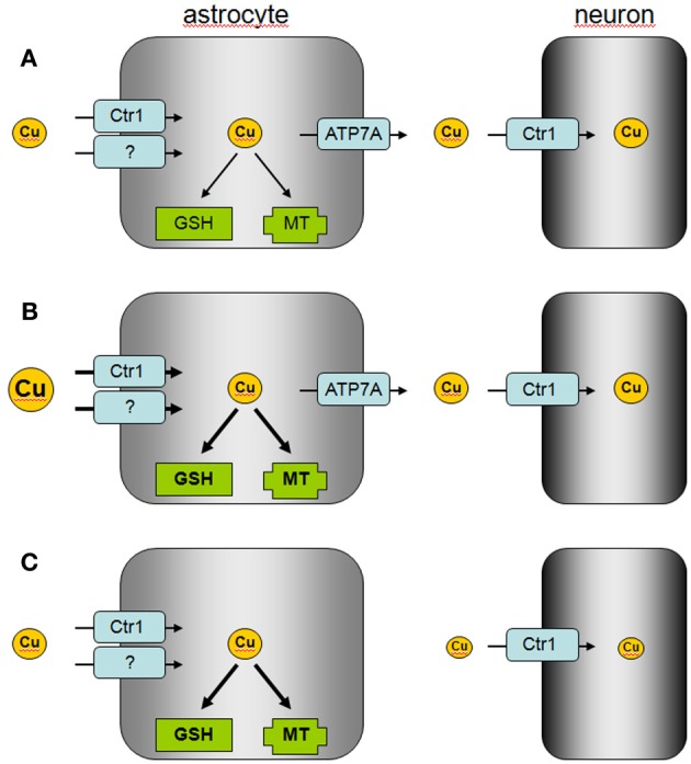
This short review will summarize the current knowledge on the uptake, storage, and export of copper ions by astrocytes and will address the potential roles of astrocytes in copper homeostasis in the normal and diseased brain. Astrocytes in culture efficiently accumulate copper by processes that include both the copper transporter Ctr1 and Ctr1-independent mechanisms. Exposure of astrocytes to copper induces an increase in cellular glutathione (GSH) content as well as synthesis of metallothioneins, suggesting that excess of copper is stored as complex with GSH and in metallothioneins. Furthermore, exposure of astrocytes to copper accelerates the release of GSH and glycolytically generated lactate. Astrocytes are able to export copper and express the Menkes protein ATP7A. This protein undergoes reversible, copper-dependent trafficking between the trans-Golgi network and vesicular structures. The ability of astrocytes to efficiently take up, store and export copper suggests that astrocytes play a key role in the supply of neurons with copper and that astrocytes should be considered as target for therapeutic interventions that aim to correct disturbances in brain copper homeostasis.

Keywords: ATP7A; Ctr1; astroglia; copper export; metallothioneins; oxidative stress; toxicity; transport.


Figures

Figure 1
Figure 1
Copper metabolism of astrocytes. Copper is taken up into astrocytes by the copper transporter receptor 1 (Ctr1) and also by Ctr1-independent mechanisms which may include the divalent metal transporter 1 (DMT1) or members of the ZIP family of metal transporters. An ecto-cuprireductase and/or extracellular ascorbate will provide the reduced copper species for astrocytic uptake by Ctr1 or DMT1. Accumulated copper is stored in astrocytes as complex with GSH or in metallothioneins (MT). In addition, copper is shuttled to its specific cellular target, by the copper chaperones CCS to superoxide dismutase 1 (SOD1), by Cox17 to Sco1/2 and Cox11 for subsequent incorporation into cytochrome c oxidase and by antioxidant protein 1 (Atox1) to ATP7A. ATP7A transports copper into the trans-Golgi network for subsequent incorporation into copper-dependent enzymes. When the cellular copper level rises above a certain threshold, ATP7A translocates reversibly via vesicles to the plasma membrane to export copper.
Figure 2
Figure 2
Proposed model of astrocytic copper supply to neurons.(A) In the normal brain, copper is efficiently taken up by astrocytes via Ctr1 and other transporters which are so far not identified. Excess of copper is stored by astrocytes in MT or as GSH complex and copper is released via ATP7A to supply neurons with copper. (B) In copper overload conditions, excess of copper is efficiently taken up into astrocytes and stored in MT or as GSH complex to prevent copper-induced neurotoxicity. (C) In Menkes disease, astrocytes accumulate and store copper, but the mutated ATP7A is unable to mediate astrocytic copper export and copper supply to neurons.

References

    1. Arredondo M., Munoz P., Mura C. V., Nunez M. T. (2003). DMT1, a physiologically relevant apical Cu1+ transporter of intestinal cells. Am. J. Physiol. Cell Physiol. 284, C1525–C1530 10.1152/ajpcell.00480.2002 - DOI - PubMed
    1. Barnes N., Tsivkovskii R., Tsivkovskaia N., Lutsenko S. (2005). The copper-transporting ATPases, Menkes and Wilson disease proteins, have distinct roles in adult and developing cerebellum. J. Biol. Chem. 280, 9640–9645 10.1074/jbc.M413840200 - DOI - PubMed
    1. Bolaños J. P., Heales S. J. (2010). Persistent mitochondrial damage by nitric oxide and its derivatives: neuropathological implications. Front. Neuroenergetics 2:1 10.3389/neuro.14.001.2010 - DOI - PMC - PubMed
    1. Brown D. R. (2004). Role of the prion protein in copper turnover in astrocytes. Neurobiol. Dis. 15, 534–543 10.1016/j.nbd.2003.11.009 - DOI - PubMed
    1. Chen S. H., Lin J. K., Liu S. H., Liang Y. C., Lin-Shiau S. Y. (2008). Apoptosis of cultured astrocytes induced by the copper and neocuproine complex through oxidative stress and JNK activation. Toxicol. Sci. 102, 138–149 10.1093/toxsci/kfm292 - DOI - PubMed