

doi: 10.1038/aja.2013.7.
Epub 2013 Mar 18.
Neuroendocrine differentiation of prostate cancer
Affiliations
- PMID: 23503426
- PMCID: PMC3739654
- DOI: 10.1038/aja.2013.7
Item in Clipboard
Neuroendocrine differentiation of prostate cancer
Asian J Androl.
2013 May.
No abstract available
Figures

Neuroendcorine cells in benign prostate and prostatic adenocarcinoma. Left panel shows scattered neuroendocrine cells (brown staining) in benign prostate as highlighted by IHC staining with an anti-chromogranin A antibody. Middle panel shows scattered neuroendocrine cells in a case of prostatic adenocarcinoma that is hormone naive. Right panel shows abundant neuroendocrine cells in a case of castration resistant adenocarcinoma. IHC, immunohistochemistry. Scale bar=20 μm.

A model of the function of cellular heterogeneity in prostate cancer. The left panel shows a treatment naive tumor in which the majority of the tumor cells are luminal type tumor cells (yellow) with few neuroendocrine cells (pink). The middle panel shows that hormonal therapy induces apoptosis of the luminal type tumor cells and an increase in the neuroendocrine cells. The right panel shows that in the castration resistant stage, there is a marked increase in neuroendocrine cells which establish a paracrine network and stimulate proliferation of the luminal type cancer cells in an androgen-deprived environment.

SCNC of the prostate. Upper left panel shows a case of mixed tumor with components of adenocarcinoma (long arrow) and SCNC. Upper right panel shows a high power view of SCNC with tumor cells demonstrating high-grade neuroendocrine features. Lower left panel shows IHC staining of SCNC with an anti-chromogranin A antibody. Lower right panel shows IHC staining with an anti-P52 antibody. IHC, immunohistochemistry; SCNC, small cell neuroendocrine carcinoma. Scale bar=20 μm.
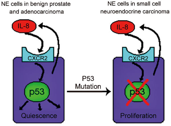
A model of the molecular pathway regulating the proliferation of neuroendocrine cells. The left panel shows a neuroendocrine cell in benign prostate and prostatic adenocarcinoma. Autocrine stimulation of CXCR2 by IL-8 activates P53 and inhibits cell proliferation. On the right is a NE tumor cell in prostatic small cell carcinoma. A p53 mutation inactivates the IL-8/CXCR2/p53 pathway and removes a major growth inhibitory signal, leading to rapid proliferation of the NE cell.
References
-
- Yuan TC, Veeramani S, Lin MF. Neuroendocrine-like prostate cancer cells: neuroendocrine transdifferentiation of prostate adenocarcinoma cells. Endocr Relat Cancer. 2007;14:531–47. - PubMed
-
- Vashchenko N, Abrahamsson PA. Neuroendocrine differentiation in prostate cancer: implications for new treatment modalities. Eur Urol. 2005;47:147–55. - PubMed
-
- Huang J, Yao JL, di Sant'agnese PA, Yang Q, Bourne PA, et al. Immunohistochemical characterization of neuroendocrine cells in prostate cancer. Prostate. 2006;66:1399–406. - PubMed
-
- Huss WJ, Gregory CW, Smith GJ. Neuroendocrine cell differentiation in the CWR22 human prostate cancer xenograft: association with tumor cell proliferation prior to recurrence. Prostate. 2004;60:91–7. - PubMed
MeSH terms
Substances
LinkOut - more resources
Full Text Sources
Other Literature Sources
Medical
