. 2013 Jul 3;32(13):1842-54.
doi: 10.1038/emboj.2013.63. Epub 2013 Mar 15.

The exoribonuclease Dis3L2 defines a novel eukaryotic RNA degradation pathway

Affiliations

The exoribonuclease Dis3L2 defines a novel eukaryotic RNA degradation pathway

Michal Malecki et al. EMBO J. .

Abstract

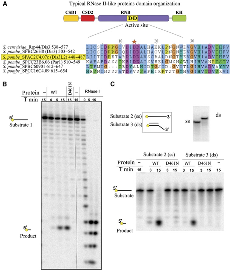
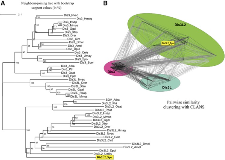
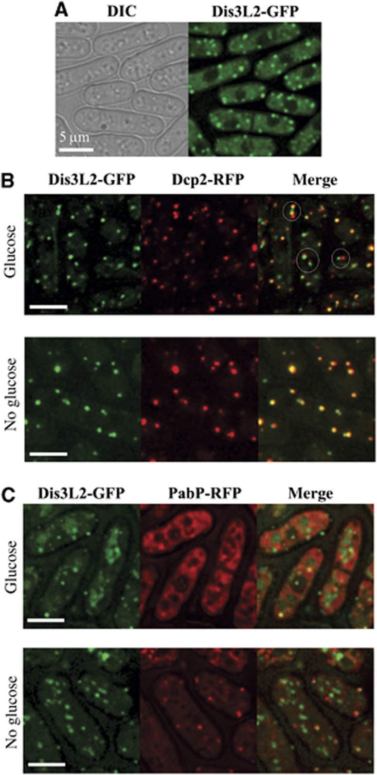
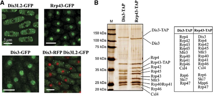
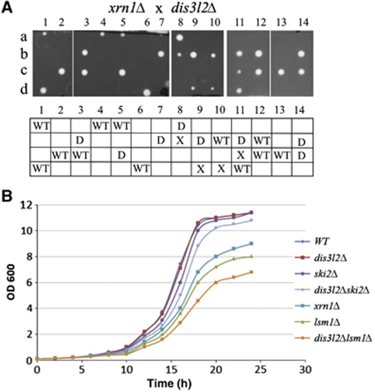
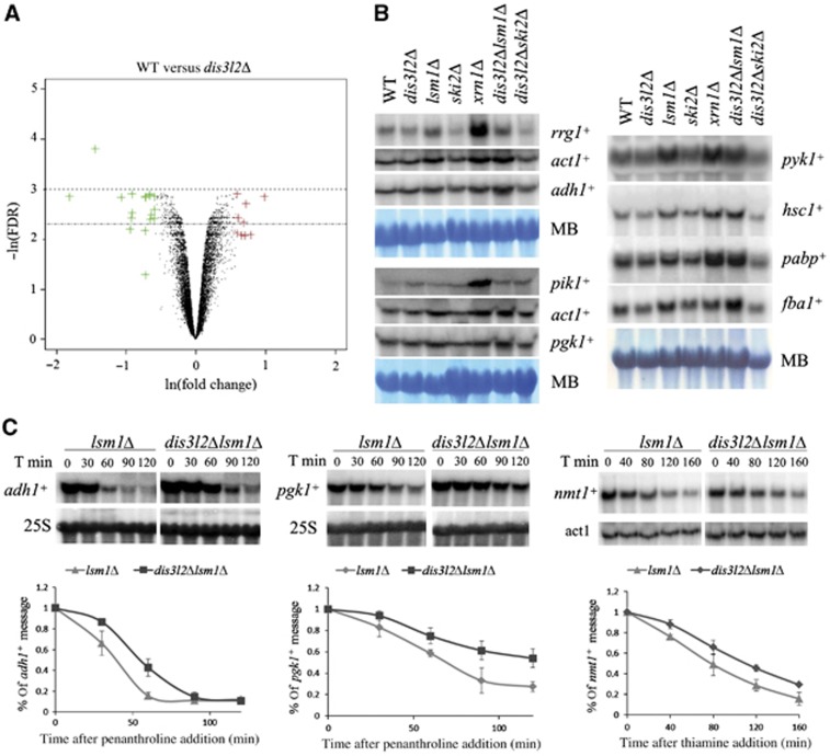
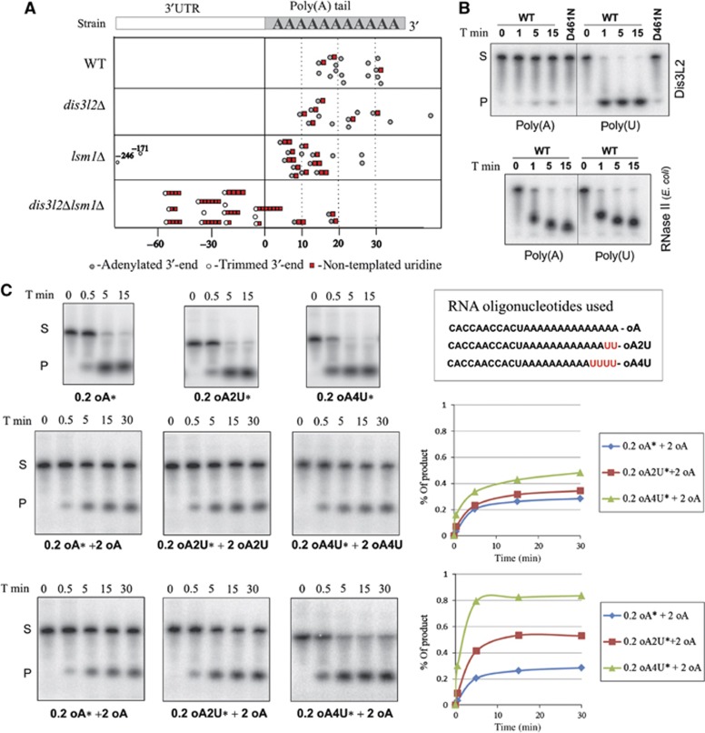
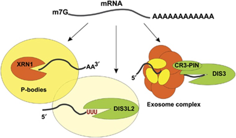
The final step of cytoplasmic mRNA degradation proceeds in either a 5'-3' direction catalysed by Xrn1 or in a 3'-5' direction catalysed by the exosome. Dis3/Rrp44, an RNase II family protein, is the catalytic subunit of the exosome. In humans, there are three paralogues of this enzyme: DIS3, DIS3L, and DIS3L2. In this work, we identified a novel Schizosaccharomyces pombe exonuclease belonging to the conserved family of human DIS3L2 and plant SOV. Dis3L2 does not interact with the exosome components and localizes in the cytoplasm and in cytoplasmic foci, which are docked to P-bodies. Deletion of dis3l2(+) is synthetically lethal with xrn1Δ, while deletion of dis3l2(+) in an lsm1Δ background results in the accumulation of transcripts and slower mRNA degradation rates. Accumulated transcripts show enhanced uridylation and in vitro Dis3L2 displays a preference for uridylated substrates. Altogether, our results suggest that in S. pombe, and possibly in most other eukaryotes, Dis3L2 is an important factor in mRNA degradation. Therefore, this novel 3'-5' RNA decay pathway represents an alternative to degradation by Xrn1 and the exosome.


Conflict of interest statement

The authors declare that they have no conflict of interest.

Figures

Figure 1
Figure 1
Fission yeast gene SPAC2C4.07c encodes an active RNase II family exonuclease with no homologue in S. cerevisiae. (A) S. pombe genome encodes five proteins with RNB domain. Sequence alignment depicts exonucleolytic active site of RNB domain of fission yeast proteins, compared with S. cerevisiae Rrp44/Dis3 sequence. Conserved aspartic acid in the central part of the active site is marked with the star. (B) Product of SPAC2C4.07c gene is an active exonuclease in vitro. Around 0.5 pmol of the wild-type protein product (WT) and the mutated version (D461N) were incubated with 2 pmol of the 20-nt 5′-radioactively labelled RNA substrate 1 (see below). After indicated times (T min), reactions were stopped and products separated on a denaturing polyacrylamide gel. The same substrate was incubated with 0.1 U of RNase ONE (Promega). Migration of the reaction substrates and products is indicated. (C) Product of SPAC2C4.07c gene is able to digest RNA in the context of secondary structures. Around 0.5 pmol of WT and mutated version (D461N) was incubated with 0.2 pmol of single-stranded substrate 2 (ss) or substrate 3 (ds), which is substrate 2 annealed with DNA oligonucleotide (see below). After the indicated times, reactions were stopped and products were separated on a denaturing polyacrylamide gel. Upper panel depicts schematic representation of the RNA substrates used for the reaction (left) and the control of the annealing of the double-stranded substrate (right) on a native polyacrylamide gel. RNA substrate 1: (GUUUUGUAUAGAAAUCAAUG); RNA substrate 2: (CCCGACACCAACCACUAAAAAAAAAAAAAA); substrate 3 (hybrid DNA/RNA): (DNA oligonucleotide: AGTGGTTGGTGTCGGG/RNA oligonucleotide: substrate 2).
Figure 2
Figure 2
Phylogenetic comparison of different Dis3 homologues in eukaryotes reveals that Dis3-like proteins can be divided into three distinct groups, corresponding to Dis3, Dis3L, and Dis3L2. Dis3 and Dis3L2 are found almost universally in Metazoa, plants, and Fungi, while proteins from the Dis3L group are found only in vertebrates and in a single invertebrate species (Nematostella vectensis). (A) A neighbour joining tree (BioNJ) of 43 eukaryotic Dis3-like proteins. Numbers correspond to percentage bootstrap support (1000 replicates) for nodes. Three main groups, corresponding to Dis3, Dis3L, and Dis3L2 proteins have 100% support, and the SPAC2C4.07c gene product of S. pombe belongs to the Dis3L2 group. (B) Clustering of the Dis3-like proteins (same set as in A) on the basis of pairwise BLAST similarity using CLANS also reveals three distinct groups of Dis3, Dis3L, and Dis3L2 sequences, with the Dis3 group forming the tightest cluster. Again, the SPAC2C4.07c gene product of S. pombe belongs to the Dis3L2 group.
Figure 3
Figure 3
Dis3L2 co-localizes with P-bodies. (A) Dis3L2 localizes in the cytoplasm and cytoplasmic foci. Cells expressing Dis3L2-GFP were grown to mid-log phase in minimal medium (EMM) and the localization of epitope-tagged protein was determined by fluorescence microscopy. (B, C) Dis3L2-GFP was examined for co-localization with Dcp2-RFP (B) or PabP-RFP (C). Cells expressing Dis3L2-GFP and Dcp2-RFP were grown to mid-log phase in minimal medium (EMM) and either immediately observed in the microscope (glucose), or deprived for glucose for 10 min and subsequently examined (no glucose) (B). Similar experiment was performed for the comparison of cells expressing Dis3L2-GFP and PabP-RFP (C) except that fission yeast was grown on full media (YES) (see Results). Examples of Dis3L2 aggregates docked to P-bodies in (B) were marked with circles. Scale bars represent 5 μm.
Figure 4
Figure 4
Dis3L2 fails to interact with the exosome complex. (A) Dis3L2 does not co-localize with the exosome complex components. Cells expressing Dis3L2-GFP, Rrp43-GFP, Dis3-GFP or both Dis3-RFP and Dis3L2-GFP were grown in the minimal media (EMM) to mid-log phase and protein localization determined by fluorescence microscopy. Scale bars represent 5 μm. (B) Dis3L2 does not co-purify with the exosome complex components. Cells expressing exosome complex components (Dis3 or Rrp43) fused with TAP tag sequence were grown to mid-log phase in rich media, subsequently tagged proteins were purified according to standard protocol (Rigaut et al, 1999). Part of each elution from the calmodulin resin was separated on SDS–PAGE gel and silver stained (gel on the left), the remaining part was subjected to mass spectrometry analysis. Table presented on the right lists all known exosome subunits and interacting proteins identified in the elutions from Dis3-TAP and Rrp43-TAP.
Figure 5
Figure 5
Genetic interactions between dis3l2+ and the components of cytoplasmic RNA degradation pathway. (A) dis3l2+ deletion is synthetically lethal with deletion of xrn1+. Haploid dis3l2+::kan cells were crossed with xrn1+::hph strain. Resulting diploids were sporulated and tetrads were dissected on YES plates. In the bottom table, the genotypes of the germinated spores are described: WT—wild-type strain, X—xrn1Δ, D—dis3l2Δ. Genotypes of the spores were analysed by their ability to grow on the selective media and by colony PCR. (B) dis3l2+ deletion enhances the growth defect of lsm1Δ strain. Overnight yeast cultures were diluted in the fresh media (EMM) to OD600 of 0.1, subsequently grown at 32°C and the OD monitored over time.
Figure 6
Figure 6
Dis3L2 is involved in mRNA degradation. (A) Volcano plot representation of microarray data. S. pombe mRNA fold change values (ln(fold change)) between wild-type and single dis3l2Δ deletion strains are plotted against the result significance (−ln(FDR)). Genes upregulated in dis3l2Δ >1.5 times are coloured in red and the ones downregulated >1.5 times coloured in green. The chosen significance window is indicated by dotted lines. (B) Northern blots of total RNA obtained from the wild-type and different deletion mutant strains. Loading was controlled by methylene blue (MB) staining. (C) Deletion of dis3l2+ in lsm1Δ strain background results in slower transcript degradation rates. lsm1Δ and dis3l2Δlsm1Δ yeast strains were grown in full media (adh1+ and pgk1+ messengers) or minimal media (nmt1+ messenger) until mid-log phase, and transcription was subsequently stopped by either 1,10-penanthroline (adh1+ and pgk1+) or thiamine (nmt1+) addition. Cells were harvested at the indicated time points after transcription blockage, total RNA was isolated and message decay investigated by Northern blots analysis. Upper part shows an example of Northern blot results, lower part graphs illustrate the comparison of the decay rates of the indicated transcripts in lsm1Δ and dis3l2Δlsm1Δ yeast strains. Data were collected in three independent experiments, error bars represent standard deviation. Source data for this figure is available on the online supplementary information page.
Figure 7
Figure 7
Dis3L2 preferentially degrades uridylated RNAs. (A) Deletion of dis3l2+ in Δlsm1 background results in the accumulation of trimmed and uridylated adh1+ transcripts. Each circle point in the table indicates the position of one adh1+ mRNA 3′-end. Point zero represents the standard polyadenylation site of adh1+ mRNA. Positive values correspond to the length of poly(A) tail attached to the polyadenylation site, and the negative values correspond to the 3′-end position in 3′-UTR if mRNA was trimmed. Uridylation is indicated by the red squares, and number of squares corresponds to the number of non-templated U residues detected at the 3′-end. (B) Dis3L2 exhibits stronger preference for poly(U) compared to poly(A) RNA substrate in vitro. Dis3L2 protein (WT) and mutated version (D461N) were incubated with the same amounts of radioactively labelled poly(A) or poly(U) RNAs. Reaction was stopped at the indicated time points and products were separated on denaturing polyacrylamide gels. The same substrates were incubated with bacterial RNase II. (C) Uridine residues added to the 3′-end can effectively target RNA substrates for degradation by Dis3L2 in vitro. Same amounts of Dis3L2 protein were incubated with the indicated amounts of different RNA substrates (0.2 or 2 pmol). Substrate sequence is indicated. Reactions with radioactive substrates (labelled with *) were supplemented with the substrates that were not radioactively labelled. Reactions were stopped at the indicated time points (T min) and products were separated on denaturing polyacrylamide gels. The graphs (on the right side) depict the accumulation of the product at different time points as calculated using Image Quant. Source data for this figure is available on the online supplementary information page.
Figure 8
Figure 8
Three pathways of mRNA degradation in the cytoplasm. In S. pombe and possibly in most other eukaryotes, mRNAs can be degraded in the 5′-3′ direction by Xrn1 exonuclease, or in the 3′-5′ direction by either the exosome complex or Dis3L2 exonuclease, which acts independently from the exosome. Our results suggest that in S. pombe Dis3L2 degradation pathway is connected with the P-bodies.

Comment in

References

    1. Alexander RD, Barrass JD, Dichtl B, Kos M, Obtulowicz T, Robert MC, Koper M, Karkusiewicz I, Mariconti L, Tollervey D, Kufel J, Bertrand E, Beggs JD (2010) RiboSys, a high-resolution, quantitative approach to measure the in vivo kinetics of pre-mRNA splicing and 3′-end processing in Saccharomyces cerevisiae. RNA 16: 2570–2580 - PMC - PubMed
    1. Amblar M, Arraiano CM (2005) A single mutation in Escherichia coli ribonuclease II inactivates the enzyme without affecting RNA binding. FEBS J 272: 363–374 - PubMed
    1. Anderson JS, Parker RP (1998) The 3′ to 5′ degradation of yeast mRNAs is a general mechanism for mRNA turnover that requires the SKI2 DEVH box protein and 3' to 5' exonucleases of the exosome complex. EMBO J 17: 1497–1506 - PMC - PubMed
    1. Anderson P, Kedersha N (2008) Stress granules: the Tao of RNA triage. Trends Biochem Sci 33: 141–150 - PubMed
    1. Astuti D, Morris MR, Cooper WN, Staals RH, Wake NC, Fews GA, Gill H, Gentle D, Shuib S, Ricketts CJ, Cole T, van Essen AJ, van Lingen RA, Neri G, Opitz JM, Rump P, Stolte-Dijkstra I, Muller F, Pruijn GJ, Latif F et al. (2012) Germline mutations in DIS3L2 cause the Perlman syndrome of overgrowth and Wilms tumor susceptibility. Nat Genet 44: 277–284 - PubMed

Publication types

MeSH terms