

Temporomandibular joint (TMJ) disc position in patients with TMJ pain assessed by coronal MRI
- PMID: 23503807
- PMCID: PMC3667529
- DOI: 10.1259/dmfr.20120199
Temporomandibular joint (TMJ) disc position in patients with TMJ pain assessed by coronal MRI
Abstract
Objectives: To assess the position of the temporomandibular joint (TMJ) disc in patients with TMJ pain and compare it with equivalent published data of asymptomatic volunteers.
Methods: The oblique coronal closed- and open-jaw MR images from 66 patients with TMJ pain were evaluated. Clinical examination followed the research diagnostic criteria for temporomandibular disorders. In all coronal images, the transverse condylar axis and the medial and lateral edges of the disc were determined using special software. Inter-rater agreement was calculated [two raters; inter-rater correlation coefficient (ICC)]. The presence of osteoarthrosis (OA) was determined by two independent raters. The influence of OA was estimated in patients (generalized estimation equation model). The results were compared with those of healthy volunteers (t-test). Differences between closed and open jaw in patients were analysed with the Wilcoxon matched-pair test.
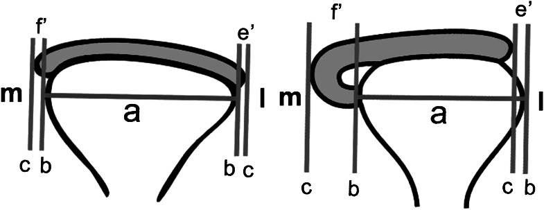
Results: The ICC was good for the transverse condylar axis (0.987) and the medial edge of the disc (0.799) and fair for the lateral edge (0.355). On average, the disc projected 5.5% to the medial side; laterally, the condyle was partially uncovered by the disc (-16.6%). In the open-jaw position, both the medial and the lateral edges shifted medially (to 17.6% vs -23.6%, Wilcoxon matched-pair test, p < 0.001). OA had no significant influence (generalized estimation equation model, p = 0.952). The disc position differed significantly from asymptomatic individuals (t-test, p < 0.001) who showed a medial disc position and full coverage of the condyle.
Conclusions: In patients with TMJ pain, the disc seems to be smaller and located less medially than in healthy volunteers. The extent of the medial shift on opening was similar.
Keywords: magnetic resonance imaging; osteoarthritis; temporomandibular joint; temporomandibular joint disc; temporomandibular joint disorders.
Figures


Similar articles
-
Analysis of magnetic resonance imaging characteristics and pain in temporomandibular joints with and without degenerative changes of the condyle.Int J Oral Maxillofac Surg. 2008 Jun;37(6):529-34. doi: 10.1016/j.ijom.2008.02.011. Epub 2008 Apr 28. Int J Oral Maxillofac Surg. 2008. PMID: 18440778
-
Temporomandibular joint internal derangement type III: relationship to magnetic resonance imaging findings of internal derangement and osteoarthrosis. An intraindividual approach.Int J Oral Maxillofac Surg. 2001 Oct;30(5):390-6. doi: 10.1054/ijom.2001.0068. Int J Oral Maxillofac Surg. 2001. PMID: 11720040
-
Relative odds of temporomandibular joint pain as a function of magnetic resonance imaging findings of internal derangement, osteoarthrosis, effusion, and bone marrow edema.Oral Surg Oral Med Oral Pathol Oral Radiol Endod. 2003 Apr;95(4):437-45. doi: 10.1067/moe.2003.95. Oral Surg Oral Med Oral Pathol Oral Radiol Endod. 2003. PMID: 12686927
-
Role of magnetic resonance imaging in the clinical diagnosis of the temporomandibular joint.Cells Tissues Organs. 2005;180(1):6-21. doi: 10.1159/000086194. Cells Tissues Organs. 2005. PMID: 16088129 Review.
-
Part II: Temporomandibular Joint (TMJ)-Regeneration, Degeneration, and Adaptation.Curr Osteoporos Rep. 2018 Aug;16(4):369-379. doi: 10.1007/s11914-018-0462-8. Curr Osteoporos Rep. 2018. PMID: 29943316 Review.
Cited by
-
Anatomical factors influencing temporomandibular joint clicking in young adults: temporomandibular joint structure disorder or lateral pterygoid muscle dysfunction?Front Bioeng Biotechnol. 2024 May 27;12:1337267. doi: 10.3389/fbioe.2024.1337267. eCollection 2024. Front Bioeng Biotechnol. 2024. PMID: 38860136 Free PMC article.
-
Selection and application of coils in temporomandibular joint MRI.Dentomaxillofac Radiol. 2020 Mar;49(3):20190002. doi: 10.1259/dmfr.20190002. Epub 2019 Sep 27. Dentomaxillofac Radiol. 2020. PMID: 31559845 Free PMC article.
-
Determining the optimal magnetic resonance imaging sequences for the efficient diagnosis of temporomandibular joint disorders.Quant Imaging Med Surg. 2021 Apr;11(4):1343-1353. doi: 10.21037/qims-20-67. Quant Imaging Med Surg. 2021. PMID: 33816173 Free PMC article.
-
Correlation between the lateral pterygoid muscle attachment type and temporomandibular joint disc position in magnetic resonance imaging.Dentomaxillofac Radiol. 2016 Oct;45(8):20160229. doi: 10.1259/dmfr.20160229. Epub 2016 Aug 30. Dentomaxillofac Radiol. 2016. PMID: 27506381 Free PMC article.
-
Assessment of articular disc displacement of temporomandibular joint with ultrasound.J Ultrasound. 2014 Oct 7;18(2):159-63. doi: 10.1007/s40477-014-0133-2. eCollection 2015 Jun. J Ultrasound. 2014. PMID: 26191103 Free PMC article.
References
-
- Larheim TA, Westesson P. TMJ imaging. In: Laskin DM, Greene CS, Hylander WL, eds. TMDs: an evidence-based approach to diagnosis and treatment. Chicago, IL: Quintessence Publishing Co, Inc; 2006
-
- Katzberg RW, Bessette RW, Tallents RH, Plewes DB, Manzione JV, Schenck JF, et al. Normal and abnormal temporomandibular joint: MR imaging with surface coil. Radiology 1986; 158: 183–189 - PubMed
-
- Tasaki MM, Westesson PL, Kurita K, Mohl N. Magnetic resonance imaging of the temporomandibular joint. Value of axial images. Oral Surg Oral Med Oral Pathol 1993; 75: 528–531 - PubMed
-
- Brooks SL, Westesson PL. Temporomandibular joint: value of coronal MR images. Radiology 1993; 188: 317–321 - PubMed
-
- Musgrave MT, Westesson PL, Tallents RH, Manzione JV, Katzberg RW. Improved magnetic resonance imaging of the temporomandibular joint by oblique scanning planes. Oral Surg Oral Med Oral Pathol 1991; 71: 525–528 - PubMed
Publication types
MeSH terms
LinkOut - more resources
Full Text Sources
Other Literature Sources
Medical
