. 2013 Apr 9;110(15):5915-20.
doi: 10.1073/pnas.1218321110. Epub 2013 Mar 15.

Atomic-level description of ubiquitin folding

Affiliations

Atomic-level description of ubiquitin folding

Stefano Piana et al. Proc Natl Acad Sci U S A. .

Abstract

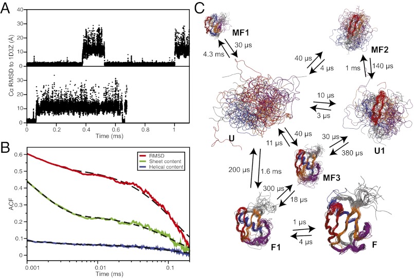
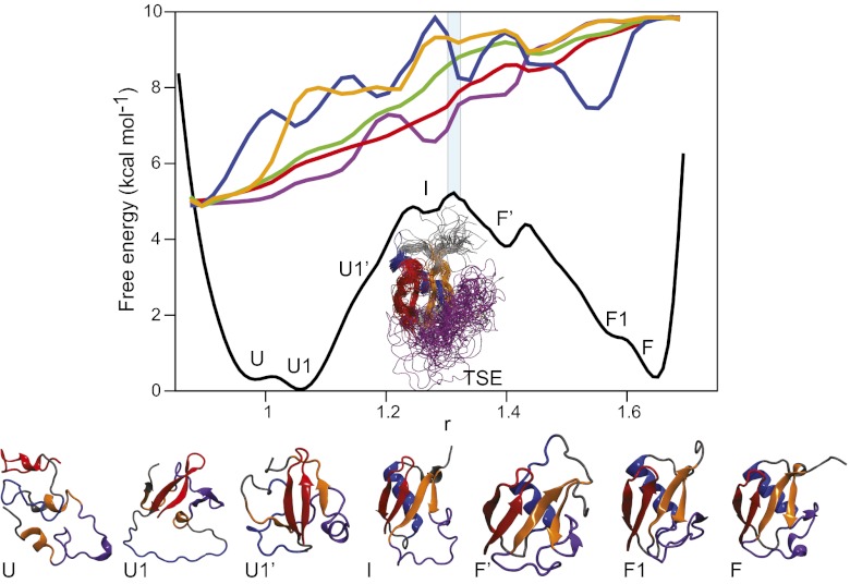
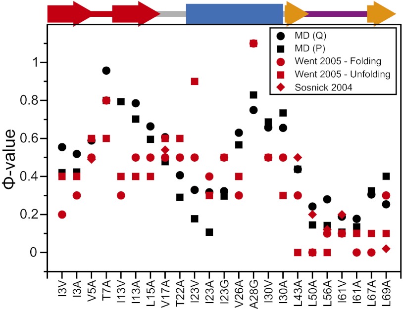
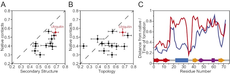
Equilibrium molecular dynamics simulations, in which proteins spontaneously and repeatedly fold and unfold, have recently been used to help elucidate the mechanistic principles that underlie the folding of fast-folding proteins. The extent to which the conclusions drawn from the analysis of such proteins, which fold on the microsecond timescale, apply to the millisecond or slower folding of naturally occurring proteins is, however, unclear. As a first attempt to address this outstanding issue, we examine here the folding of ubiquitin, a 76-residue-long protein found in all eukaryotes that is known experimentally to fold on a millisecond timescale. Ubiquitin folding has been the subject of many experimental studies, but its slow folding rate has made it difficult to observe and characterize the folding process through all-atom molecular dynamics simulations. Here we determine the mechanism, thermodynamics, and kinetics of ubiquitin folding through equilibrium atomistic simulations. The picture emerging from the simulations is in agreement with a view of ubiquitin folding suggested from previous experiments. Our findings related to the folding of ubiquitin are also consistent, for the most part, with the folding principles derived from the simulation of fast-folding proteins, suggesting that these principles may be applicable to a wider range of proteins.


Conflict of interest statement

The authors declare no conflict of interest.

Figures

Fig. 1.
Fig. 1.
Equilibrium folding simulations of ubiquitin. (A) Traces of the Cα RMSD (for residues 2–71) from the native structure for the two simulations where reversible folding is observed. (B) Autocorrelation function of the Cα RMSD (red), the number of helical residues (blue), and the number of residues in β sheets (green). The dashed black lines are biexponential fits to the autocorrelation functions with characteristic times of 1.5 µs and 120 µs. (C) Kinetic model of the folding free-energy surface. For each state, 20 random structures are displayed, superimposed using Theseus (91), and scaled according to state population. Hairpin 1 is colored in red, hairpin 2 in orange, the α helix in blue, and the C-terminal loop containing the 310 helix and the fifth β sheet in purple. The kinetic model was obtained from an eight-state fit to the time autocorrelation function of 400 random Cα–Cα contacts (71). A higher number of states results in negligible improvement on the quality of the fit. The transition times, modified to enforce detailed balance (92), are also reported here. Two unstructured unfolded-state clusters were generated by the fitting procedure and are here merged in a single state (U) to simplify the representation.
Fig. 2.
Fig. 2.
Folding free-energy surface of ubiquitin. The folding free-energy surface of ubiquitin projected along an optimal one-dimensional reaction coordinate. Representative structures for each basin on the folding free-energy surface are reported in a cartoon representation colored according to the scheme of Fig. 1. Basin U corresponds to the completely unstructured protein, while basin F corresponds to the native-state structure (average Cα RMSD from the X-ray structure, 0.8 Å). The letter codes of the macrostates determined in the kinetic clustering analysis of Fig. 1 are used to identify the corresponding basins observed in the one-dimensional projection. Some of the basins are not observed in the macrostate analysis because their population is too low (state I) or because they exchange too rapidly with other states (states F′ and U1′). The average values of a number of structural properties (blue, number of helical residues; orange, number of sheet residues; green, Cα RMSD; purple, contact order; red, fraction of native contacts) are also reported as a function of the reaction coordinate. To facilitate the comparison between different properties, all of the structural properties have been normalized so that they have the same values in the unfolded state U and in the folded state F.
Fig. 3.
Fig. 3.
Calculated and experimental Φ-values. The Φ-values for folding were calculated from the simulation using the contact approximation (Methods) and a Q-based reaction coordinate (black circles). For comparison, Φ-values were also calculated using a different P-based reaction coordinate (black squares) (71). Experimental values from Went et al. (red circles and squares) (44) and Sosnick et al. (red diamonds) (43) are reported for comparison. The native secondary structure is reported at the top of the graph (color scheme as in Fig. 2). The Φ-values for large hydrophilic or charged residues were also calculated, but they were found to deviate substantially from experiment, most likely because of a failure of the simple contact approximation, and are not reported here.
Fig. 4.
Fig. 4.
Global order of events in ubiquitin folding. (A) Relative formation order of the secondary structure and the long-range native contacts, as calculated from the integral over the transition path (19). The integrals calculated for several fast-folding proteins (19) are also reported on the same graph for comparison. (B) Same as A, but here reporting the relative formation order of native topology (93, 94) and long-range native contacts. (C) For each residue, the probability of forming local native structure in the unfolded state (red) is reported together with the order of formation of native structure along the transition path (blue) (19); Fig. S3 shows a comparable analysis of the order of events for individual folding pathways. The secondary structure of ubiquitin is reported at the bottom of the graph for comparison.

Comment in

  • A "slow" protein folds quickly in the end.
    Best RB. Best RB. Proc Natl Acad Sci U S A. 2013 Apr 9;110(15):5744-5. doi: 10.1073/pnas.1303539110. Epub 2013 Apr 1. Proc Natl Acad Sci U S A. 2013. PMID: 23572570 Free PMC article. No abstract available.

References

    1. Kubelka J, Hofrichter J, Eaton WA. The protein folding ‘speed limit’. Curr Opin Struct Biol. 2004;14(1):76–88. - PubMed
    1. Liu F, et al. An experimental survey of the transition between two-state and downhill protein folding scenarios. Proc Natl Acad Sci USA. 2008;105(7):2369–2374. - PMC - PubMed
    1. Kubelka J, Chiu TK, Davies DR, Eaton WA, Hofrichter J. Sub-microsecond protein folding. J Mol Biol. 2006;359(3):546–553. - PubMed
    1. Horng JC, Moroz V, Raleigh DP. Rapid cooperative two-state folding of a miniature alpha-beta protein and design of a thermostable variant. J Mol Biol. 2003;326(4):1261–1270. - PubMed
    1. Nauli S, Kuhlman B, Baker D. Computer-based redesign of a protein folding pathway. Nat Struct Biol. 2001;8(7):602–605. - PubMed