. 2013 Feb;8(1):56-74.
doi: 10.1016/j.nantod.2012.12.008.

Solid-State and Biological Nanopore for Real-Time Sensing of Single Chemical and Sequencing of DNA

Affiliations

Solid-State and Biological Nanopore for Real-Time Sensing of Single Chemical and Sequencing of DNA

Farzin Haque et al. Nano Today. 2013 Feb.

Abstract

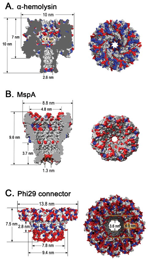
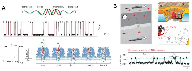
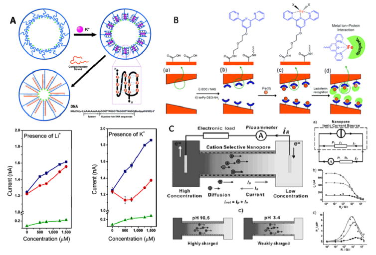
Sensitivity and specificity are two most important factors to take into account for molecule sensing, chemical detection and disease diagnosis. A perfect sensitivity is to reach the level where a single molecule can be detected. An ideal specificity is to reach the level where the substance can be detected in the presence of many contaminants. The rapidly progressing nanopore technology is approaching this threshold. A wide assortment of biomotors and cellular pores in living organisms perform diverse biological functions. The elegant design of these transportation machineries has inspired the development of single molecule detection based on modulations of the individual current blockage events. The dynamic growth of nanotechnology and nanobiotechnology has stimulated rapid advances in the study of nanopore based instrumentation over the last decade, and inspired great interest in sensing of single molecules including ions, nucleotides, enantiomers, drugs, and polymers such as PEG, RNA, DNA, and polypeptides. This sensing technology has been extended to medical diagnostics and third generation high throughput DNA sequencing. This review covers current nanopore detection platforms including both biological pores and solid state counterparts. Several biological nanopores have been studied over the years, but this review will focus on the three best characterized systems including α-hemolysin and MspA, both containing a smaller channel for the detection of single-strand DNA, as well as bacteriophage phi29 DNA packaging motor connector that contains a larger channel for the passing of double stranded DNA. The advantage and disadvantage of each system are compared; their current and potential applications in nanomedicine, biotechnology, and nanotechnology are discussed.

Keywords: DNA packaging; MspA; bacteriophage phi29; bionanotechnology; connector; ion channel; liposomes; membrane channel; nanobiotechnology; nanomedicine; nanomotor; nanostructure; single channel conductance; solid state pore; stoichiometry quantification; synthetic nanopores; viral assembly; α-hemolysin.


Figures

Figure 1
Figure 1. Structure of three biological nanopores
Side and top views of (A) heptameric α-hemolysin toxin from Staphylococcus aureus (PDB ID: 3ANZ)[7]; (B) octameric MspA porin from Mycobacterium smegmatis[18,20]; (C) dodecameric connector channel from bacteriophage phi29 DNA packaging motor[33]. In the figures, acidic (red), basic (blue), and other (white) amino acids are shown. Figures reproduced with permissions from: (B) Ref.[20], © The National Academy of Sciences of the USA; (C) Ref. [33], © Nature Publishing Group.
Figure 2
Figure 2. Application of phi29 connector channel for translocation of dsDNA and sensing of single chemicals
(A) DsDNA translocation through membrane-embedded phi29 connector induced numerous current blockades. Insert: magnified image of a single translocation event. (B) Histogram of current blockade percentage induced by linear 2 kbp dsDNA. (C) Comparison of dwell times for translocation of 38 bp and 5.5 kbp dsDNA. One-way traffic in DNA translocation through a single connector channel in a lipid bilayer, examined under a ramping potential (D–E); and switching polarity (F). DNA is present in both cis- and trans-chambers. (G) Illustration of conjugation of chemical ligands to channel wall resulted in the reduction of channel size as indicated by uniform stepwise blockage of channel current. (H) Analysis of current blockage events induced by thioesters. Histogram of permanent binding events for the binding of thioesters groups containing ethane, thymine, and benzene respectively. Figures reproduced with permissions from: (A) Ref. [33], © Nature Publishing Group; (B) Ref. [35], © The Royal Society of Chemistry; (C) Ref. [33], © Nature Publishing Group; (D–F) Ref. [34], © American Chemical Society; (G–H) Ref. [38], © American Chemical Society.
Figure 3
Figure 3. DsDNA translocation through graphene nanopores
Illustration (left) and data (right) from (A) Dekker lab[41]; (B) Golovchenko lab[59]; (C) Drndic lab[58]; and (D) Bashir lab[60]. Figures reproduced with permissions from: (A) Refs.[41], © American Chemical Society, Refs.[153], © Nature Publishing Group; (B) Refs.[59], © Nature Publishing Group; (C) Refs.[58], © American Chemical Society; (D) Refs.[60], © American Chemical Society.
Figure 4
Figure 4. DNA translocation through hybrid nanopores
(A) α-hemolysin heteroheptamer with a 3 kbp dsDNA attached via a 12-nucleotide oligomer to one protein subunit. (B) Insertion of α-hemolysin protein pore into the solid state nanopore in three phases. (C) Current trace through a hybrid nanopore showing the baseline conductance directly after insertion (left) and upon addition of poly(dA)100 (middle). On the right is an expanded view of a typical event. (D) Schematic representation of the insertion of a DNA origami nanopore into a solid-state nanopore. (E) Typical events for the bare nanopore (blue) and the hybrid nanopore (red). (F) Current histograms indicating DNA translocations for the bare (blue) and hybrid nanopore (red). Figures reproduced with permissions from: (A–C) Ref. [66], © Nature Publishing Group; (D–F) Ref.[68], © American Chemical Society.
Figure 5
Figure 5. Proposed exonuclease-sequencing with a chimera of α-hemolysin and exonucleoase
(A) An exonuclease (pale blue) attached to the top of an α-hemolysin pore through a genetically encoded (deep blue), or chemical, linker sequentially cleaves dNMPs (gold) off the end of a DNA strand. A dNMP’s identity (A, T, G or C) is determined by the level of the current blockade it causes when driven into an aminocyclodextrin adaptor (red) lodged within the pore. (B) Single-channel recording from the WT-(M113R/N139Q)6(M113R/N139Q/L135C)1-am6amDP1bCD pore upon addition of mono-nucleotides showing dGMP, dTMP, dAMP and dCMP discrimination, with colored bands added to represent the residual current distribution for each nucleotide. (C) Corresponding residual current histogram of nucleotide binding events, including Gaussian fits. Figures reproduced with permissions from (A) Ref. [1], © Nature Publishing Group; (B-C) Refs. [96], © Nature Publishing Group.
Figure 6
Figure 6
Strategies for reading DNA at single-nucleotide resolution using phi29 DNA polymerase to ratchet template DNA across (A) MspA nanopore and (B) α-hemolysin pore. Figures reproduced with permissions from: Ref. [23], © Nature Publishing Group; (B) Ref. [97], © Nature Publishing Group.
Figure 7
Figure 7. Nanowire–nanopore field-effect transistors (FET) sensor
(A) Schematic of the sensor setup. Insert: magnified view at the nanopore. (B) Schematic illustration of the sensing circuit. (C) The real-time change of ionic current and FET conductance signals at 2.4 V voltage. Figures reproduced with permissions from Ref. [110], © Nature Publishing Group.
Figure 8
Figure 8. Electron tunneling approach for single base detection
(A) Electron tunneling approach using a benzamide recognition group to identify single bases within a short DNA oligomer d(CCACC). A characteristic signal given by the shortest tunneling path is highlighted for the connection to the single A in d(CCACC). (B) Schematic of concurrent tunneling detection and ionic current detection of DNA molecules in a SiN nanopore platform. Figures reproduced with permissions from: (A) Ref [111], © Nature Publishing Group; (B) Ref. [112], © American Chemical Society.
Figure 9
Figure 9. Medical diagnostics and sensing applications
(A) Data showing α-hemolysin pore based detection of miRNA (miR-155) using programmable oligonucleotide probe (P155). The hybridization of probe with miRNA produced characteristic multi-level current signals upon translocating through α-hemolysin nanopores. (B) Gold-coated SiN nanopore functionalized with a mixed SAM of SC15EG3 matrix thiols and NTA receptor thiols for stochastic sensing of proteins. Charged analyte molecules (red) are electrokinetically driven through the pore and are detected by transient blockades in the ion current. When the pore is equipped with a specific receptor site (green), the blockade time reflects the binding time of the analyte (ligand) to the receptor. Figures reproduced with permissions from: (A) Ref [135], © Nature Publishing Group; (B) Ref. [76], © Nature Publishing Group.
Figure 10
Figure 10. Examples of biomimetic application of solid-state nanopores
(A) Top: Schematic representation of G4 DNA undergoing conformational change in the presence of K+, and after adding the complementary DNA, a closely packed double-strand DNA was formed, all processes are shown within the nanopore; Bottom: Current-concentration (I-C) characters of the single-conical PET nanopore before and after the G4 DNA molecule modified onto the nanopore inner space. (B) Schematic representation of nanopore-based lactoferrin sensor via the biorecognition of metal-chelating ligand. (C) Left: concentration gradient caused ion diffusion across the cation-selective nanopore; Right: supplying the electrical power produced by the nanopore system to an electric resistance RL, three lines represent different concentration gradient respectively. Figures reproduced with permissions from: (A) Ref. [148], © American Chemical Society; (B) Ref. [149], © American Chemical Society; (C) Ref. [150] © WILEY-VCH Verlag GmbH & Co.KGaA.

References

    1. Branton D, Deamer DW, Marziali A, Bayley H, Benner SA, Butler T, et al. Nat Biotechnol. 2008;26:1146. - PMC - PubMed
    1. Venkatesan BM, Bashir R. Nature Nanotechnology. 2011;6:615. - PubMed
    1. Healy K. Nanomedicine. 2007;2:459. - PubMed
    1. Majd S, Yusko EC, Billeh YN, Macrae MX, Yang J, Mayer M. Current Opinion in Biotechnology. 2010;21:439. - PMC - PubMed
    1. Iqbal SM, Bashir R. Nanopores: Sensing and Fundamental Biological Interactions. Springer; London: 2011.