

Protective effect of alpha-melanocyte-stimulating hormone (α-MSH) on the recovery of ischemia/reperfusion (I/R)-induced retinal damage in a rat model
- PMID: 23504281
- PMCID: PMC3675276
- DOI: 10.1007/s12031-013-9998-3
Protective effect of alpha-melanocyte-stimulating hormone (α-MSH) on the recovery of ischemia/reperfusion (I/R)-induced retinal damage in a rat model
Abstract
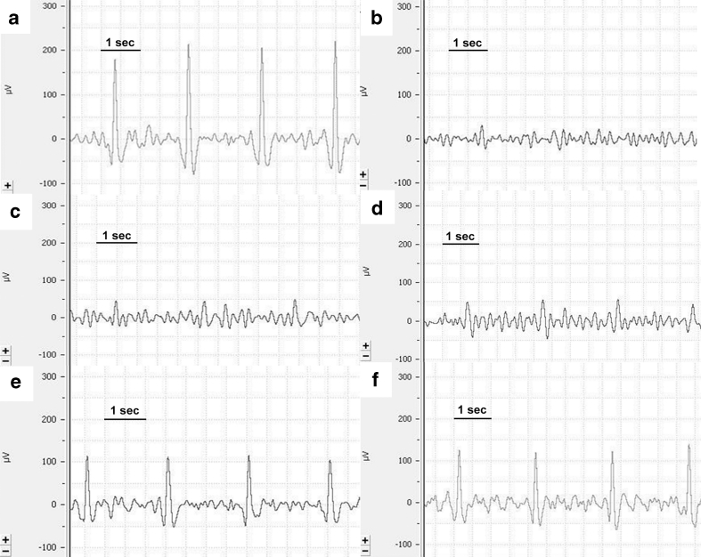
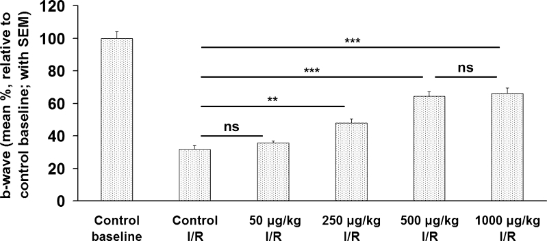
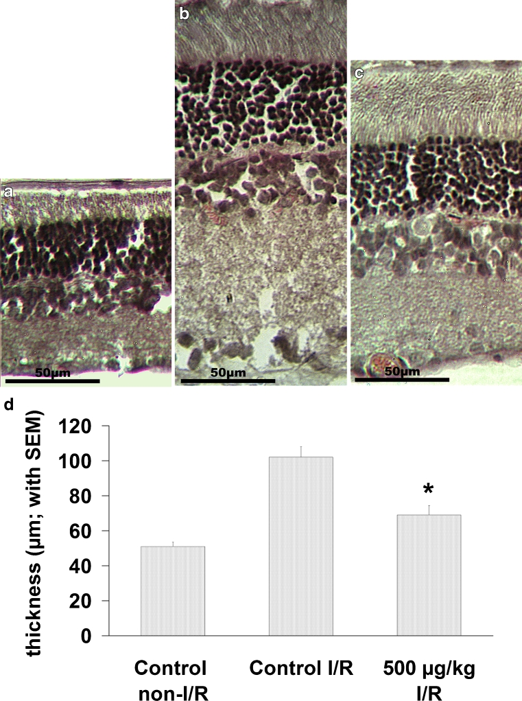
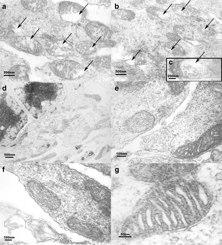
The present study demonstrates capacity of α-MSH to augment recovery from ischemia/reperfusion (I/R)-induced retinal damage in vivo and correlation of its protective effects with expression of heme oxygenase-1 (HO-1). Used techniques include ocular ischemia and reperfusion, electroretinography, histology, electron microscopy, and molecular-biological techniques. The results demonstrate the α-MSH-mediated inhibition of I/R-induced functional deterioration of the retina. Outcomes suggest that the protective effects of α-MSH occur mainly through HO-1-dependent pathways but HO-1-independent mechanisms may also contribute to protection. The observation that post-ischemic treatment with α-MSH exhibits therapeutic efficacy in the same range as pre-ischemic treatment, is a novel result. This outcome suggests a highly conserved protective role for α-MSH as a major stress response mechanism--and offers the possibility for development of novel therapeutic strategies utilizing this hormone, in particular in treatment of conditions resulting from I/R injury, such as deterioration of retinal microcirculation. The merit of the study lies in the fact that I/R injury contribute significantly to the severity of retinopathies. However, currently there are no evidence-based treatments for retinal I/R injury available for clinical use. Our finding suggests that α-MSH may have a very wide range of uses in the prevention of I/R-mediated pathologies.
Figures
Similar articles
-
Protective Effect of Prunus Cerasus (Sour Cherry) Seed Extract on the Recovery of Ischemia/Reperfusion-Induced Retinal Damage in Zucker Diabetic Fatty Rat.Molecules. 2017 Oct 21;22(10):1782. doi: 10.3390/molecules22101782. Molecules. 2017. PMID: 29065463 Free PMC article.
-
Delayed administration of alpha-melanocyte-stimulating hormone or combined therapy with BAY 11-7085 protects against gut ischemia-reperfusion injury.Shock. 2003 Nov;20(5):469-75. doi: 10.1097/01.shk.0000091205.08003.fd. Shock. 2003. PMID: 14560113
-
Α-Melanocyte-Stimulating Hormone Protects Early Diabetic Retina from Blood-Retinal Barrier Breakdown and Vascular Leakage via MC4R.Cell Physiol Biochem. 2018;45(2):505-522. doi: 10.1159/000487029. Epub 2018 Jan 25. Cell Physiol Biochem. 2018. PMID: 29402864
-
alpha-Melanocyte-stimulating hormone and acute renal failure.Curr Opin Nephrol Hypertens. 1998 Jul;7(4):413-7. doi: 10.1097/00041552-199807000-00011. Curr Opin Nephrol Hypertens. 1998. PMID: 9690041 Review.
-
Therapeutic Potential of Heme Oxygenase-1/carbon Monoxide System Against Ischemia-Reperfusion Injury.Curr Pharm Des. 2017;23(26):3884-3898. doi: 10.2174/1381612823666170413122439. Curr Pharm Des. 2017. PMID: 28412905 Review.
Cited by
-
Improved Survival and Retinal Function of Aging ZDF Rats in Long-Term, Uncontrolled Diabetes by BGP-15 Treatment.Front Pharmacol. 2021 Apr 16;12:650207. doi: 10.3389/fphar.2021.650207. eCollection 2021. Front Pharmacol. 2021. PMID: 33935754 Free PMC article.
-
Endothelin-1-induced hypertrophic alterations and heme oxygenase-1 expression in cardiomyoblasts are counteracted by beta estradiol: in vitro and in vivo studies.Naunyn Schmiedebergs Arch Pharmacol. 2018 Apr;391(4):371-383. doi: 10.1007/s00210-018-1462-z. Epub 2018 Jan 21. Naunyn Schmiedebergs Arch Pharmacol. 2018. PMID: 29354880 Free PMC article.
-
Alpha-Melanocyte-stimulating Hormone Induces Vasodilation and Exerts Cardioprotection Through the Heme-Oxygenase Pathway in Rat Hearts.J Cardiovasc Pharmacol. 2017 May;69(5):286-297. doi: 10.1097/FJC.0000000000000472. J Cardiovasc Pharmacol. 2017. PMID: 28195947 Free PMC article.
-
Alpha-Melanocyte Stimulating Hormone Protects against Cytokine-Induced Barrier Damage in Caco-2 Intestinal Epithelial Monolayers.PLoS One. 2017 Jan 19;12(1):e0170537. doi: 10.1371/journal.pone.0170537. eCollection 2017. PLoS One. 2017. PMID: 28103316 Free PMC article.
-
Activation of the Nrf2/HO-1 antioxidant pathway contributes to the protective effects of Lycium barbarum polysaccharides in the rodent retina after ischemia-reperfusion-induced damage.PLoS One. 2014 Jan 6;9(1):e84800. doi: 10.1371/journal.pone.0084800. eCollection 2014. PLoS One. 2014. PMID: 24400114 Free PMC article.
References
-
- Bazzani C, Guarini S, Botticelli AR, et al. Protective effect of melanocortin peptides in rat myocardial ischemia. J Pharmacol Exp Ther. 2001;297:1082–1087. - PubMed
Publication types
MeSH terms
Substances
LinkOut - more resources
Full Text Sources
Other Literature Sources
