. 2013 May 3;288(18):12866-79.
doi: 10.1074/jbc.M112.413393. Epub 2013 Mar 15.

Role of RNF4 in the ubiquitination of Rta of Epstein-Barr virus

Affiliations

Role of RNF4 in the ubiquitination of Rta of Epstein-Barr virus

Ya-Chun Yang et al. J Biol Chem. .

Abstract

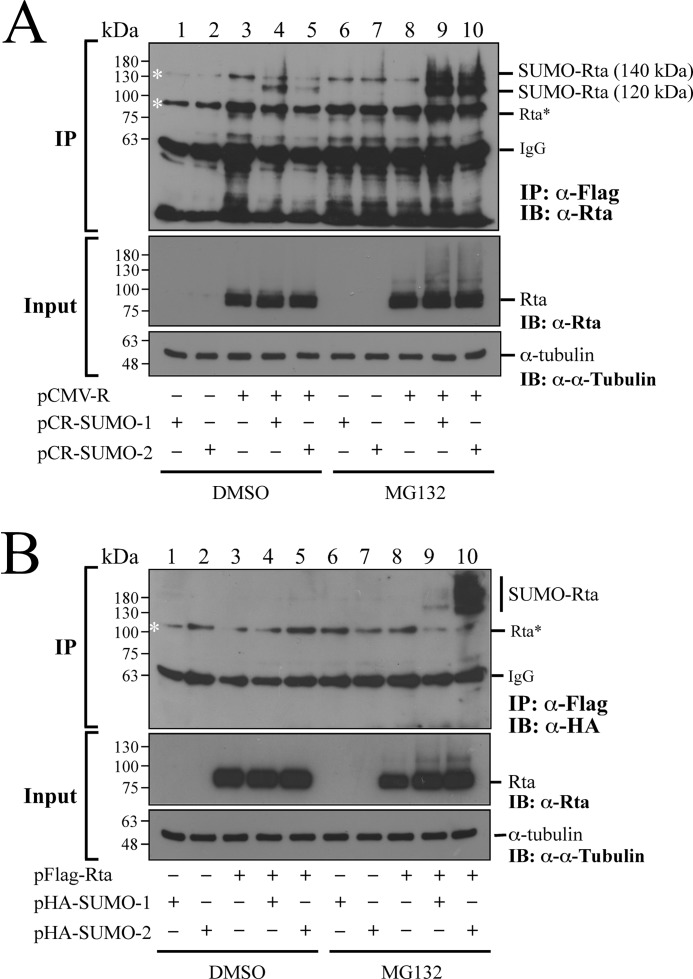
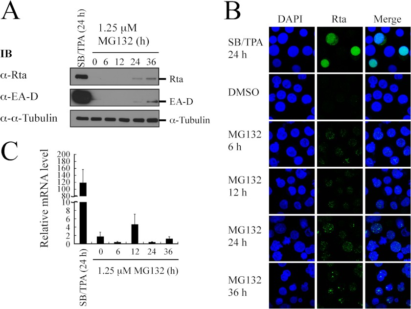
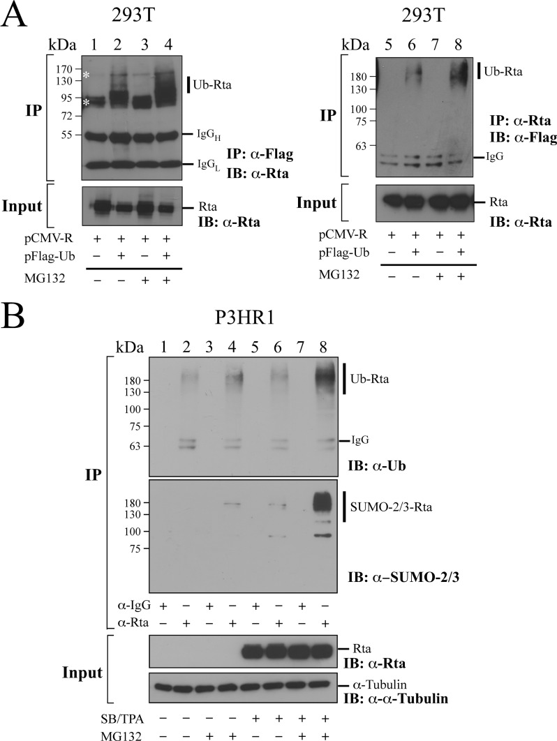
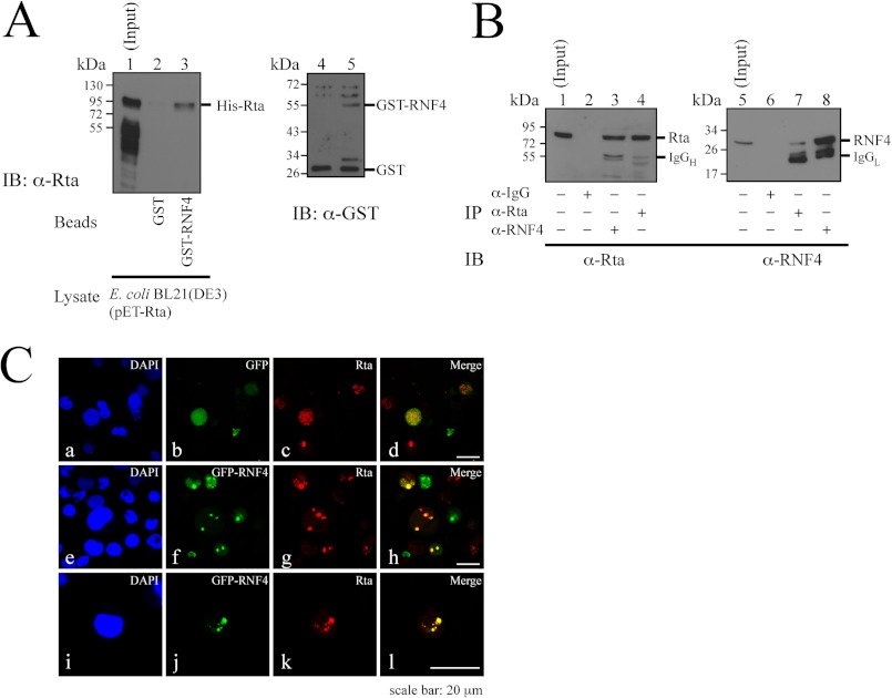
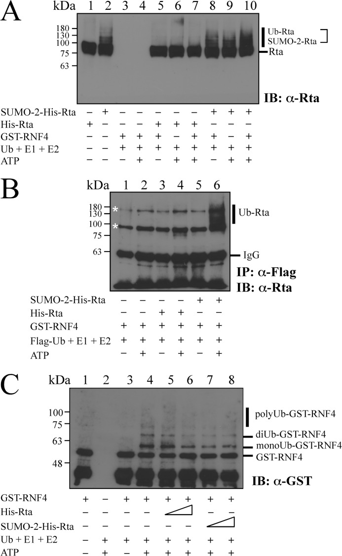
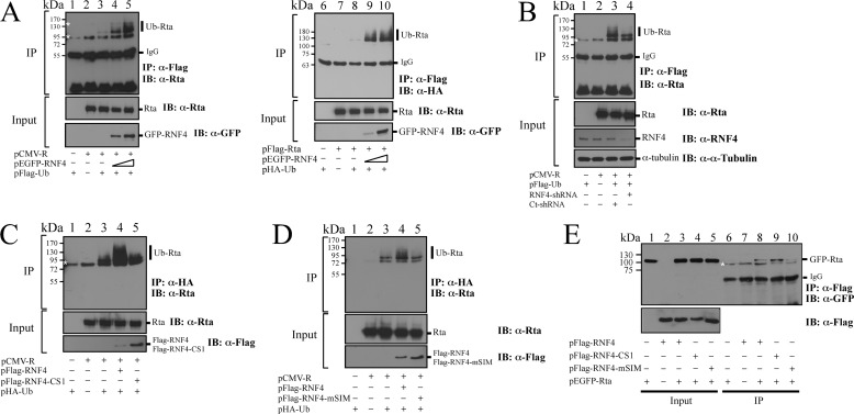
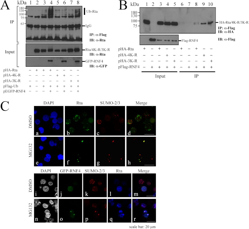
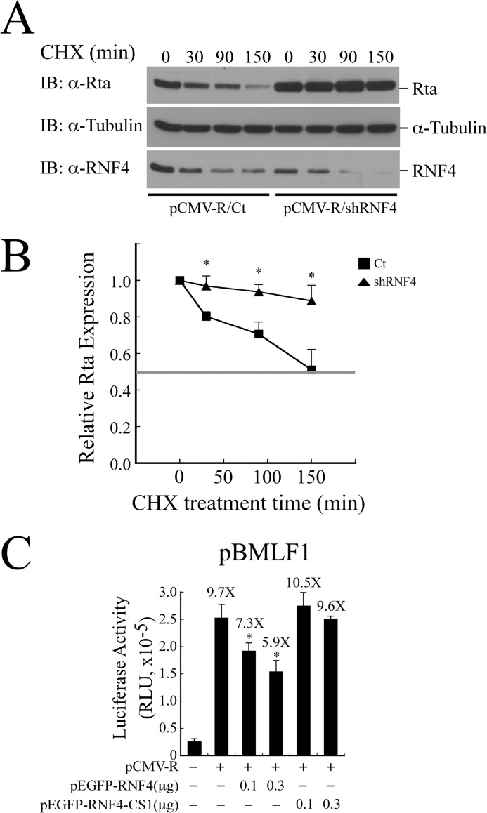
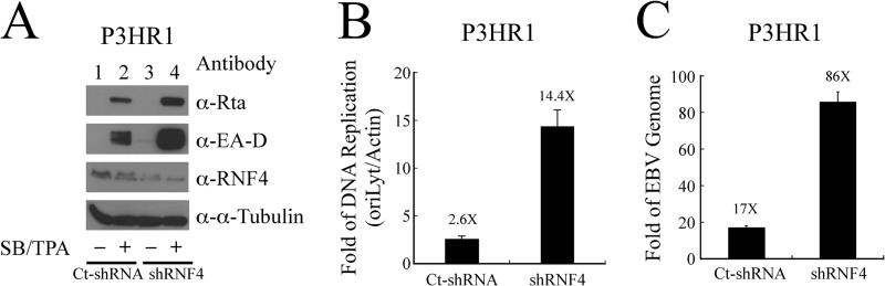
Epstein-Barr virus (EBV) encodes a transcription factor, Rta, which is required to activate the transcription of EBV lytic genes. This study demonstrates that treating P3HR1 cells with a proteasome inhibitor, MG132, causes the accumulation of SUMO-Rta and promotes the expression of EA-D. GST pulldown and coimmunoprecipitation studies reveal that RNF4, a RING-domain-containing ubiquitin E3 ligase, interacts with Rta. RNF4 also targets SUMO-2-conjugated Rta and promotes its ubiquitination in vitro. Additionally, SUMO interaction motifs in RNF4 are important to the ubiquitination of Rta because the RNF4 mutant with a mutation at the motifs eliminates ubiquitination. The mutation of four lysine residues on Rta that abrogated SUMO-3 conjugation to Rta also decreases the enhancement of the ubiquitination of Rta by RNF4. This finding demonstrates that RNF4 is a SUMO-targeted ubiquitin E3 ligase of Rta. Finally, knockdown of RNF4 enhances the expression of Rta and EA-D, subsequently promoting EBV lytic replication and virions production. Results of this study significantly contribute to efforts to elucidate a SUMO-targeted ubiquitin E3 ligase that regulates Rta ubiquitination to influence the lytic development of EBV.

Keywords: DNA Viruses; E3 Ubiquitin Ligase; Epstein-Barr Virus; Protein Degradation; RNF4; Rta; Sumoylation; Ubiquitination.


Figures

FIGURE 1.
FIGURE 1.
Accumulation of SUMO-modified Rta after MG132 treatment. A, 293T cells were transfected with pCR-SUMO-1 (lanes 1 and 6), pCR-SUMO-2 (lanes 2 and 7), or pCMV-R (lanes 3 and 8) or cotransfected with pCMV-R and pCR-SUMO-1 (lanes 4 and 9) or pCR-SUMO-2 (lanes 5 and 10) in the presence of 5 μm MG132 (lanes 6–10) or DMSO (lanes 1–5). At 24 h after transfection, the cells were washed with PBS containing 10 mm N-ethylmaleimide. B, 293T cells were transfected with pHA-SUMO-1 (lanes 1 and 6), pHA-SUMO-2 (lanes 2 and 7), pFLAG-Rta (lanes 3 and 8) or cotransfected with pFLAG-Rta and pHA-SUMO-1 (lanes 4 and 9) or pFLAG-Rta and pHA-SUMO-2 (lanes 5 and 10) and then treated with MG132 (lanes 6–10) or DMSO (lanes 1–5). In A, SUMO-Rta was immunoprecipitated (IP) using anti-FLAG antibody and detected by immunoblotting (IB) using anti-Rta antibody. In B, SUMO-Rta was immunoprecipitated using anti-FLAG antibody and detected using anti-HA antibody. Asterisks indicate bands detected nonspecifically. Rta* indicates where the Rta band is supposed to locate.
FIGURE 2.
FIGURE 2.
Expression of EA-D after MG132 treatment. A, P3HR1 cells were treated with 1.25 μm MG132 for 36 h. Cells treated with sodium butyrate (SB) and TPA (SB/TPA) for 24 h were used as a positive control. Proteins in the lysate were detected by immunoblotting (IB) using anti-Rta, anti-EA-D, and anti-α-tubulin antibodies. B, indirect immunofluorescence analysis was performed using P3HR1 cells that were treated with MG132 for 36 h. Cells treated with SB/TPA or DMSO for 24 h were used as a control. Cells were incubated with anti-Rta monoclonal antibody. DAPI staining revealed the nucleus. Cells were observed under a confocal laser-scanning microscope. C, total RNA from P3HR1 cells that had been treated with MG132 was analyzed by qRT-PCR. The level of BRLF1-BZLF1 expression was normalized relative to those of actin. The data shown are the averages and S.D. of the results from two independent experiments, and each sample in the experiment was prepared in duplicate.
FIGURE 3.
FIGURE 3.
Ubiquitination of Rta in vivo. A, 293T cells were transfected with pCMV-R (lanes 1, 3, 5, and 7) or cotransfected with pCMV-R and pFLAG-Ub (lanes 2, 4, 6, and 8). Cells were treated (lanes 3, 4, 7, and 8) or untreated (lanes 1, 2, 5, and 6) with MG132 for 12 h after transfection for 24 h. Proteins in the cell lysate was immunoprecipitated (IP) using anti-FLAG antibody (lanes 1–4) or anti-Rta antibody (lanes 5–8) after denaturing the proteins in the lysates at 95 °C. The proteins were then detected by immunoblotting (IB) using anti-Rta antibody (lanes 1–4) or anti-FLAG antibody (lanes 5–8). B, P3HR1 cells were treated (lanes 5, 6, 7, and 8) or untreated (lanes 1, 2, 3, and 4) with sodium butyrate and TPA. After incubation for 24 h, cells were treated with MG132 (lanes 3, 4, 7, and 8) or DMSO (lanes 1, 2, 5, and 6) for 12 h. Proteins in cell lysates were immunoprecipitated using anti-Rta antibody and detected by immunoblot analysis with anti-SUMO-2/3 antibody, anti-ubiquitin (Ub), and anti-Rta antibody. Anti-IgG (lanes 1, 3, 5, and 7) was used in IP as a negative control. Asterisks indicate nonspecific bands. Ub-Rta, ubiquitinated Rta; IgGH, the heavy chain of IgG; IgGL, the light chain of IgG.
FIGURE 4.
FIGURE 4.
Interaction between Rta and RNF4. A, bacterially expressed GST and GST-RNF4 were bound to glutathione-Sepharose beads. The beads were then mixed with a lysate from E. coli BL21(DE3)(pET-Rta). Proteins bound to GST-glutathione-Sepharose (lane 2) and GST-RNF4-glutathione-Sepharose (lane 3) beads were analyzed by immunoblotting (IB) using anti-Rta antibody. Lane 1 was loaded with 1% of the cell lysate. GST proteins that were bound to the beads were eluted and analyzed by immunoblot analysis using anti-GST antibody (lanes 4 and 5). B, P3HR1 cells were treated with TPA and sodium butyrate. Proteins in the lysate were subsequently immunoprecipitated (IP) using anti-IgG (lanes 2 and 6), anti-Rta (lanes 4 and 7), or anti-RNF4 (lanes 3 and 8) antibodies. Immunoblotting was performed using anti-Rta (lanes 1–4) and anti-RNF4 (lanes 5–8) antibodies. Input lanes were loaded with 1% of the lysates. IgGH, heavy chain of IgG; IgGL, light chain of IgG. C, P3HR1 cells were transfected with pEGFP-C1 (a–d) or pEGFP-RNF4 (e–l) and then treated with 5 μm MG132, sodium butyrate, and TPA for 24 h. Cells were incubated with anti-Rta monoclonal antibody. DAPI staining revealed the nucleus. Cells were observed under a confocal laser-scanning microscope. d, h, and l are merged images.
FIGURE 5.
FIGURE 5.
Enhancement of Rta ubiquitination by RNF4 in vitro. A, conjugation of ubiquitin to Rta was analyzed in vitro using purified Rta, an Rta mixture that contains SUMO-2-conjugated Rta, and GST-RNF4 in the presence of ubiquitin (Ub), ubiquitin E1-activating enzyme, and ubiquitin E2 enzyme (UbcH5a). Ubiquitinated proteins were examined by immunoblotting (IB) using anti-Rta antibody. B, similar in vitro Ub assay was conducted using FLAG-ubiquitin (FLAG-Ub), purified Rta, SUMO-2-conjugated Rta, E1, E2, and GST-RNF4. Thereafter, Ub-Rta was immunoprecipitated (IP) using anti-FLAG antibody and then detected by immunoblotting using anti-Rta antibody. C, additionally, the ubiquitination of RNF4 was examined in vitro with purified GST-RNF4, Rta, or SUMO-2-conjugated Rta in the reaction mixture containing ubiquitin (Ub), ubiquitin E1-activating enzyme, UbcH5a, or ATP. Ubiquitinated proteins including monoubiquitinated, diubiquitinated, and polyubiquitinated GST-RNF4 were detected by immunoblot analysis using anti-GST antibody. Asterisks indicate bands detected nonspecifically.
FIGURE 6.
FIGURE 6.
RNF4 and the ubiquitination of Rta. A, 293T cells were cotransfected with pCMV-R, pEGFP-RNF4, and pFLAG-Ub (lanes 1–5). At 24 h after transfection cells were treated with 5 μm MG132 for another 12 h. Proteins in the lysate were immunoprecipitated (IP) using anti-FLAG antibody. Proteins bound to FLAG-M2 beads were analyzed by immunoblot analysis (IB) using anti-Rta and anti-GFP antibodies. Similarly, 293T cells were cotransfected with plasmids that express FLAG-Rta, GFP-RNF4, and FLAG-Ub (lanes 6–10). At 24 h after transfection, cells were treated with 5 μm MG132 for another 12 h. Proteins in the lysate were immunoprecipitated using anti-FLAG antibody. Proteins bound to FLAG-M2 beads were analyzed by immunoblot analysis using anti-HA, anti-Rta, and anti-GFP antibodies. B, shown is the effect of knockdown of RNF4 on the ubiquitination of Rta. 293T cells were cotransfected with plasmids that express FLAG-Ub, Rta, and RNF4 shRNA or control shRNA (Ct-shRNA) and then treated with 5 μm MG132 for 12 h. Ubiquitinated Rta was immunoprecipitated using anti-FLAG antibody and detected by immunoblotting using anti-Rta antibody. C, 293T cells were cotransfected with pHA-Ub, pCMV-R, and pFLAG-RNF4 or pFLAG-RNF4-CS1. At 24 h after transfection, cells were treated with 5 μm MG132 for another 12 h. Proteins in the cell lysate were immunoprecipitated using anti-HA antibody. Immunoprecipitated proteins were detected by immunoblotting with anti-Rta antibody. D, a similar experiment as described in C was conducted except that pFLAG-RNF4-CS1 was replaced with pFLAG-RNF4-mSIM. E, interaction of Rta with RNF4 mutants is shown. 293T cells were cotransfected with pEGFP-Rta and plasmids that express FLAG-RNF4, FLAG-RNF4-CS1, or FLAG-RNF4-mSIM. A coimmunoprecipitation assay was performed using anti-FLAG antibody, and immunoprecipitated proteins were detected by immunoblotting with anti-GFP antibody (lanes 6–10). Asterisks indicate bands detected nonspecifically. Ub-Rta, ubiquitinated Rta.
FIGURE 7.
FIGURE 7.
SUMO-dependent ubiquitination of Rta by RNF4. A, 293T cells were cotransfected with pFLAG-Ub, pEGFP-RNF4, and pHA-Rta (lanes 2–4), pHA-4K-R (lanes 5 and 6), or pHA-3K-R (lanes 7 and 8). At 24 h after transfection, cells were treated with 5 μm MG132 for another 12 h. Proteins in the lysate were immunoprecipitated (IP) using anti-FLAG antibody. Proteins were then detected by immunoblotting (IB) using anti-Rta and anti-GFP antibodies. B, interaction of RNF4 with Rta is shown. 293T cells were cotransfected with pFLAG-RNF4 and plasmids that express HA-Rta, HA-3K-R, or HA-4K-R. A coimmunoprecipitation assay was performed using anti-FLAG antibody, and immunoprecipitated proteins were detected by immunoblotting using anti-HA antibody. C, P3HR1 cells were transfected with plasmids pEGFP-RNF4 (a–r), and then cells were treated with 5 μm MG132 or DMSO for 12 h after lytic induction by TPA and sodium butyrate. Cells were incubated with anti-Rta monoclonal antibody and anti-SUMO-2 polyclonal antibody. DAPI staining revealed the nucleus. Thereafter, cells were observed under a confocal laser-scanning microscope. d, h, m, and r are merged images.
FIGURE 8.
FIGURE 8.
Effect of RNF4 on the stability and transactivation activity of Rta. A, 293T cells were transfected with plasmids pCMV-R and control shRNA (Ct-shRNA) or RNF4 shRNA. Cycloheximide (CHX) was added at 40 h after transfection to inhibit protein synthesis. Whole cell lysate was prepared at 0, 30, 90, and 150 min after transfection. Proteins in the lysate were detected by immunoblotting (IB) using anti-Rta, anti-α-tubulin, and anti-RNF4 antibodies. B, a densitometric analysis of Rta level normalized to α-tubulin was plotted using Image J software. Data are presented as the mean with S.D. and represent three independent experiments. The intensity corresponding to 50% of the initial value is indicated by the horizontal line. C, 293T cells were cotransfected with the reporter plasmid pBMLF1 and pCMV-R, pEGFP-RNF4, or pEGFP-RNF4-CS1. Luciferase activities were monitored 24 h post-transfection. Each transfection experiment was performed three times, and each sample in the experiment was prepared in duplicate. The value from each experiment was analyzed statistically with the least square means method. *, p < 0.05.
FIGURE 9.
FIGURE 9.
Role of RNF4 on EBV lytic development. A, P3HR1 cells were infected by the lentivirus that contains RNF4 shRNA (shRNF4) or control shRNA (Ct-shRNA) under the selection of puromycin. Thereafter, cells were treated with sodium butyrate and TPA (SB/TPA) for 48 h, and proteins in the lysate were examined by immunoblotting using anti-Rta, anti-EA-D, anti-RNF4, and anti-α-tubulin antibodies. B, moreover, cells that were harvested from A were lysed, and EBV lytic DNA replication assay was assayed by qPCR. The amount of EBV DNA was normalized with the amount of actin DNA that was determined in the same assay. C, the lentiviral-transduced P3HR1 cells were treated with TPA and sodium butyrate for 5 days. EBV DNA from viral particles that were released into the culture medium was determined by qPCR after the DNA extraction. The copy number of EBV genome was calculated by using maxi-EBV that had been isolated from E. coli as a standard.

Similar articles

Cited by

References

    1. Pickart C. M., Fushman D. (2004) Polyubiquitin chains. Polymeric protein signals. Curr. Opin. Chem. Biol. 8, 610–616 - PubMed
    1. Grabbe C., Husnjak K., Dikic I. (2011) The spatial and temporal organization of ubiquitin networks. Nat. Rev. Mol. Cell Biol. 12, 295–307 - PMC - PubMed
    1. Chau V., Tobias J. W., Bachmair A., Marriott D., Ecker D. J., Gonda D. K., Varshavsky A. (1989) A multiubiquitin chain is confined to specific lysine in a targeted short-lived protein. Science 243, 1576–1583 - PubMed
    1. Thrower J. S., Hoffman L., Rechsteiner M., Pickart C. M. (2000) Recognition of the polyubiquitin proteolytic signal. EMBO J. 19, 94–102 - PMC - PubMed
    1. Komander D. (2009) The emerging complexity of protein ubiquitination. Biochem. Soc. Trans. 37, 937–953 - PubMed

Publication types

MeSH terms

LinkOut - more resources